XI
Radiol Bras. 2010 Jan/Fev;43(1):XI–XII
Ceci Obara Kurimori1
, Marcelo Bordalo-Rodrigues2
, Giovanni Guido Cerri3
Study developed at Instituto de Ortopedia e Traumatologia (IOT) and Instituto de Radiologia (InRad) – Hospital das Clínicas da Faculdade de Medicina da Uni-versidade de São Paulo (HC-FMUSP), São Paulo, SP, Brazil. 1. MD, Assistant Physician at Instituto de Ortopedia e Traumatologia – Hospital das Clínicas da Faculdade de Medicina da Universidade de São Paulo (IOT/HC-FMUSP), São Paulo, SP, Brazil. 2. MD, Director, Instituto de Ortopedia e Traumatologia – Hospital das Clínicas da Faculdade de Medicina da Universidade de São Paulo (IOT/HC-FMUSP), São Paulo, SP, Brazil. 3. Titular Professor, Division of Radiology, Head of Group of Radiology, Instituto de Radiologia – Hospital das Clínicas da Faculdade de Medicina da Universidade de São Paulo (InRad/HC-FMUSP), São Paulo, SP, Brazil. Mailing address: Dr. Marcelo Bordalo Rodrigues. Avenida Doutor Eneas de Carvalho Aguiar, 255, Pinheiros. São Paulo, SP, Brazil, 05403-001. E-mail: [email protected]
0100-3984 © Colégio Brasileiro de Radiologia e Diagnóstico por Imagem
Which is your diagnosis?
•
Qual o seu diagnóstico?
Kurimori CO, Bordalo-Rodrigues M, Cerri GG. Which is your diagnosis? Radiol Bras. 2010;43(1):XI–XII.
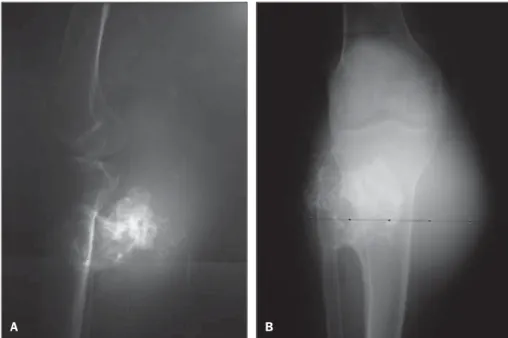
Male, 77-year-old patient. Incidental finding at radiography of left lower limb, with a progressive volumetric increase of the posterior face of the leg, after a fall from a roof two years ago.
Figure 1. Radiography – lateral (A) and anteroposterior (B) views.
A B
Figure 3. Magnetic resonance imaging – sagittal, T1-weighted (A) and T2-weighted (B) sequences, and axial, T2-weighted sequence without intravenous gadolinium injection (C).
A B C
Figure 2. Computed tomography with axial (A) and sagittal (B) reconstructions.
A
XII Radiol Bras. 2010 Jan/Fev;43(1):XI–XII
Images description
Figure 1. Radiography – lateral (A) and anteroposterior (B) views demonstrating exostosis with irregular margins in associa-tion with soft tissues growth intermingled with chondroid calcifications in the poste-rior aspect of the leg.
Figure 2. Computed tomography with axial (A) and sagittal (B) reconstructions demonstrating exostosis in the posterior face of the proximal tibial and fibular meta-physis, with irregular surface and volumi-nous soft parts component intermingled with chondroid calcifications.
Figure 3. Magnetic resonance imaging – sagittal, T1-weighted (A) T2-weighted (B) sequences, and axial, T2-weighted se-quence without intravenous gadolinium injection (C) demonstrating exostosis in the posterior face of the proximal tibial metaphysis, with irregular ill-defined sur-faces in association with a voluminous soft-tissue mass with high signal intensity on the T2-weighted image, with lobulated margins and internal septa with low signal intensity (cartilaginous cap).
Diagnosis: Malignant degeneration to chondrosarcoma from a tibial pediculated osteochondroma.
COMMENTS
Osteochondroma is the most common bone tumor, representing about 15% of all bone tumors and 20% of benign bone tu-mors. The radiological aspect of this entity is typical, reflecting its macroscopic ap-pearance composed of medullary and
cor-tical bone projecting from the adjacent bone and covered by a hyaline cartilaginous cap. Such tumors may be either solitary or multiple, the latest ones being generally as-sociated with hereditary multiple exostosis. Some complications may be associated with osteochondromas as follows: bone deformity, fractures (particularly those re-lated to pedicure-lated osteochondromas), vascular alterations (pseudoaneurysms, vascular displacement and occlusion), neu-rologic compression and bursae formation. Malignant transformation is, however, the most feared complication related to os-teochondromas, occurring in approxi-mately 1% of cases of solitary lesions and with highest prevalence in hereditary mul-tiple exostoses (3%-5% of cases)(1,2).
Chondrosarcomas secondary to osteo-chondromas are generally solitary and low grade lesions.
Lesions that begin to enlarge and be-come painful after the skeletal maturity are suspect for malignant transformation, con-sidering that osteochondromas rarely de-velop after skeletal maturity(1).
Generally, these lesions are found be-tween the second and third decades of life, with higher prevalence in men, with slow and indolent growth, clinically presenting with pain and regional edema. The most frequent sites are the metaphyseal region of long bones, particularly femur and hu-merus(3).
Radiological findings suggestive of ma-lignancy include(1,4):
1. Development of osteochondroma in mature skeleton.
2. Irregular or indistinct surface. 3. Focal radiolucency within the lesion. 4. Erosion or destruction of adjacent bone.
5. Significant soft tissue mass contain-ing irregular calcifications.
Magnetic resonance imaging plays a useful role in the differentiation between osteochondromas and low-grade chondro-sarcomas, the later ones generally present-ing a massive, lobulated soft tissue compo-nent, with high signal intensity on T2-weighted images and septa with low signal intensity with post-contrast enhance-ment(2,3,5).
The cartilaginous cap thickness repre-sents an important criterion in the identifi-cation of malignant degeneration, so thick-ness > 1.5 cm after skeleton maturity should raise the suspicion of malignancy(1).
REFERENCES
1. Murphey MD, Choi JJ, Kransdorf MJ, et al. Im-aging of osteochondroma: variants and compli-cations with radiologic-pathologic correlation. Radiographics. 2000;20:1407–34.
2. Masciocchi C, Sparvoli L, Barile A. Diagnostic imaging of malignant cartilage tumors. Eur J Radiol. 1998;27(Suppl 1):S86–90.
3. Chaabane S, Bouaziz MC, Drissi C, et al. Peri-osteal chondrosarcoma. AJR Am J Roentgenol. 2009;192:W1–6.
4. Gomes ACN, Silveira CRS, Paiva RGS, et al. Condrossarcoma em paciente com osteocondro-matose múltipla: relato de caso e revisão da lite-ratura. Radiol Bras. 2006;39:449–51.