Magnetic resonance imaging evaluation of perianal
fistulas: iconographic essay*
Avaliação por ressonância magnética das fístulas perianais: ensaio iconográfico
Claudio Marcio Amaral de Oliveira Lima1, Flávia Pegado Junqueira2, Mônica Cristina Salazar Rodrigues2, César Augusto Salazar Gutierrez2, Romeu Côrtes Domingues3, Antonio Carlos Coutinho Junior4
Fistula in ano is an uncommon condition that has a tendency to recur despite seemingly appropriate surgery. Recurrent fistula in ano is usually caused by infection that was missed during surgical exploration. Magnetic resonance imaging has been shown to accurately demonstrate the anatomy of the perianal region as well as the fistula’s relationship with the pelvic diaphragm and ischiorectal fossa, allowing the classification of fistulas into five types. Magnetic resonance imaging depicts infectious foci in the perianal region better than any other investigation modality, including surgical exploration. Magnetic resonance image-guided surgery helps to reduce postoperative recurrence by 75% in patients with complex disease.
Keywords: Magnetic resonance imaging; Fistula; Perianal; Sphincter; Abscess; Infection.
A fístula perianal é uma condição incomum com tendência a recorrência, que usualmente é decorrente de infecção prévia não observada à cirurgia. A ressonância magnética mostra com acurácia a anatomia da re-gião e a relação da fístula com o diafragma pélvico e a fossa isquiorretal, classificando-a em cinco tipos. A ressonância magnética é superior a qualquer outra modalidade para a detecção de focos infecciosos na re-gião perianal, incluindo a exploração cirúrgica. Tem a capacidade de guiar o procedimento cirúrgico, reduzindo a taxa de recorrência em 75% em pacientes com doença complexa.
Unitermos: Imagem por ressonância magnética; Fístula; Perianal; Esfíncter; Abscesso; Infecção. Abstract
Resumo
* Study developed at Centro de Diagnóstico por Imagem Fá-tima Digittal, Nova Iguaçu, RJ, and at Clínica de Diagnóstico Por Imagem (CDPI), Rio de Janeiro, RJ, Brazil.
1. MD, Radiologist at Centro de Diagnóstico por Imagem Fá-tima Digittal, Nova Iguaçu, RJ, Brazil.
2. MDs., Radiologists at Clínica de Diagnóstico Por Imagem (CDPI), Rio de Janeiro, RJ, Brazil.
3. MD, Radiologist, Director for Clínicas Multi-Imagem and Clínica de Diagnóstico Por Imagem (CDPI)/MD.X, Rio de Janeiro, RJ, Brazil.
4. MD, Radiologist at Clínica de Diagnóstico Por Imagem (CDPI)/MD.X, Rio de Janeiro, RJ, and at Centro de Diagnóstico por Imagem Fátima Digittal, Nova Iguaçu, RJ, Brazil.
Mailing address: Dr. Claudio Marcio Amaral de Oliveira Lima. CDPI/MD.X Barra Medical Center. Avenida das Américas, 6205, Loja G e subsolo, Barra da Tijuca. Rio de Janeiro, RJ, Brazil, 22793-080. E-mail: [email protected] / [email protected] Received March 17, 2010. Accepted after revision May 4, 2010.
MRI PROTOCOLS
Previous preparation was not required for the study that was performed with an abdominal surface coil, in a Magnetom Avanto 1.5 T equipment (Siemens Medical System; Erlangen, Germany) with, respec-tively, T2-weighted sequences in the sag-ittal plane (FOV: 180 mm; thickness: 3.0 mm; GAP: 10%; matrix: 320 × 70; NEX: 3; TA: 2:34 min; TR: 2970; TE: 114), axial plane (FOV: 180 mm; thickness: 3.0 mm; GAP: 0%; matrix: 320 × 70; NEX: 3; TA: 3:43 min; TR: 3660; TE: 114) and coronal plane (FOV: 180 mm; thickness: 3.0 mm; GAP: 10%; matrix: 320 × 70; NEX: 3; TA: 2:33 min; TR: 2500; TE: 114); and T1-weighted sequences in the axial plane (FOV: 240 mm; thickness: 4.0 mm; GAP: 10%; matrix: 256 × 100; NEX: 3; TA: 3:19 min; TR: 516; TE: 6,9); and STIR sequence (FOV: 180 mm; thickness: 3.0 mm; GAP: 10%; matrix: 256 × 126; NEX: 3; TA: 2:57 min; TR: 4790; TE: 24; TI: 150). After in-travenous contrast agent injection,
T1-Lima CMAO, Junqueira FP, Rodrigues MCS, Gutierrez CAS, Domingues RC, Coutinho Junior AC. Magnetic resonance im-aging evaluation of perianal fistulas: iconographic essay. Radiol Bras. 2010;43(5):330–335.
and tendency towards recurrence. Clini-cally, it manifests with local pain resulting from the inflammatory process, although it may be completely asymptomatic in some patients(1–3).
Magnetic resonance imaging (MRI) is an effective and essential imaging method in the evaluation of fistula in ano, and the multiplanar images acquisition has demon-strated to be extremely useful in the surgi-cal planning, considering its high reliabil-ity in the reproduction of perianal anatomy, allowing the characterization and classifi-cation of the fistula based on its relation with the pelvic diaphragm and the anal sphincter, evaluating its extent and identi-fying foci of infection that otherwise would go unnoticed at surgery, contributing for a successful surgical approach and avoiding the disease recurrence(1–4).
Considering the frequency and rel-evance of this disease, the authors describe the main findings observed in the evalua-tion and classificaevalua-tion of fistula in ano by MRI, in the form of an iconographic essay.
INTRODUCTION
weighted sequences were performed with fat suppression in the axial plane (FOV: 230 mm; thickness: 3.0 mm; GAP: 30%; matrix: 256 × 100; NEX: 2; TA: 1:47 min; TR: 511; TE: 7,9) and coronal plane (FOV: 230 mm; thickness 3.0 mm; GAP: 30%; matrix: 256 × 100; NEX: 2; TA: 1:47 min; TR: 511; TE: 7,9).
DISCUSSION
Fistula in ano is an uncommon but rel-evant condition, with high morbidity, af-fecting approximately 10 in 100,000 indi-viduals, with highest frequency in men(4). The anal canal is essentially a cylinder surrounded by two muscles, the internal anal sphincter (IAS) and the external anal sphincter (EAS), respectively composed of smooth and striated musculature. The EAS is the continuation of the levator ani muscle and the IAS is the terminal segment of the intestinal circular muscle(5). The inter-sphincteric space (ISS) lies between the IAS and the EAS, with a thin fat layer con-taining flaccid tissue. Laterally to the sphincteric complex is the ischioanal fossa, containing fat and a transverse network of fibroelastic connective tissue(5). The pres-ence of anal glands within the surrounding tissues is variable, and in about two thirds of the population they are located in the ISS. Such glands were first related to fis-tula in ano by Chiari, who suggested that the glands were the source of infection (cryptoglandular theory)(5). Currently, most studies are in agreement with this hypoth-esis(5).
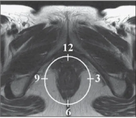
By definition, a fistula is an abnormal connection between two epithelial sur-faces. The anatomic path of a fistula will be determined by the location of the in-fected anal gland and the adjacent anatomi-cal planes. Surgeons describe the location and direction of the fistulous tract by us-ing the image of a clock face (Figure 1), with a view of the anal region when the patient is in the lithotomy position, corre-sponding to the view of the anal canal on MRI images in the axial plane, thus facili-tating the correlation of imaging findings with those observed during the surgical procedure. Typically, there is an aperture to the anal canal (proximal orifice) at the level of the jagged line, that originally is the
gland drainage site; from there the fistula can reach the skin of the perianal region by a variety of tracts and even penetrate and involve with variable degrees the EAS and adjacent tissues(4,5).
The relation between the fistulas and the perianal musculature is well demonstrated through a combination of images in the different planes, as well as by the different combinations of sequences. The utilization of gadolinium provides excellent contrast between the fistulous tracts and adjacent tissues. MRI is currently the most appropri-ate method for the diagnosis and classifi-cation of such lesions, which is performed according to the pathway between the in-ternal and exin-ternal apertures of the fis-tula(4–6).
The selection of the coil type depends on individual preference. The utilization of the abdominal surface coil does not require any previous patient preparation and also is very well tolerated. Endoanal coils are poorly tolerated by symptomatic patients and, because of the limited field of view, they may not allow the observation of the full extent of the fistulas. For these reasons and also for the limited availability and acceptance of the endoluminal coil, the abdominal surface coil is used as the stan-dard in cases of fistula in ano(3–5).
The role of MRI in the diagnosis of fis-tula in ano and its complications has been increasing. The imaging findings demon-strate a great agreement with surgical find-ings, more than at any other imaging method, and the techniques in the several planes demonstrate very well the relation between the fistulous tract with the sphinc-teric complex and the ischioanal fossa.
Another important advantage is its capabil-ity of the method to demonstrate clinically occult abscesses(4).
Abscesses present high signal intensity on T2- and STIR sequences and iso- or slightly hyperintense signal on T1-weighted sequences, because of the great protein content; eventually such increased content may demonstrate a decrease in sig-nal intensity on T2-weighted sequences. The presence of gas within the abscess is demonstrated by the signal absence on all sequences. The fistulous tracts are seen as tubuliform structures, presenting signal characteristics similar to those of the ab-scess. After intravenous gadolinium injec-tion, contrast uptake is observed at the thickened abscess and fistulous tract walls, besides a usually hypointense content on T1-weighted sequences.
Fistulas in ano classification
The fistulas were already known by Hippocrates and have been described along the centuries, receiving special attention in the 19th century, when, in 1835, Frederick Samon founded the Benevolent Dispensary for the Relief of the Poor Afflicted with Fistula and Other Diseases of the Rectum and Lower Intestines, which has become the worldwide famous St. Mark’s Hospital in London, England. Much of what is known about fistulas in ano originates from the work of surgeons in that hospital(4).
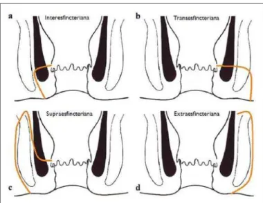
In 1934, Milligan and Morgan catego-rized the fistulas location into below, above and within the puborectal muscle, drawing attention to the great likelihood of postop-erative incontinence in the case of inappro-priate surgical approach of a high fistula. Such classification was modified and re-fined by other authors, including Parks et al.(7), who have analyzed a consecutive se-ries of 400 patients of the St. Mark’s Hos-pital, classifying the fistulas into four groups according to the cryptoglandular hypothesis, as follows: intersphincteric, trans-sphincteric, suprasphincteric and extrasphincteric(5,6).
The ISS is the pathway with least resis-tance for infection dissemination, thus rais-ing the suspicion of intersphincteric fistula, observed in 45% of the cases studied by Parks et al., in which the infectious process is limited to the ISS and does not penetrate
the ES that represents a natural barrier to the infection dissemination. In a more in-tense infectious process, the fistulas may cross the ES and reach the ischioanal fossa, resulting in trans-sphincteric type fistula, observed in 30% of the original cases stud-ied by Parks et al. In other circumstances, the fistula may extend to a plane above the ISS, partially surrounding the puborectal muscle and crossing the plateau of the leva-tor ani muscle to reach the perianal skin, resulting in suprasphincteric type fistula, observed in 20% of the cases by Parks et al.(7). They also have found a fourth type of fistula in 5% of the cases, representing an extension of a primary pelvic disease that also crosses the levator ani muscle and goes down through the ischioanal fossa reach-ing the skin, classified as extrasphincteric type. The classification proposed by Parks et al. was much criticized because of the specific nature of the hospital and also be-cause it did not include the submucosal fis-tulas, very superficial and not involving the sphincteric structures(5).
In 2000, Morris et al.(4), in a study with more than 300 patients at the St. James’s University Hospital (Leeds, England), cor-related the Parks et al. classification with MRI findings in the axial and coronal planes, classifying the fistulas into five types as follows: simple linear inter-sphincteric, intersphincteric with abscess or secondary tract, sphincteric, trans-sphincteric with abscess or secondary tract in the ischioanal fossa and supra/extra-sphincteric. This classification does not deal only with the demonstration of the primary fistulous tract, but also with the secondary branching and associated ab-scesses. This is an easily acceptable system, considering that it is based on anatomic landmarks in the axial and coronal planes that are very familiar to radiologists, with excellent applicability and reproducibility, as it presents the best correlation with ini-tial surgical evaluation(4). Many fistulas begin as a simple primary tract, but un-treated infections may result in branching (secondary tracts), with the ischioanal fossa
being the most common site. The horizon-tal branching of the fistulas is described as “horseshoe shaped” with two tracts in the ISS(5).
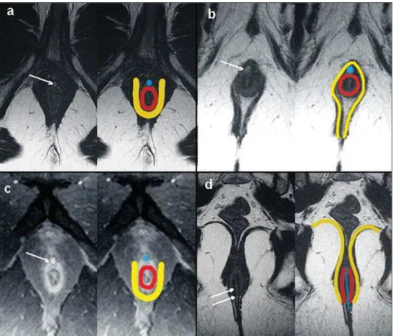
Type 1: simple linear intersphincteric fistula (Figure 2) – The fistulous tract
ex-tends itself from the anal canal to the perineal skin, with a tract confined between the sphincters, with no branching beyond the sphincteric complex. It is by far the most common fistula type . The ischioanal fossae are unaffected(4,5).
Type 2: intersphincteric fistula with abscess or secondary tract (Figure 3) –
Such fistulas are also confined to a plane between the sphincters, and so do their sec-ondary tracts and/or abscesses. The second-ary tracts may be horseshoe-shaped, cross-ing the midline, or may branch in an ipsi-lateral intersphincteric plane(4,5).
Type 3: trans-sphincteric fistula (Fig-ure 4) – The fistulous tract crosses the two
layers of the sphincteric complex, arching to the skin through the ischioanal fossa. These fistulas also differentiate by the site
Figure 2. Simple linear intersphinc-teric fistula (type 1). T1-weighted (a)
Figure 3. Intersphincteric fistula with ab-scess or secondary tract (type 2). T2-weighted sequences in the axial plane (a), sagittal plane (c) and coronal plane (d) and T1-weighted sequence in the axial plane with fat suppression after gadolinium injec-tion (b) show abscess located in the pos-terior intersphincteric space (arrow). On (c) and (d) one can observe the relationship between the abscess and the external sphincter. Schematic drawing: abscess (blue), internal sphincter (red) and exter-nal sphincter (yellow).
of entry into the anal canal, which corre-sponds to the middle third of the anal ca-nal, i.e., at the level of the jagged line, as demonstrated on the images in the coronal plane. As they affect the whole sphincteric complex, surgical treatment may eventually lead to fecal incontinence(4–6).
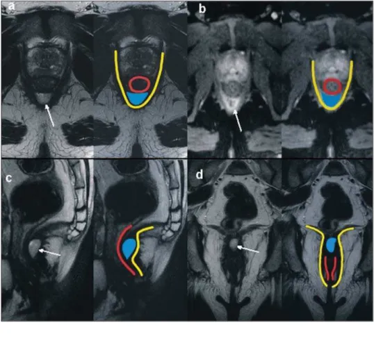
Type 4: trans-sphincteric fistula with formation of abscess or secondary tract within the ischioanal fossa (Figure 5) –
Like the type 3 fistulas, the classification into type 4 requires the demonstration that an infectious process occurs through the ES. The abscess may manifest as an expan-sion along the primary fistulous tract. The tract or associated abscess necessarily in-volve the ischioanal or ischiorectal fossa and, in some cases, takes over a “dumbbell” shape, crossing the ES(4,5).
Type 5: suprasphincteric or extra-sphincteric fistula (Figure 6) – If the
fis-tula extends through the ISS, above the levator ani muscle up to the ischiorectal fossa and from there to the skin, it is clas-sified as suprasphincteric fistula; represent-ing the extension of a primary pelvic dis-ease that crosses the levator ani muscle and goes down the ischiorectal fossa reaching the skin, it is classified as extrasphincteric fistula. Such fistulas are difficult to be managed and approached, considering the necessity of additional evaluation for detec-tion of pelvic sepsis. Contrast-enhanced coronal images clearly demonstrate failures in the levator ani muscle, with horseshoe-shaped tracts being also a possible find-ing(4,5).
CONCLUSION
MRI is an effective and fundamental imaging method for the evaluation of fis-tulas in ano, and the multiplanar images acquisition has demonstrated to be ex-tremely useful, so the method is considered as the most appropriate for the diagnosis and classification of such lesions (Figure 7). Additional studies have demonstrated that the evaluation by means of MRI is better than the initial surgical exploration in the prediction of outcomes and, as the St. James’ University Hospital is utilized, a significant correlation with better out-comes is observed(4,5).
Figure 5. Trans-sphincteric fistula with abscess or secondary tract within the ischiorectal fossa (type 4). T2-weighted sequence in the axial plane (a), T1-weighted sequence with fat suppression in the axial plane after gadolinium injection (b), T2-weighted (c) and STIR sequence (d) in the coronal plane demonstrat-ing two fistulous tracts: the primary, trans-sphincteric on the left lateral wall of the anal canal, where one observes an associated abscess (arrow), and the secondary intersphincteric tract at right (arrow head). Schematic drawing: fistulous tract and abscess (blue), internal sphincter (red) and external sphincter (yellow).
Figure 7. Schematic drawing of the types of fistula in ano: internal sphincter (black); external sphincter (white); fistulous tracts (orange).
REFERENCES
1. Zagrodnik II DF, Schneider M. Fistula-in-ano. E-medicine. [acessado em 15 de fevereiro de 2010]. Disponível em: http://emedicine.medscape.com/ article/190234-overview
2. Lunnis PJ, Armstrong P, Barker PG, et al. Mag-netic resonance imaging of anal fistulae. Lancet. 1992;340:394–6.
3. Stoker J, Halligan S, Bartram CI. Pelvic floor im-aging. Radiology. 2001;218:621–41.
4. Morris J, Spencer JA, Ambrose NS. MR imaging classification of perianal fistulas and its implica-tions for patient management. Radiographics. 2000;20:623–35.
5. Halligan S, Stoker J. Imaging of fistula in ano. Radiology. 2006;239:18–33.
6. Zimmerman DDE. Diagnostic and treatment of transsphincteric perianal fistulas [Doctoral The-sis]. Rotterdam, The Netherlands: Erasmus Uni-versity; 2003.