Magnetic resonance imaging in lower urinary tract
endometriosis: iconographic essay*
Ressonância magnética na endometriose do trato urinário baixo: ensaio iconográfico
Cláudio Márcio Amaral de Oliveira Lima1, Elisa Pompeu Dias Coutinho2, Érica Barreiros Ribeiro3, Marisa Nassar Aidar Domingues4, Flávia Pegado Junqueira4, Antonio Carlos Coutinho Junior2
Endometriosis is defined as the presence of functional endometrial tissue outside the endometrial cavity and myometrium. Although this is a frequent disease with multifactorial causes, involvement of the lower urinary tract is rare. Magnetic resonance imaging is highly sensitive, specific and accurate in the diagnosis of endometriosis in the lower urinary tract, especially for allowing the identification of lesions obscured by adhesions or with subperitoneal extension. The present iconographic essay presents the main magnetic resonance imaging findings of the lower urinary tract involvement by endometriosis.
Keywords: Magnetic resonance imaging; Endometriosis; Infertility; Genitourinary tract; Bladder; Urethra.
Endometriose é definida como a presença de tecido endometrial funcionante fora da cavidade endometrial e do miométrio. É uma doença comum, de causas multifatoriais, porém o envolvimento do trato urinário baixo é raro. A ressonância magnética tem elevada sensibilidade, especificidade e acurácia no diagnóstico da endometriose do trato geniturinário baixo, principalmente por permitir a identificação das lesões de permeio a aderências e a avaliação da extensão das lesões subperitoneais. Neste estudo são ilustrados, sob a forma de ensaio iconográfico, os principais achados à ressonância magnética do envolvimento por endometriose do trato urinário baixo.
Unitermos: Imagem por ressonância magnética; Endometriose; Infertilidade; Trato geniturinário; Bexiga; Uretra.
Abstract
Resumo
* Study developed at Clínicas de Diagnóstico Por Imagem (CDPI) and Multi-Imagem, Rio de Janeiro, RJ, and Centro de Diagnóstico por Imagem Fátima Digittal, Nova Iguaçu, RJ, Bra-zil.
1. MD, Radiologist, Trainee at Clínicas de Diagnóstico Por Imagem (CDPI) and Multi-Imagem, Radiologist at Hospital Na-val Marcílio Dias (HNMD), Rio de Janeiro, RJ, Brazil.
2. MDs, Radiologists at Clínicas de Diagnóstico Por Imagem (CDPI) and Multi-Imagem, Rio de Janeiro, RJ, and Centro de Diagnóstico por Imagem Fátima Digittal, Nova Iguaçu, RJ, Bra-zil.
3. MD, Radiologist, Trainee at Clínicas de Diagnóstico Por Imagem (CDPI) and Multi-Imagem, Rio de Janeiro, RJ, Radiolo-gist at Centro de Diagnóstico por Imagem Fátima Digittal, Nova Iguaçu, RJ, Brazil.
4. MDs, Radiologists at Clínicas de Diagnóstico Por Imagem (CDPI) and Multi-Imagem, Rio de Janeiro, RJ, Brazil.
Mailing address: Dr. Cláudio Márcio Amaral de Oliveira Lima. Rua Silva Rabelo, 154/503, Ed. Barcelona, Méier. Rio de Janei-ro, RJ, Brazil, 20735-080. E-mail: [email protected] / cmao-lima @gmail.com
Received February 14, 2009. Accepted after revision May 4, 2009.
vaginal ultrasonography in demonstrating the involvement of the bladder muscular layer and defining the relation of the lesion and the adjacent structures in the surgical planning(8).
The present iconographic essay aims to demonstrate the main MRI findings in the involvement of the lower urinary tract by deep endometriosis.
MRI PROTOCOLS
Specific protocols must be followed for MRI acquisition to evaluate patients under suspicion of endometriosis in the lower uri-nary tract.
At the “Clínicas de Diagnóstico Por Imagem e Multi-Imagem” the examination is performed during the menstrual period because of the higher likelihood of identi-fying hemorrhage foci, thus facilitating the tissue characterization of such lesions. Moderate bladder repletion is required, considering that the identification of small lesions becomes more difficult with a
com-Lima CMAO, Coutinho EPD, Ribeiro EB, Domingues MNA, Junqueira FP, Coutinho Jr AC. Magnetic resonance imaging in lower urinary tract endometriosis: iconographic essay. Radiol Bras. 2009;42(3):193–197.
Endometriosis is defined as the pres-ence of functional endometrial tissue out-side the endometrial cavity and myo-metrium(2–4). It is a frequent multifactorial disease, affecting from 7% to 10% of the general population. Genitourinary involve-ment is observed in 1% to 3% of cases, most frequently in women between 25 and 40 years of age(5). Deep endometriosis is a specific entity, histologically defined as invasion of the peritoneum by ectopic foci at a depth of > 5 mm, generally involving uterosacral ligaments, bowel, rectovaginal septum, and the urinary tract(6).
The clinical diagnosis is difficult and based on history, physical examination, laparoscopy and biopsy of suspicious
le-sions(6,7). Magnetic resonance imaging
(MRI) is a noninvasive method that allows multiplanar evaluations, with high spatial resolution and tissue characterization ca-pacity, without use of ionizing radiation or iodinated contrast agents. MRI presents high accuracy for the diagnosis of vesical lesions, with higher sensitivity than
trans-INTRODUCTION
considerable discomfort or pain for the patients, in spite of the menstrual period. No bowel preparation was required.
The imaging protocol icludes the fol-lowing sequences: T1-weighted (TR: 589; TE: 10; FOV: 240; slice thickness: 3 mm; interval: 10%; matrix: 256 × 256) in the axial plane, T1-weighted with fat suppres-sion (TR: 706; TE: 14; FOV: 250; slice thickness: 5 mm; interval: 45%; matrix: 230 × 256) in the sagittal and axial planes, T2-weighted (TR: 3610; TE: 108; FOV: 240; slice thickness: 3.5 mm; interval: 10%; matrix: 384 × 326) in the sagittal, coronal and axial planes. After intravenous gadolinium injection, T1-weighted se-quences with fat suppression are performed in the sagittal and axial planes. Contrast agent injection may be useful in the detec-tion of lesions on the abdominal wall, since peritoneal/serous surfaces surrounding foci of deep endometriosis can enhance(8) most probably because of the intense inflamma-tory process associated with endometriosis. In cases of ureteral involvement, uro-reso-nance techniques may be utilized for a bet-ter anatomical characbet-terization of urebet-tero- uretero-hydronephrosis as a result of the larger field of view and urographic effect.
MRI FINDINGS
The involvement of the vesicouterine space is demonstrated as nodular forma-tions with low signal on T2- weighted se-quences, generally attached to the anterior uterine surface, forming an obtuse angle with the bladder wall(4,5) (Figure 1).
Vesical endometriosis may be of an in-trinsic nature, primarily involving muscu-lature (detrusor), or extrinsic, when lesions in the vesicouterine space involving the
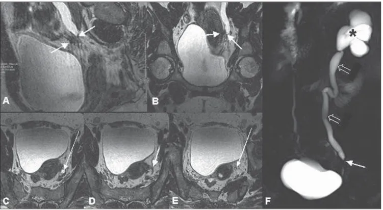
serous/peritoneal surfaces infiltrate the vesical walls. A predominantly hypoin-tense, diffuse or focal thickening of the vesical wall related to the fibrotic compo-nent may be observed on T2-weighted se-quences, substituting the characteristic sig-nal of the detrusor musculature. Eventually, intermingled, hyperintense foci may be ob-served on T2-weighted sequences, corre-sponding to ectatic endometrial glands with or without hematic contents(5,6) (Figure 2). Bladder mucosa invasion is not frequent in cases of vesical endometriosis, and MRI can demonstrate alteration even in asymp-tomatic patients with normal cystoscopy(9). MRI plays a significant role in the di-agnosis of vesical endometriosis (88% sen-sitivity, 98.9% specificity, 88% positive predictive value, 98.9% negative predictive value, and 97.9% accuracy), particularly for allowing the identification of submu-cosal lesions, even in cases with normal
cystoscopy and associations with other subperitoneal foci and /or intermingled with extensive adhesions(2). In accordance with the literature, in the present essay a highly variable signal intensity was ob-served with a high frequency of hemor-rhagic foci intermingled with these lesions, probably because these examinations were performed during the menstrual period, and also for the remarkable predilection for the posterior vesical wall and dome.
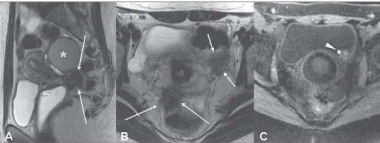
Figure 2. Sagittal (A), and axial (B) T2-weighted images, and axial, T1-weighted image with fat suppression (C) demonstrate hypointense focal thickening of the right posterolateral wall of the bladder on T2-weighted sequence (long arrows), with intermingled minute bleeding foci visualized on T1-weighted sequence with fat suppression (arrowhead). There is also an extraperitoneal, irregular, ill-defined, hypointense mass on T2-weighted sequence (fibrotic component), involving the posterior compartment, highlightingthe uterosacral ligaments, the retrocervical region and the rectovaginal septum (short arrows).
nective tissue and probably originates from ovary, broad ligament or uterosacral in-volvement. The presence of fibrotic/cica-tricial tissue without a true endometriotic involvement of the ureter may also be clas-sified as extrinsic involvement(5,6).
Frequently, endometriosis involves the pelvic ureter (Figure 3). Most affected women are in the premenopausal period(5,6). Ureteral obstruction (Figure 4) may be slow and progressive, eventually causing renal failure(10). Intrinsic involvement of the
ure-ter is rare, with periureure-teral fibrosis, either in association or not with bleeding foci, being the most frequent finding.
tents or not(10). Many times, foci of deep en-dometriosis are simultaneously observed in different sites, among them the retrocer-vical region, the uterosacral ligaments, the rectovaginal and vesicovaginal septa and other hollow viscus(4–6) (Figure 6).
The gold standard for management of endometriosis is complete resection of these lesions. Therefore, the preoperative evaluation plays a relevant role, generally being limited to clinical and sonographic data(6,7).
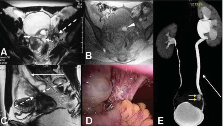
MRI plays a relevant role in the diagno-sis of lower urinary tract endometriodiagno-sis, with high sensitivity, specificity, accuracy and positive predictive value, mainly for al-lowing the identification of subperitoneal lesions and/or lesions intermingled with ex-Figure 4. Sagittal (A), coronal (B) and axial (C,D,E) T2-weighted images and uro-resonance image (F) showing distal ureteral stenosis, related to an irregular, ill-defined, hypointense image on T2-weighted sequence, involving the left pelvic ureter (short arrows) and determining significant pyelocalyceal (asterisk) and upstream ureteral (open arrows) dilatation, compatible with extrinsic ureteral endometriosis. The images on axial T2-weighted sequence demonstrate the ure-teral dilatation (C) (plane above stenosis), and anatomic configuration of ureter (E) (plane below stenosis) (long arrows). Renal dynamic study after gadolinium injection (not shown) demonstrates late concentration and renal elimination of the venous contras medium.
tensive adhesions, besides demonstrating and evaluating the extent of subperitoneal and/or visceral lesions that cannot be visu-alized at laparoscopy and/or cystoscopy.
Acknowledgements
To Dr. Romeu Côrtes Domingues, for encouragement and for making this and other scientific studies possible, and to Dr. Emerson L. Gasparetto, for his valuable help.
REFERENCES
1. Wong-You-Cheong JJ, Woodward PJ, Manning
Figure 6. Sagittal (A), axial (B) T2-weighted images; and axial T1-weighted image with fat suppression (C) showing focal, hypointense thickening on T2-weighted sequence of the left postero-lateral bladder wall (short arrows), with minimum intermingled bleeding foci (arrow head). Additionally, there is also a retroperi-toneal irregular, ill-defined, hypointense mass on T2-weighted sequence, involving the right uterosacral ligament (long arrows) without evidence of a defined cleavage plane between the uterine cervix and the anterior rectal wall. Also, an ovarian endometrioma can be observed (asterisk).
MA, et al. From the archives of the AFIP: Inflam-matory and nonneoplastic bladder masses: radio-logic-pathologic correlation. Radiographics. 2006;26:1847–68.
2. Bazot M, Darai E, Hourani R, et al. Deep pelvic endometriosis: MR imaging for diagnosis and prediction of extension of disease. Radiology. 2004;232:379–89.
3. Generao SE, Keene KD, Das S. Endoscopic di-agnosis and management of ureteral endometrio-sis. J Endourol. 2005;19:1177–9.
4. Coutinho Jr AC, Lima CMAO, Coutinho EPD, et al. Ressonância magnética na endometriose pél-vica profunda: ensaio iconográfico. Radiol Bras. 2008;41:129–34.
5. Deval B, Danoy X, Buy JN, et al. Bladder
en-dometriosis. Apropos of 4 cases and review of the literature. Gynecol Obstet Fertil. 2000;28:385–90.
6. Comiter CV. Endometriosis of the urinary tract. Urol Clin North Am. 2002;29:625–35. 7. Woodward PJ, Sohaey R, Mezzetti TP Jr.
En-dometriosis: radiologic-pathologic correlation. Radiographics. 2001;21:193–216.
8. Ghattamaneni S, Weston MJ, Spencer JA. Imag-ing in endometriosis. ImagImag-ing. 2007;19:345–68. 9. Del Frate C, Girometti R, Pittino M, et al. Deep retroperitoneal pelvic endometriosis: MR imag-ing appearance with laparoscopic correlation. Radiographics. 2006;26:1705–18.