Diagn Interv Radiol 2015; 21:4–9
© Turkish Society of Radiology 2015
Uterine sarcomas: clinical presentation and MRI features
Pedro Santos, Teresa Margarida Cunha
A B D O M I N A L I M A G I N G
R E V I E WABSTRACT
Uterine sarcomas are a rare heterogeneous group of tumors of mesenchymal origin, accounting for approximately 8% of uterine malignancies. They comprise leiomyosarcoma, endometrial stromal sarcoma, undifferentiated endometrial sarcoma, and adenosarcoma. Compared with the more com-mon endometrial carcinomas, uterine sarcomas behave more aggressively and are associated with a poorer prognosis. Due to their distinct clinical and biological behavior, the Interna-tional Federation of Gynecology and Obstetrics introduced a new staging system for uterine sarcomas in 2009, categoriz-ing uterine carcinosarcoma as a variant of endometrial carci-noma, rather than a pure sarcoma. Magnetic resonance im-aging (MRI) has a developing role in the assessment of these malignancies. Features such as tumor localization, irregular or nodular margins, necrosis, rapid growth, intense contrast enhancement, and restriction at diffusion-weighted imaging can suggest the diagnosis and help differentiate from more common leiomyomas and endometrial carcinoma. MRI is therefore extremely useful in preoperative detection and staging and, consequently, in determination of appropriate management. This pictorial review aims to discuss the clinical features of uterine sarcomas, as well as their most common appearances and distinct characteristics in MRI.
U
terine sarcomas are a rare heterogeneous group of tumors of mes-enchymal origin, accounting for approximately 8% of uterine malignancies (1), although they were thought to represent only 2% to 3% of all uterine tumors in the past (2). This increased incidence may be the result of improved diagnosis, as well as a true increase in an ageing population (1).These malignancies may originate from the smooth muscle in myo-metrium (leiomyosarcoma), from the endometrial stroma (endometrial stromal sarcoma [ESS] and undifferentiated endometrial sarcoma [UES]) or both (adenosarcoma) (3). According to the Gynecologic Oncology Group, uterine sarcomas can be classified into two categories: nonep -ithelial and mixed ep-ithelial-nonep-ithelial, depending on the type of cancerous cell and its presumed tissue of origin (4).
The clinical presentation of uterine sarcomas is nonspecific and de-pendent of histologic subtype. Classically, they present as a rapidly growing pelvic mass, which may be accompanied by vaginal bleeding and abdominal or pelvic pain (1, 5).
Leiomyosarcoma is the most common histological variant of uterine sarcomas and is considered an aggressive tumor associated with poor prognosis, with a five-year survival rate ranging from 18.8% to 68%. ESS is relatively indolent, associated with long-term survival, but character-ized by late recurrences (14%–60% of women). In contrast, UES has a very aggressive behavior and poor prognosis, with a five-year survival rate of 25%–55%. Adenosarcomas are rare mixed tumors (glandular and mesenchymal origin) with relatively low malignant potential and slow-growth pattern, with a five-year survival rate above 80% (6).
The recognition of their distinct clinical and biological behavior when compared to endometrial carcinoma, which tend to behave more ag-gressively and are associated with a poorer prognosis, led the Interna-tional Federation of Gynecology and Obstetrics (FIGO) to develop a new staging system for uterine sarcomas in 2009 (Tables 1 and 2). One important feature of the new staging system is that carcinosarcoma (for-merly referred to as “malignant mixed Müllerian tumor”) is no longer considered as part of uterine sarcomas, being classified as a dedifferenti-ated or metaplastic form of endometrial carcinoma (7).
The distinction among different subtypes of uterine sarcomas and other uterine tumors (especially leiomyoma and endometrial carcino-ma) cannot be made on clinical grounds. Therefore, imaging, particular -ly MRI, has a developing role in the assessment of these malignancies, being useful in the evaluation of pelvic masses at presentation, adequate staging (assessment of invasion depth, spread to adjacent organs and lymph nodes), and consequently, determination of appropriate man -agement.
From the Department of Radiology (P.S. pedrofrsantos@gmail. com), Hospital de Santa Maria, Lisbon, Portugal; the Department of Radiology (T.M.C.), Instituto Português de Oncologia de Lisboa Francisco Gentil, Lisbon, Portugal.
Received 4 March 2014, revision requested 27 April 2014, revision received 3 June 2014, accepted 8 July 2014.
Published online 28 October 2014. DOI 10.5152/dir.2014.14053
Leiomyosarcoma
Clinical features
Leiomyosarcoma is the most com-mon histological subtype of uterine sarcomas. The great majority arise de novo, but rarely (in 0.2% of cases) it
may result from a sarcomatous trans-formation in a benign leiomyoma (2). It is characterized by an aggressive behavior (even when confined to the uterus), with a five-year survival rate ranging from 18.8% to 68%, which varies widely according to different stages. Low-grade and serosal involve-ment seem to be significant prognostic factors. The reported risk of recurrence varies from 45% to 73% (1, 6, 8).
Most leiomyosarcomas occur in
women over 40 years of age, with a median age of 60 years. There is a two-fold incidence of leiomyosarcoma in African-American women; long-term tamoxifen use and prior pelvic radia-tion seem to be associated with a small increase in risk (4). Signs and symp -toms are similar to those occurring with leiomyomas, and include abnor-mal vaginal bleeding (56%), palpable pelvic mass (54%) and pelvic pain (22%). Less frequently, they can pres -ent as hemoperitoneum (due to tumor rupture), or symptoms resulting from extra-uterine extension or metasta-ses (8). Besides that, although “rapid growth” of a presumed leiomyoma is
considered a suspicious finding, the definition of the latter remains contro-versial. Therefore, preoperative distinc-tion between benign leiomyomas and malignant leiomyosarcomas is very difficult (if not impossible) based sole -ly on clinical features, and remains a challenge for clinicians (1, 8).
MRI features
Leiomyosarcomas are often difficult to differentiate from leiomyomas (the most common myometrial tumor), based on clinical features and even en-dometrial biopsy or dilatation and frac-tional curettage. Furthermore, recent advances in leiomyoma management (i.e., development of uterus-preserving treatments, such as gonadotropin-re-leasing hormone analogues, uterine arterial embolization, and focused ul-trasound surgery) have significantly raised the importance of pretreatment imaging diagnosis of uterine sarcomas (3, 9, 10).
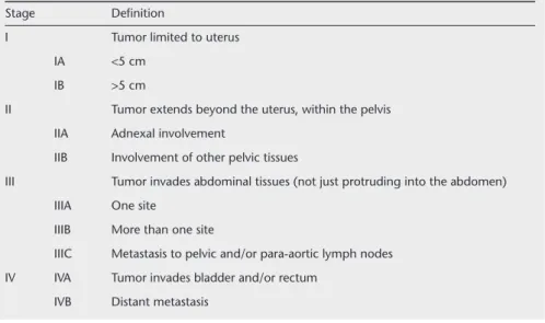
On MRI, leiomyosarcomas com-monly manifest as large infiltrating myometrial mass of heterogeneous hypointensity on T1-weighted images, with irregular and ill-defined margins. On T2-weighted images, they usually show intermediate-to-high signal tensity, with central hyperintensity in-dicative of extensive necrosis (present in >50% of cases) (Fig. 1a, 1b). Hemor -rhage is common, and foci of calcifi-cations may be present. After contrast administration, they present early het-erogeneous enhancement, due to the aforementioned areas of necrosis and hemorrhage (Fig. 1c) (1–3, 10, 11).
Leiomyosarcomas are generally larg-er and show more rapid growth than leiomyomas (3). On the other hand, common benign leiomyomas can show areas of increased signal intensi-ty on T2-weighted images as well, due to various types of degeneration or cel-lular histologic subtypes (9, 10). Some authors have suggested the presence of irregular margins, necrosis, and rapid growth as the most suggestive features of malignancy (2).
Diffusion-weighted imaging (DWI) has the potential to delineate malig-nant lesions as hyperintense areas with excellent tissue contrast, providing quantitative measurements of apparent diffusion coefficient (ADC) values (Fig. 2). Tamai et al. (10) reported significant differences in mean ADC values of
leio-Table 1. Staging for uterine leiomyosarcoma (7)
Stage Deinition
I Tumor limited to uterus
IA <5 cm
IB >5 cm
II Tumor extends beyond the uterus, within the pelvis
IIA Adnexal involvement
IIB Involvement of other pelvic tissues
III Tumor invades abdominal tissues (not just protruding into the abdomen)
IIIA One site
IIIB More than one site
IIIC Metastasis to pelvic and/or para-aortic lymph nodes
IV IVA Tumor invades bladder and/or rectum
IVB Distant metastasis
Table 2. Staging for uterine endometrial stromal sarcoma and adenosarcoma (7)
Stage Deinition
I Tumor limited to uterus
IA Tumor limited to endometrium/endocervix with no myometrial invasion
IB Less than half or half myometrial invasion
IC More than half myometrial invasion
II Tumor extends beyond the uterus, within the pelvis
IIA Adnexal involvement
IIB Involvement of other pelvic tissues
III Tumor invades abdominal tissues (not just protruding into the abdomen)
IIIA One site
IIIB More than one site
IIIC Metastasis to pelvic and/or para-aortic lymph nodes
IV IVA Tumor invades bladder and/or rectum
myosarcomas, compared with normal myometrium and degenerated leiomy-omas, without any overlap. Namimoto et al. (9) showed that overlap in ADC values between leiomyosarcomas and ordinary leiomyomas (attributed to the “T2 blackout effect,” i.e., hypointensi -ty on DWI caused by hypointensi-ty on T2-weighted images) could be resolved with the evaluation of tumor-myome-trium contrast ratio on T2-weighted images. Thomassin-Naggara et al. (12) reported that by combining the analy-sis of T2 signal intensity, b1000 images and ADC map, MRI achieved 92.4% accuracy in distinguishing benign and uncertain or malignant myometrial tu-mors. Therefore, they concluded that DWI may limit misdiagnosis of uterine sarcomas as benign leiomyomas, and should be the first criterion to help ra-diologists characterize a unique uterine tumor. Based on these results, the use of DWI must be recommended in the setting of myometrial lesions, especial-ly when high signal intensity is seen on T2-weighted images.
Endometrial stromal sarcoma
Clinical features
According to the World Health Orga-nization classification, an endometrial stromal tumor is composed of cells that resemble endometrial stromal cells of the proliferative endometrium (13). ESS corresponds to the former “low-grade ESS”, and UES replaces the term “high-grade ESS”. This new classification is a result of the recognition of the very dis-tinct biological behaviors and clinical outcomes of these tumors (3, 4, 6, 8).
ESS is a rare tumor, accounting for 0.2% of all malignant uterine tumors and 10%–15% of uterine malignancies with a mesenchymal component (3, 6, 8). It is considered to be a low-grade, well-differentiated tumor without sig-nificant cellular atypia (4). ESS is there -fore a relatively indolent lesion, gen-erally with a favorable prognosis, with five- and 10-year survival rates of 98% and 89% for stage I disease, which cor -responds to the majority of patients at presentation (3, 8). However, the out -come is largely dependent on the extent of the tumor at presentation, and stage seems to be the most significant indica-tor for survival (3, 6, 8). For stages II and III, five-year survival significantly drops to 50% and 65%, respectively (3). ESS is also characterized by late recurrences (14%–60% of patients), even in patients with stage I disease (6, 8). Therefore, long-term follow-up is mandatory.
ESS occurs more commonly in wom-en betwewom-en 40 and 55 years of age (8).
There has been a reported association with tamoxifen and estrogen use, al-though data are limited (3, 14). They usually present with abnormal vaginal bleeding, pelvic pain, and dismenor-rhea; however, as many as 25% of pa -tients are asymptomatic (1, 3, 8).
According to the new FIGO staging for uterine sarcomas, ESS is staged in the same manner as adenosarcoma (Table 2).
MRI features
Imaging (and particularly MRI) may be a powerful tool in the management of these tumors, allowing characteriza-tion of the initial stage and also pro-viding differentiation from the more common endometrial carcinoma (3).
ESS more frequently appears as pol-ypoid endometrial mass, with low sig-nal on T1-weighted images and heter -ogeneously increased high T2 signal (3, 11, 15). It typically shows myo -metrial involvement, either sharply
Figure 1. a–c. Leiomyosarcoma in a 52-year-old woman. Sagittal T1-weighted image (a), T2-weighted image (b), and gadolinium-enhanced T1-weighted image with fat suppression (c) show marked uterine enlargement due to a heterogeneous myometrial tumor. The lesion demonstrates central hyperintensity on T1-weighted image (a) attributable to extensive hemorrhage, a central area of high signal on T2-weighted image (b)
representing cystic necrosis, and early intense enhancement in solid areas of the tumor (c,arrow), as compared with normal myometrium. Irregular central zones of low signal intensity (asterisk) suggest extensive tumor necrosis. Endometrial cavity is pushed anteriorly by the tumor (b, arrow).
a b c
Figure 2. a, b. Leiomyosarcoma in a 54-year-old woman. Axial DWI on b1000 (a) demonstrates a hyperintense mass. The mass appears hypointense on ADC map (b,asterisk), with the normal myometrium seen as an area of hyperintensity (arrows).
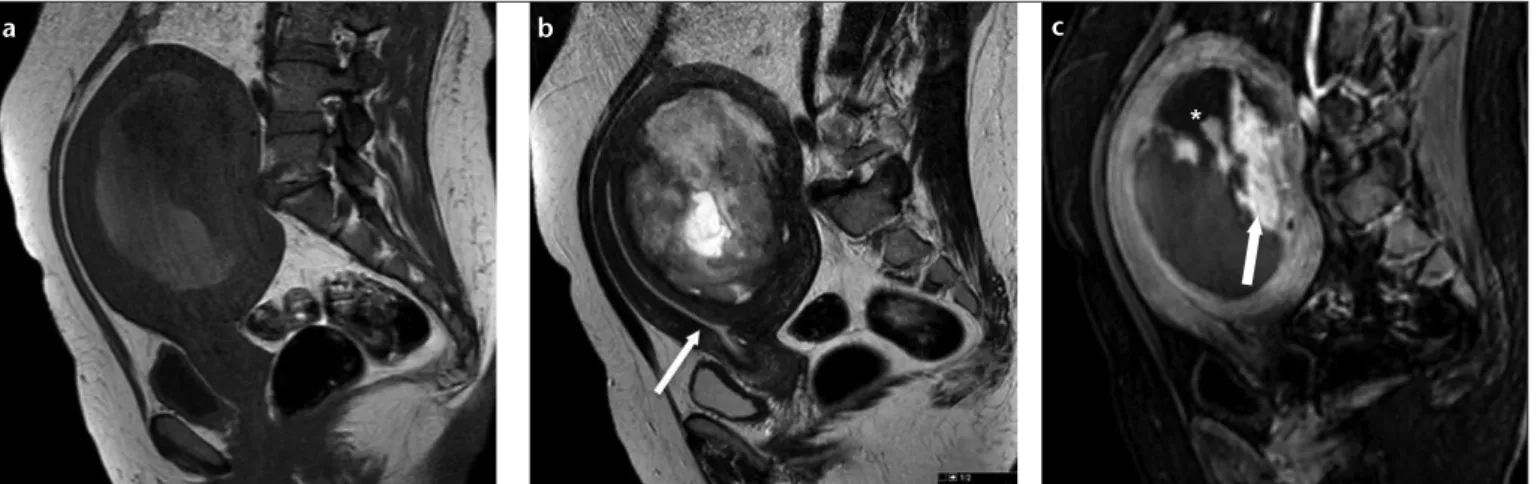
demarcated or in a more diffuse and destructive manner (the latter is far more common with UES) (Fig. 3a). These tumors have a tendency for lym-phatic and vascular invasion, showing worm-like extension bands of low sig-nal intensity within areas of myome-trial involvement on T2-weighted im -ages (“bag of worms”), corresponding to preserved bundles of myometrium (Fig. 4) (2, 3, 11, 15–17). After contrast administration, enhancement is mod-erate and commonly heterogeneous (Fig. 3b) (3, 15).
Compared to endometrial carcino-ma, ESS usually shows larger size, more contrast enhancement, irregular mar-gin, nodular extension into the myo-metrium, and marginal nodularity due to tumor extension along vessels and lymphatics (2, 3, 15, 16).
Rarely, ESS can appear as a myome-trial mass mimicking intramural leio-myoma with cystic degeneration. In these cases, intramyometrial ESS can be differentiated based on their rapid and invasive growth, lower degree of enhancement, lymphatic and vascu-lar invasion, higher incidence of ne-crosis, peripheral hypointense rim on T2-weighted images, and enhanced marginal irregularity (3, 11, 15, 18).
Lymph node metastases are seen in 10% of patients, being more frequent locally within the pelvis or vagina, fol-lowed by the lung parenchyma (3).
Undifferentiated endometrial sarcoma
Clinical features
UES, previously referred to as “high-grade ESS”, is an aggressive tumor lack-ing specific differentiation, and show-ing no endometrial stromal features. It is characterized by myometrial in-vasion, severe nuclear polymorphism, high mitotic activity, and/or tumor cell necrosis (4, 8). UES shows an ag -gressive behavior, with five-year sur-vival rates of 25%–55% (6). The most significant prognostic factor seems to be the presence of vascular invasion, decreasing five-year survival to as low as 17% (19). Local recurrences and dis -tant metastases (due to hematogenous spread) are also associated with high mortality and poor outcome (8, 14).
UES tends to occur in an older age group, when compared to ESS, with
a mean age of 61 years at the time of diagnosis (2, 11). Signs and symptoms resemble those of leiomyosarcoma and include vaginal bleeding, palpable pel-vic mass, and pelpel-vic pain (16).
MRI features
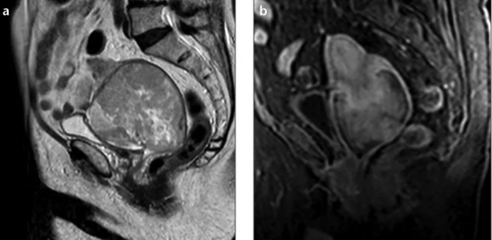
UES typically appears as a large pol -ypoid mass in an expanded endome-trial cavity, showing heterogeneous signal intensity on both T1- and T2-weighted images (Fig. 5a). The lat -ter is attributable to the high frequen-cy of hemorrhage and necrosis within the tumor (11, 14, 17). UES tends to infiltrate the myometrium in a more destructive and extensive manner than ESS, due to marked vascular and lymphatic invasion (3, 11). Contrast
enhancement is generally heteroge-neous, and iso- or hyperintense when compared with normal myometrium, allowing differentiation from endo-metrial carcinoma (Fig. 5b). Hyperen -hancement, the presence of irregular margins, multiple marginal tumor nodules, intramyometrial worm-like extension, and multiple nodular mass formation, are more frequently seen in UES than ESS (2, 3, 15).
Adenosarcoma
Clinical features
Adenosarcoma is a rare uterine ma-lignancy, accounting for 5.5%–9% of all uterine sarcomas (6). It is a mixed tumor, composed of a benign (but oc-casionally atypical) epithelial compo
-Figure 3. a, b. Endometrial stromal sarcoma in an 82-year-old woman. Sagittal T2-weighted image
(a) and sagital T1-weighted image with fat suppression, after contrast administration (b) show a very large lesion centered at cervix region, iniltrating uterine body superiorly and superior half of the vagina inferiorly. The tumor shows multiple foci of hyperintense signal on T2-weighted image due to extensive necrosis, as well as moderate and mildly heterogeneous contrast enhancement.
a b
Figure 4. a, b. Sagittal (a) and axial (b) T2-weighted images show an endometrial stromal sarcoma in a 64-year-old woman. The lesion shows heterogeneous signal with extensive nodular invasion into the myometrium and marked marginal irregularity and nodularity (attributable to tumor extension along vessels and lymphatics).
nent, and a malignant stromal (sarco-matous, usually low-grade) element (4, 6, 16). Adenosarcoma is considered a mixed Müllerian tumor, intermediate between adenofibroma and carcinosar-coma; it was even suggested that some adenofibromas are in fact well-differ-entiated adenosarcomas (3, 6, 8). It is a slow-growing tumor, with low ma-lignant potential and good prognosis. Majority of women are diagnosed at stage I (>60%), with an overall five-year survival over 80% (3, 4, 6). More than 70% of adenosarcomas occur in the en -dometrium, but they can also be found in the myometrium (probably from ad-enomyosis), cervix, and extra-uterine
tissues such as the ovaries. Extra-uter-ine location is more common in adoles-cents and young women (6, 8).
Adenosarcoma with sarcomatous overgrowth is defined as an adenos-arcoma with the sadenos-arcomatous com-ponent constituting more than 25% of the tumor; it occurs in 8%–54% of uterine adenosarcomas, and 30% of ovarian adenosarcomas. It carries a worse prognosis, with mortality reach-ing 50% at five years. Other proposed factors associated with a poorer out-come and higher risk of recurrence are advanced age, myometrial invasion (found in 15% of cases), and lympho -vascular extension (3, 4, 6, 8, 16). Late
recurrence can be seen in one-third of the women at five years, therefore, long-term imaging follow-up is man-datory (3).
Adenosarcoma most commonly presents with abnormal vaginal bleed-ing; some women also complain of pelvic pain, palpable pelvic mass, or vaginal discharge (8).
According to the new FIGO staging for uterine sarcomas, adenosarcoma is staged in the same manner as ESS (Table 2).
MRI features
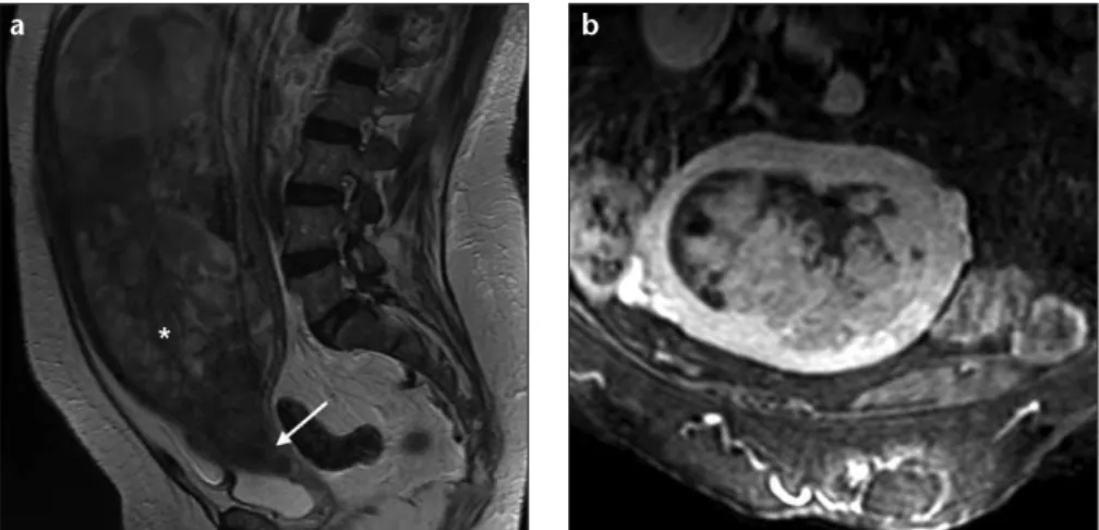
Adenosarcoma is typically seen as a large well-demarcated polypoid mass arising within the endometrial cavity and protruding through the cervical os, causing marked enlargement of the uterus with a thin myometrium (3, 20, 21). This polypoid mass usually shows a multiseptated cystic appearance, with multiple heterogeneous solid components that fill the endometrial cavity, and may mimic the appearance of gestational trophoblastic disease (Fig. 6a) (3, 20). On T2-weighted imag -es, small hyperintense foci may be seen scattered within the mass, representing glandular epithelial components or ne-crosis (8, 22). After administration of gadolinium, there is heterogeneous en-hancement, with solid components of the mass showing enhancement simi-lar to that of the myometrium (Fig. 6b) (3, 20, 21).
Adenosarcoma with sarcomatous overgrowth may present with myome-trial invasion and areas of hemorrhage and necrosis. However, on DWI, they usually show relatively low signal in-tensity on high b-value, reflecting its low grade nature (21, 22).
Conclusion
Uterine sarcomas are a rare and ag -gressive heterogeneous group of tumors of mesenchymal origin, with nonspe-cific clinical features. MRI is useful in lesion detection and characterization, as well as assessment of disease stag-ing. Although the radiologic findings of these lesions can overlap, characteristic features (summarized in Table 3) can help narrow the differential diagnosis and guide adequate treatment selection and follow-up. In addition to morpho-logical features, DWI seems to be a po-tentially useful tool in characterization of large uterine lesions.
Figure 5. a, b. Undifferentiated endometrial sarcoma in a 36-year-old woman. Sagittal T2-weighted image (a) and T1-weighted image after gadolinium administration (b) show marked uterine enlargement due to a large polypoid heterogeneous tumor, with some nodular marginality
(arrow). The lesion shows intense and heterogeneous contrast uptake (uncommon for endometrial carcinoma), with a hypointense area (asterisk) suggestive of necrosis.
a b
Figure 6. a, b. Adenosarcoma in a 76-year-old woman. Sagittal T2-weighted image (a) and oblique coronal T1-weighted image with fat suppression, after contrast administration (b) show a very large polypoid mass with heterogeneous high signal intensity arising within the endometrial cavity and protruding into the cervical os (arrow), causing marked enlargement of the uterus. The tumor demonstrates a multicystic appearance (asterisk), with solid areas demonstrating enhancement similar to myometrium.
Conflict of interest disclosure
The authors declared no conflicts of interest.
References
1. Wu TI, Yen TC, Lai CH. Clinical presenta-tion and diagnosis of uterine sarcoma, in-cluding imaging. Best Pract Res Clin Obstet Gynaecol 2011; 25:681–689.[CrossRef]
2. Sala E, Rockall AG, Freeman SJ, Mitchell DG, Reinhold C. The added role of MR imaging in treatment stratification of patients with gynecologic malignancies: what the radiologist needs to know. Ra-diology 2013; 266:717–740. [CrossRef]
3. Shaan SH, Jagannathan JP, Krajewski K, O’Regen KN, George S, Ramaiya NH. Uter -ine sarcomas: then and now. AJR Am J Roentgenol 2012; 199:213–223. [CrossRef]
4. Memarzadeh S, Mundt AJ, Berek JS. Uter -ine sarcoma: Classification, clinical man-ifestations, and diagnosis. In: UpToDate, Goff B, Falk SJ, eds. 2012.
5. Seddon BM, Davda R. Uterine sarcomas – recent progress and future challenges. Eur J Radiol 2011; 78:30–40. [CrossRef]
6. Tse KY, Crawford R, Ngan HYS. Staging of uterine sarcomas. Best Pract Res Clin Obstet Gynaecol 2011; 25:733–749.[CrossRef]
7. FIGO Committee on Gynecologic Oncol -ogy. FIGO staging for uterine sarcomas. Int J Gynaecol Obstet 2009; 106:277. 8. D’Angelo A, Prat J. Uterine sarcomas: a review.
Gynecol Oncol 2010; 116:131–139. [CrossRef]
9. Namimoto T, Yamashita Y, Awai K, et al. Com-bined use of T2-weighted and diffusion-weight -ed 3-T MR imaging for differentiating uterine sarcomas from benign leiomyomas. Eur Radiol 2009; 19:2756–2764. [CrossRef]
10. Tamai K, Koyama T, Saga T, et al. The utility of diffusion-weighted MR imaging for differentiating uterine sarcomas from benign leiomyomas. Eur Radiol 2008; 18:723–730. [CrossRef]
11. Rha SE, Byun JY, Jung SE, et al. CT and MRI of Uterine Sarcomas and their mimickers. AJR Am J Roentgenol 2003; 181:1369–1374. [CrossRef]
12. Thomassin-Naggara I, Dechoux S, Bon -neau C, et al. How to differentiate benign from malignant myometrial tumours using MR imaging. Eur Radiol 2013; 23:2306–2314.[CrossRef]
13. World Health Organization classification of tumours. Pathology and genetics of tumours of the breast and female genital organs. In Tavassoli FA, Devilee P, Eds. Lyon: IARC Press, 2003.
14. Lenhard SM, Untch M, Himsl I, et al. The high-grade endometrial sarcoma: a rare entity. Arch Gynecol Obstet 2006; 274:56–59. [CrossRef]
15. Ueda M, Otsuka M, Hatakenaka M, et al. MR imaging findings of uterine endo-metrial stromal sarcoma: differentiation from endometrial carcinoma. Eur Radiol 2001; 11:28–33. [CrossRef]
16. Amant F, Coosemans A, Debiec-Rychter M, Timmerman D, Vergote I. Clinical management of uterine sarcomas. Lancet Oncol 2009; 10:1188–1198.[CrossRef]
17. Ueda M, Otsuka M, Hatakenaka M, Torii Y. Uterine endometrial stromal sarcoma located in uterine myometrium: MRI ap-pearance. Eur Radiol 2000; 10:780–782.
[CrossRef]
18. Furukawa R, Akahane M, Yamada H, et al. Endometrial stromal sarcoma located in the myometrium with a low-intensity rim on T2-weighted images: report of three cases and literature review. J Magn Reson Imaging 2010; 31:975–979.[CrossRef]
19. Abeler VM, Royne O, Thoresen S, Daniel -sen HE, Nesland JM, Kristen-sen GB. Uter -ine sarcomas in Norway. A histopatho-logical and prognostic survey of a total population from 1970 to 2000 includ -ing 419 patients. Histopathology 2009; 54:355–364. [CrossRef]
20. Szklaruk J, Tamm EP, Choi H, Varavithya V. MR imaging of common and uncom-mon large pelvic masses. Radiographics 2003; 23:403–424.[CrossRef]
21. Yoshizako T, Wada A, Kitagaki H, Ishika -wa N, Miyasaki K. MR imaging of uterine adenosarcoma: case report and litera-ture review. Magn Reson Med Sci 2011; 10:251–254. [CrossRef]
22. Takeuchi M, Matsuzaki K, Yoshida S, et al. Adenosarcoma of the uterus: magnetic resonance imaging characteristics. Clin Imaging 2009; 33:244–247. [CrossRef]
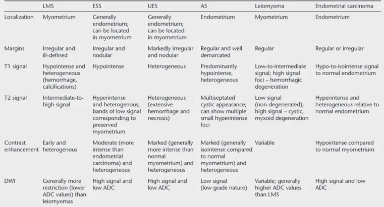
Table 3.MRI features of uterine sarcomas, leiomyoma, and endometrial carcinoma
LMS ESS UES AS Leiomyoma Endometrial carcinoma
Localization Myometrium Generally Generally Endometrium Myometrium Endometrium endometrium; endometrium;
can be located can be located in myometrium in myometrium
Margins Irregular and Irregular and Markedly irregular Regular and well Regular Regular or irregular ill-deined nodular and nodular demarcated
T1 signal Hypointense and Hypointense Heterogeneous Predominantly Low-to-intermediate Hypo-to-isointense signal heterogeneous hypointense, signal; high signal to normal endometrium
(hemorrhage, heterogeneous foci – hemorrhagic
calciications) degeneration
T2 signal Intermediate-to- Hyperintense Heterogeneous Multiseptated Low signal Hyperintense and high signal and heterogenous; (extensive cystic appearance; (non-degenerated); heterogeneous relative to
bands of low signal hemorrhage and can show multiple high signal – cystic, normal endometrium corresponding to necrosis) small hyperintense myxoid degeneration
preserved foci
myometrium
Contrast Early and Moderate (more Marked (generally Marked (generally Variable Hypointense compared enhancement heterogenous intense than more intense than isointense compared to normal myometrium
endometrial normal to normal carcinoma) and myometrium) and myometrium) and heterogeneous heterogeneous heterogeneous
DWI Generally more High signal and High signal and Low signal Variable; generally High signal and low restriction (lower low ADC low ADC (low grade nature) higher ADC values ADC
ADC values) than than LMS
leiomyomas