Neurology Discipline, Cerebrovascular Diseases Division (Atherosclerosis League) of the Santa Casa de São Paulo- Faculty of Medical Sciences, São Paulo SP, Brazil (SCSP): 1Associate Professor, responsible for the Cerebrovascular Diseases Division, SCSP; 2Instructor Professor, SCSP; 3Voluntary Doctor, Cerebrovascular Diseases Division, SCSP; 4Assistant Professor of the Social Medicine Department, SCSP.
Received 3 September 2003, received in final form 13 February 2004. Accepted 19 March 2004.
Dr. Rubens José Gagliardi - Avenida Angelica 916/305 - 01228-000 São Paulo SP - Brasil. E-mail: [email protected]
SPONTANEOUS EXTRACRANIAL CAROTID
ATHEROSCLEROSIS EVOLUTION IN
ASYMPTOMATIC INDIVIDUALS
A three-year prospective study
Rubens José Gagliardi
1, Ibsen Thadeu Damiani
2, Rogério Menoncello
3,
Manoel Carlos Sampaio de Almeida Ribeiro
4ABSTRACT - Objective: To evaluate the spontaneous evolution of extracranial carotid atherosclerosis in asymp-tomatic patients who did not present the main risk factors associated to the disease. Method: A prospec-tive study including patients of both genders, age ranging from 40 to 70 years, not presenting any signs and symptoms of cerebrovascular disease and without the main atherosclerosis risk factors were included. Patients who were using or had used medication during the follow-up period that could potentially influ-ence in the spontaneous course of atherosclerosis were excluded. The evaluation of the plaque and degree of stenosis were acquired using mode B, 7.5 MHz Doppler ultrasonography (USG). The follow-up was car-ried out for 36 months, with clinical, neurological, and USG exams repeated in a period of 6 to 8 months. Ninety-six individuals (48 women) completed the study with the presence of plaque, and 52 (26 women) with a degree of stenosis. Results:As to the degree of stenosis, 25% of the patients had worsening, 69% remained stable and 6% improved. When only the presence or absence of plaque was considered, 20% showed worsening (plaque developed during follow-up), 7% improved (disappearance of plaque), and 73% remained stable. No differences were found between the male and female patients. Conclusion:These results confirm the dynamic characteristics of plaque. In asymptomatic individuals without specific treat-ment, spontaneous improvement may occur, however, rarely. These findings may contribute as an assess-ment criterion when a decision is to be made in high-risk patients.
KEY WORDS: atherosclerosis, carotid, stroke, plaque, stenosis.
Evolução espontânea da aterosclerose carotídea extra craniana em indivíduos assintomáticos: estudo prospectivo de três anos
RESUMO - Objetivo: avaliar a evolução espontânea da aterosclerose carotídea. Método: estudo prospec-tivo com pessoas de ambos os sexos, idade de 40 a 70 anos, sem sinais e sintomas de doença cerebrovas-cular e sem os principais fatores de risco para aterosclerose. Foram excluídos os doentes que estavam em uso ou os que, durante o período de acompanhamento, usaram medicações que potencialmente pudessem influir no curso espontâneo da aterosclerose. As avaliações da placa e do grau de estenose foram obtidas por ultrassonografia com Doppler (USG). Investigou-se separadamente a presença de placa e o grau de estenose. O seguimento foi feito por 36 meses, com exame clínico, neurológico e novo USG repetidos com intervalo de 6 a 8 meses. Completaram o estudo 96 indivíduos (48 mulheres) quanto à presença da placa e 52 (26 mulheres) quanto ao grau de estenose. Resultados: Quanto ao grau de estenose, 25% dos indi-víduos pioraram, 69% permaneceram estáveis e 6% melhoraram. Quando se considerou apenas a presença ou ausência da placa, 20% pioraram (desenvolveram placa durante o seguimento), 7% melhoraram (desa-parecimento da placa) e 73% permaneceram estáveis. Não houve diferença em relação ao sexo. Conclusão: Estes resultados confirmam as características dinâmicas da placa. Em indivíduos assintomáticos e sem trata-mento específico, a melhora espontânea ocorre, porém é pequena. Estes dados podem contribuir para auxi-liar na tomada de uma decisão, em doentes de alto risco.
Atherosclerosis of the carotid arteries is the main cause of stroke or transient ischemic attack (TIA) in adults1. It may provoke thrombosis and/or
emboly usually related to serious obstructive processes. Atherosclerotic lesions may be found since infancy and worsen with aging2. The
preva-lence is low in the 40-year age range, and is pres-ent in approximately 80% in the groups over 60 years of age3,4. The process of this disease is
dynam-ic, in constant evolution, with a possibility of accu-mulation of new elements in the atheroma, regres-sion of the same, degeneration or tissue restruc-ture, causing, as a final result, a tendency of pro-gressive worsening5,6. This worsening can be seen
as the increase of the plaque and/or in the degree of stenosis. The reduction of the plaque and/or in the degree of stenosis is currently one of the great challenges in the treatment and prevention of atherosclerotic diseases. Modification and stabiliza-tion of the plaque has been proposed as one of the benefits of the treatment with lipid reducers7
and inhibitors of the angiotensin-converting enzyme (I-ACE)8. Recent studies7,9 have
demon-strated that plaque regression is related to the reduction of lipid content and that worsening is related to an increase in lipid content. In this process of evolution there is also a strong relation-ship to inflammatory and infectious events5,10-12. A
likely small increase of the fibrotic components and atrophy of smooth muscle cells are factors that may be related to plaque regression7,9. Atherogenesis
involves the tunica intima and begins through an endothelial dysfunction with progression to the subendothelial space5,7,9. Several studies have
eval-uated the regression of the plaques with lipids reduction13,14and blood pressure8and the role of
drugs with antiinflammatory properties and endothelial remodeling5,8,15,16. There are studies
that stress the importance of the role of the tuni-ca media and of the tunituni-ca adventitia in athero-genesis, especially in the remodeling process of the vessel walls7,9,17.
There is a direct relationship between the risk of stroke and the degree of stenosis, according the trials ACAS (“Asymptomatic Carotid Atherosclerosis Study”)18, NASCET (“North American Symptomatic Carotid Endarterectomy Trial”)19 and CSTCG
(“European Carotid Surgery Trialists Collaborative Group”)20, though this is not the only element
capable of triggering ischemic complications5,7.
Currently, several atherogenic risk factors are known that, if controlled, may contribute to the reduction of disease incident, and slow down or
improve its evolution. Few studies are dedicated to analyzing the spontaneous evolution of the plaque, independent of the presence of risk fac-tors, its maintenance or correction.
This present study aims to address the follow-ing aspect: study the spontaneous evolution of carotid atherosclerosis, in patients without the classic risk factors, who do not use medication, that, theoretically, could interfere in the course of the disease.
METHOD
A prospective study realized in patients followed-up in the Cerebrovascular Disease Clinic of the Neurology Discipline of the Santa Casa de São Paulo-Faculty of Medical Sciences. Hospital admission of the patients occurred in the period between 05/1997 and 05/1999 and the study was concluded in 07/2002. Caucasian patients of both genders were included, with ages ranging from 40 to 70 years, not presenting carotid disease symp-toms, without the main atherogenic risk factors: high blood pressure, dyslipidemia, diabetes, inactivity, and use of tobacco. Patients excluded were in continuous use, for more than three months, of anticoagulants, plaque antiaggregants, corticoids, anti-inflammatories, immuno-suppresants, calcium channel blockers, anticonvulsants, anxiolytics, statins, fibrates, hypotensives, and/or free rad-ical removers, because the prolonged use of these drugs could eventually alter the course of the atherosclerosis. The plaques were analyzed using the measures of plaque thickness and degree of vessel stenosis, individ-ually established by Doppler ultrasonography (USG) of the carotid arteries, using the SD-800 Phillips module, with a 7.5 Mhz linear transducer. The follow-up was done in a period of 36 months, with clinical, neurological, and USG exams repeated in a period of 6 to 8 months, for every patient. Patients who did not complete the 36-month follow-up were excluded, as well as those who developed disease during follow-up and used one or more of the drugs mentioned above for a period longer than 3 months, or who developed, in a persistent man-ner, for more than 3 months, one of the mentioned atherogenic risk factors. Within these criteria, 96 indi-viduals completed the study with the presence or absence of plaque (measure), and 52 in relation to the degree of stenosis. Forty-four patients did not present stenosis in the first ultrasonography exam; these patients were not included in the stenosis degree analysis group. Consequently, this group was limited to 52 patients out of the entire patient group.
Because the evolution of atherosclerosis is slow, sta-tistical analysis was done at the end of the follow-up peri-od in order to avoid data overlapping from analyses of intermediate observations.
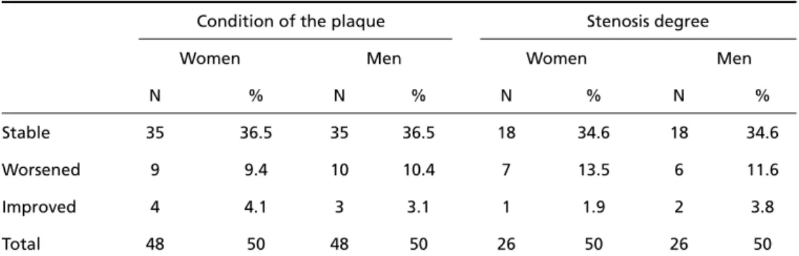
indi-Table 3. Comparative spontaneous evolution of the atherosclerotic plaque and stenosis degree in the carotid artery, between asymptomatic men and women after the 36-month follow-up.
Condition of the plaque Stenosis degree
Women Men Women Men
N % N % N % N %
Stable 35 36.5 35 36.5 18 34.6 18 34.6
Worsened 9 9.4 10 10.4 7 13.5 6 11.6
Improved 4 4.1 3 3.1 1 1.9 2 3.8
Total 48 50 48 50 26 50 26 50
Obs: In the statiscal analysis, the p value was not significant neither for the condition of the plaque nor the stenosis degree.
Table 1. Spontaneous evolution of stenosis degree after the 36-month follow-up in 52 asymptomatic patients.
Degree of Stenosis Frequency % 95% CI
Increase 13 25 14.5-39.2
Reduction 3 6 1.5-16.9
Total 52 100
Table 2. Spontaneous evolution of the atherosclerotic plaque after the 36-month follow-up in 96 asymptomatic patients.
Plaque condition Frequency % 95% CI
Worsened 19 20 12.6-29.4
Improved 7 7 3.2-14.9
Total 96 100
ner, always from the individual patient, without associating the data between different patients. The first exam was considered as the basis for all of the comparisons and was the established con-trol of each patient. From the results of the indi-vidual variations, percentages of improvement, deterioration, or stability were established for the entire study group. On average, each patient underwent 6 ultrasonography exams. For plaque measure evaluation, vessel wall thickening was considered.
As to the degree of stenosis (Table 1), the study showed that 25% worsened, 69% remained sta-ble and 6% showed improvement in the degree of stenosis. When only the presence or absence of plaque was considered (Table 2), the findings were similar, 20% of the patients who did not have plaque in the beginning of the study developed plaque during follow-up (worsened), 7% of the patients had plaque disappearance during the 36 months of follow-up, and the remaining patients (73%) remained stable. There was no significant difference between men and women Table 3.
DISCUSSION
There are few studies in the specialized litera-ture that evaluate the evolution of the atheroscle-rotic plaques of the carotid arteries in humans asymptomatic of cerebrovascular disease. Despite the fact of being an important parameter for mak-ing a clinical decision, there are few studies with this characteristic. Most of the studies were real-ized in coronaries or in symptomatic patients, dif-ferent than the approach of this present study.
The analysis of the carotids was done using Doppler ultrasonography, which is a reliable diag-nostic method, free from risk, and allowing a
com-viduals with clinical deterioration or evolution (increase) of the plaque, and the proportion of individuals with spontaneous improvement (reduction or disappearance of plaque). Regarding the degree of carotid stenosis, two variables were also considered after the 36-month peri-od: the proportion of individuals with the increase of stenosis and the proportion of individuals with the reduction of stenosis.
Data were processed using the EPI-INFO v. 6.04b soft-ware and all of the estimations were calculated using a confidence interval (CI) of 95%.
The Ethics and Research Committee of the Santa Casa de São Paulo approved this study.
RESULTS
man-parative study with a good safety index1,6. Other
methods that may be considered more precise, such as digital angiography, angio-resonance, and angio-tomography, have important limitations; such as high cost and procedure risk, that makes regular use not viable. The main parameters that are usually analyzed by ultrasonography are the presence of atherosclerotic plaque, its physico-chemical and morphological characteristics, and its degree of stenosis. The latter characteristic is one of the important elements used to determine the risk of the patients developing ischemic events, from plaque rupture and consequent thrombo-sis18,19,20. Other elements also of significant
impor-tance that contribute to the vulnerability of the plaque, its breaking and thrombosis are the con-stitution of the plaque, especially in relation to the amount of lipids and calcium, and the thickness of the fibrous cover5,7. Several inflammatory
mecha-nisms have been implicated in the evolution of the plaques, such as macrophage and T-lymphocyte acti-vation, which release cytokines (interleukins Iβ, 2, 6, 8), tumor necrosis factor, and interferon gam-ma, responsible for triggering reactions that bring about the escalation of atherosclerotic processes, that may provoke plaque destabilization10.
Infec-tions, especially from Chlamydia pneumonia, have also been associated to the appearance and/or wor-sening of atherosclerosis11. The association between
lipid oxidation and atherogenesis and the progres-sion of carotid atherosclerosis, in humans, has been much emphasized2. According to KAPS (Kuopio Atherosclerosis Prevention Study), the most impor-tant predictive factors for determining atheroscle-rotic progression are elevated serum levels of 7β -hydroxycholesterol, the major product of choles-terol oxidation in lipoproteins and cell membranes, lipid hydroxyperoxidase in the LDL, the oxidative susceptibility of the VLDL and LDL autoantibodies2.
Drugs with anti-inflammatory action, including statins and the I-ACES, have been used with good expectations in plaque reduction5,15,16.
The possible roles of these factors, which inter-act in the atherosclerotic evolution, were not a spe-cific aim for evolution in this study. We tried to focus on the behavior of the plaque in the spontaneous evolution, which could be a base for prompt clin-ical decisions. In order to accomplish this, the pres-ence of carotid plaque was evaluated and its degree of stenosis in asymptomatic individuals, without the classic risk factors for atherosclerosis. Plaque behav-ior and degree of stenosis were separately analyzed
because both are important and should be indi-vidually analyzed as risk factors for cerebrovascu-lar diseases. This methodological criterion might allow for the analysis of the spontaneous evolu-tion of the degree of stenosis, regardless of the action of these factors. Symptomatic individuals were excluded because they present a great chance of presenting a vulnerable atherosclerotic plaque, with an increased risk of rupture and progression. Based on this same principle, patients undergoing drug therapy that could potentially alter the course of atherosclerosis were excluded. In this study, the age was limited between 40 and 70 years, with the goal of homogenizing the study group, because it is known that atherosclerosis is rare in individ-uals under the age of 40, and has accentuated progression in individuals over the age of 70 years3,4.
We included only white patients, because it is known that atherosclerosis has different behavior in different races; furthermore, the inclusion of only one race had the goal of homogenizing the study group.
As to the degree of stenosis (Table 1), our results showed that 25% worsened during the period the study was conducted, 69% remained stable and 6% showed improvement in the degree of stenosis. When only the presence or absence of plaque was considered (Table 2), the findings were similar, being that 20% of the patients worsened 7% of the patients had plaque disappearance during the 36 months of follow-up, and the remaining patients (73%) remained stable. There was no significant difference between men and women (Table 3); however, it is important to emphasize that a few subgroups formed by this division are fairly small, not permitting a confident analysis of this finding, which would require a bigger study group. Hen-nerice et al.6, using a methodology similar to the
present study, found more optimistic results than in ours, verifying progression in 30% of the cases, stability in 51% of the cases, and improvement in 19% of the cases. Akins et al.21, in a study about
intracranial atherosclerotic evolution in 21 patients, using angiography, in a 26.7 month follow-up peri-od, found results very similar to those of Hennerice et al.6, concluding that, on average, 40% of the
The morphological and/or physical characteristics were not evaluated in this initial study, which would require an increased number of cases in order for sub-group division. It is known that cal-cified plaques are those that less regress, rarely ulcerating, and the soft plaques have greater vari-ability and instvari-ability6.
These results confirm the dynamic characteris-tics of the plaque and may be useful in aiding in the decision making process, especially in high-risk patients, in view of the necessity to choose between a therapeutic or a surgical approach. This informa-tion could be useful as a parameter for compari-son in the follow-up of high-risk patients that could be candidates for intervention or surgical pro-cedures.
Acknowledgments - We are grateful to the Support Center for Scientific Publications of Santa Casa de São Paulo - Faculty of Medical Sciences for the editorial assistance.
REFERENCES
1. Spence JD, Eliasziw M, DiCicco M, Hackam DG, Galil R, Lohmann TM. Carotid plaque area: a tool for targing and evaluation vascular preven-tive therapy. Stroke 2002;33:2916-2922.
2. Salonen JT, Nyyssonen K, Salonen R, et al. Lipoprotein oxidation and progression of carotid atherosclerosis. Circulation 1997;95:840-845. 3. Salonen R, Salonen J. Progression of carotid atherosclerosis and its
determinants: a population based ultrasonography study. Atherosclerosis 1990;81:33-40.
4. Gagliardi RJ, Rossi HJZ, Rosenthal A. Incidence of atherosclerosis in carotid artery of asyntomatic out-patients in São Paulo, Brazil: a prospec-tive populational study. Cerebrovasc Dis 1996;6(Suppl):40. 5. Ross R. Atherosclerosis: an inflammatory disease. N Engl J Med
1999;340:115-126.
6. Hennerici M, Trockel U, Rautenberg W, Kladetzky RG. Spontaneous pro-gression and repro-gression of small carotid atheroma. Lancet 1985;1:1415-1419. 7. Corti R, Fuster V, Fayad ZA, et al. Lipid lowering by simvastatin induces regression of human atherosclerotic lesions two years’ follow-up by high-resolution noninvasive magnetic resonance imaging. Circulation 2002;106:2884.
8. Cohn JN. ACE inhibition and vascular remodeling of resistance ves-sels: vascular compliance and cardiovascular implications. Heart Dis 2000;2:S2–S6.
9. Michel JB. Contrasting outcomes of atheroma evolution intimal accu-mulation versus medial destruction. Arterioscl Thromb Vasc Biol 2001; 21:1389.
10. Geng YJ, Libby P. Progression of atheroma: a struggle between death and procreation. Arterioscl Thromb Vasc Biol. 2002;22:1370-1380. 11. Rosenfeld ME, Blessing E, Lin TM, Moazed TC, Campbell LA. Chlamydia,
inflammation, and atherogenesis. J Infect Dis 2000;181:S492-S497. 12. Gagliardi RJ, Silveira DR, Caffaro RA, et al. Is Chlamydia pneumoniae a
risk factor for vulnerable carotid atherosclerosis plaque? Neurology 2003;60(Suppl 1):A134
13. Aragane K, Kojima K, Fuginami K, Kamei J, Kusunoki J. Effect of F-1394, an acyl-CoA:cholesterol acyltransferase inhibitor, on atheroscle-rosis induced by high cholesterol diet in rabits. Atheroscleatheroscle-rosis 2001;158:139-145.
14. Chiwata T, Aragame K, Fuginami K, et al. Direct effect of an acyl-CoA: cholesterol acyltransferase inhibitor, F-1394, on atherosclerosis in apolipoprotein E and low density lipoprotein receptor double knock-out mice. Br J Pharmacol 2001;133:1005-1012.
15. Rosenson RS. Pluripotential mechanisms of cardioprotection with HMG-CoA reductase inhibitor therapy. Am J Cardiovasc Circ 2001;1:411-420. 16. Rosenson RS, Tangney C. Coronary events with lipid-lowering
thera-py: the AFCAPS/TexCAPS Trial. JAMA 1999;281:414-419.
17. Helft G, Worthley SG, Fuster V, et al. Progression and regression of ath-erosclerotic lesions: monitoring with serial noninvasive magnetic res-onance imaging. Circulation2002;105:993-998.
18. Toole JF, on behalf of the ACAS Executive Committee. ACAS recom-mendations for carotid endarterectomy. Lancet 1996;347:121. 19. North American Symptomatic Carotid Endarterectomy Trial
Colla-borators. Beneficial effect of carotid endarterectomy in symptomatic patients with high-grade carotid stenosis. N Engl J Med 1991;325:445-453.
20. European Carotid Surgery Trialist Collaborative Group. MRC European Carotid Surgery Trial: interim results for symptomatic patients with severe (70-99%) or with mild (0-20%) carotid stenosis. Lancet 1991; 337:1235-1243.