Evaluation of the apical seal after
intraradicular retainer removal with
ultrasound or carbide bur
Avaliação do selamento apical após remoção
de retentor intra-radicular com ultra-som ou
instrumento cortante rotatório
Abstract: There are situations in which intraradicular retainers have to be removed and replaced. The objective of this research was to evaluate the apical seal after the removal of a custom cast post and core with a carbide bur or with an ultrasound apparatus. Twenty ive roots of extracted human incisors were used. They were endodontically treated and prepared to receive the posts. The posts and cores were cast with 2 types of dental al-loys, CuAlZn and PdAg, and were cemented with zinc phosphate cement. After 24 hours, they were removed using the two above mentioned techniques. Then, the roots had their external surface made impermeable by two layers of cyanoacrylate adhesive, leaving only the cervical area for dye penetration. The teeth were immersed in rhodamine for 24 hours. They were then cut and observed under an optical microscope and analyzed with appropriate software (Imagelab). The results were submitted to ANOVA, and they evidenced that, regarding the alloy factor, PdAg posts presented a larger mean iniltra-tion value (2.23 ± 0.48 mm) as compared to the posts made of CuAlZn (1.39 ± 0.48 mm) (p = 0.025). Regarding the technique factor, there was no signiicant difference (p = 0.9) between the removal of the intraradicular retainer using ultrasound (1.99 ± 0.62 mm) or using a rotating cutting instrument (1.62 ± 0.62 mm). Under these experimental condi-tions, it was possible to conclude that the degree of apical leakage was directly related to the alloy type, and it was present in both techniques used.
Descriptors: Dental leakage; Post and core technique; Dental alloys; Dental instruments; Ultrasonics.
Resumo: Há situações que exigem a remoção ou substituição de retentores intra-radicu-lares. O objetivo deste trabalho foi avaliar o selamento apical do material obturador após a remoção de pino intra-radicular metálico fundido com instrumento cortante rotatório ou com aparelho de ultra-som. Utilizaram-se 25 raízes de incisivos humanos extraídos, que foram endodonticamente tratadas e preparadas para receberem os pinos. Os retento-res intra-radicularetento-res foram fundidos com 2 tipos de ligas odontológicas, CuAlZn e PdAg, e foram cimentados com cimento de fosfato de zinco. Após 24 horas, foram removidos com as 2 técnicas citadas. Em seguida, os dentes foram impermeabilizados com cianoa-crilato de etila, permitindo a entrada do corante apenas por via cervical. Os dentes foram imersos em rodamina por 24 horas, depois foram clivados e observados em microscó-pio óptico de luz e analisados em “software” próprio (Imagelab). Os resultados foram submetidos à análise de variância e mostraram que, para o fator liga, os pinos de PdAg geraram uma média de iniltração maior (2,23 ± 0,48 mm) do que os pinos de CuAlZn (1,39 ± 0,48 mm), com p = 0,025; para o fator técnica, não houve diferença signiicante, com p = 0,9, entre a remoção com ultra-som (1,99 ± 0,62 mm) e a remoção com instru-mento cortante rotatório (1,62 ± 0,62 mm). Nestas condições experimentais, foi possível concluir que o grau de iniltração apical está relacionado com o tipo de liga, e a iniltração esteve presente em ambas as técnicas utilizadas.
Descritores: Iniltração dentária; Técnica para retentor intra-radicular; Ligas dentárias; Instrumentos odontológicos; Ultra-som.
Tomie Nakakuki Campos(a)
Cleber Henrique Inoue(b)
Elcio Yamamoto(c)
Ângela Toshie Araki(d)
Lena Katekawa Adachi(e)
Jose Eduardo Chorres Rodriguez(e)
(a) Associate Professor; (b)Undergraduate
Student; (c)MSc; (d)PhD Student; (e)PhDs
– Department of Dental Prosthesis, School of Dentistry, University of São Paulo.
Corresponding author:
Tomie Nakakuki Campos
Faculdade de Odontologia da USP, Depto. de Prótese Dentária Av. Prof. Lineu Prestes, 2227, Cidade Universitária São Paulo - SP - Brazil CEP: 05508-900 E-mail: [email protected]
Introduction
One of the objectives of endodontic treatment is to obtain an adequate root seal, to avoid commu-nication between the external environment and the lateral and apical periodontal tissues, creating favor-able conditions for repair. Several researches have evaluated this aspect, comparing the materials and methods for root sealing.7,14,17
The apical seal obtained after endodontic thera-py can be disturbed during preparation of the dowel space and cementation of an intraradicular retainer. Leakage after dental preparation can be compen-sated for by cementation of an intraradicular re-tainer, according to Wu et al.23 (1998). On the other hand, Usumez et al.21 (2004) assessed microleakage in endodontically treated teeth with different dowel systems and concluded that some systems should be further evaluated because of their unacceptable level of leakage.
There are situations in which the intraradicular retainer needs to be removed and replaced due to in-correct prosthetic procedures, resulting in marginal iniltration, or because of inadequate planning.
Removal of the intraradicular retainer is a deli-cate procedure and it should be done carefully to avoid such problems as root fracture or perfora-tions. The most commonly employed techniques are the use of rotating cutting instruments;9 the applica-tion of tracapplica-tion forces to the retainer with devices like bag-posts;19 trepan burrs;22 and the small giant.2 These methods produce excessive stress on the den-tal structure as a result of the use of strong forces, making the retainer removal a risky procedure in some situations.13 Nowadays, many researches have been focusing on the ultrasound technique for the removal of intraradicular posts.1,3,8,10
Frequently, there are cases in which the end-odontic treatment is considered acceptable based on an X-ray exam presenting no periapical lesion. The question is whether it would be necessary to perform endodontic treatment again, after post removal.
The hypothesis is that the heat generated by the removal of the intra-radicular retainer by means of a bur could compromise the apical seal, and the vi-bration added to the heat produced by ultrasound could worsen the damage. There were no studies in
literature focusing on this aspect. Hence, the aim of the present study was to determine the direct rela-tionship between the use of two techniques of in-traradicular retainer removal, bur or ultrasound, and their effects on the apical seal of endodontically treated teeth.
Material and Methods
Samples were distributed in 5 groups:
control: root illing, preparation for intraradicu-lar retainer, and sealing of the apical ends of the roots by means of two layers of cyanoacrylate adhesive;
intraradicular retainer (Goldent) removed with carbide burs;
intraradicular retainer (Goldent) removed with ultrasound;
intraradicular retainer (Pors-on 4) removed with carbide burs;
intraradicular retainer (Pors-on 4) removed with ultrasound.
Twenty ive human superior central incisors were selected from the teeth bank of the School of Den-tistry, University of São Paulo (FOUSP). After being cleaned, the teeth were submersed in physiological solution (0.9% NaCl) during 72 hours to hydrate.
A coronal section of the teeth was removed using a carborundum compound disk under water irriga-tion, thus producing a root of 15 mm in length.
The samples were cleaned and shaped up to ile size 50 at the apex with 0.5% sodium hypochlorite irrigation, K iles (Dentsply, Petrópolis, RJ, Brazil) and endo-PTC (Polidental, São Paulo, SP, Brazil). The canals were dried with paper points and sealed with gutta-percha points and N-rickert cement (Biodinâmica, Ibiporã, Paraná, Brazil).16
The canals were prepared with a Gates-Glidden drill (Maillefer, Ballaigues, Switzerland) and a #3 Peeso drill (Maillefer, Ballaigues, Switzerland) to re-ceive a post of 10 mm in height. The inal preparation was executed with a drill (#302302, Edenta, Haupt-strasse, Switzerland). Cotton pellets were placed in-side the root preparations and the teeth were closed with Cimpat® (Septodont, São Paulo, SP, Brazil), a temporary illing. The specimens were stored under 100% humidity and at 37°C, during 10 days.
1.
The root preparations were washed with a 0.5% sodium hypochlorite solution, dried with paper points and isolated with solid Vaseline. The intrara-dicular retainers were cast in a CuAlZn alloy (Gold-ent, L.A., AJE, São Paulo, SP, Brazil) or in a PdAg alloy (Pors-on 4, Degussa, Hanau, Germany). The teeth were washed and dried again. The cast posts were adapted and cemented with zinc phosphate ce-ment (S.S.White, Rio de Janeiro, RJ, Brazil).
The samples were stored at a temperature of 37°C under 100% humidity for 24 hours.
The posts were removed using 2 methods: posts cut with a carbide bur (#329, JET, Wheel-ing, IL, USA) and high speed rotation;4
cement around the posts removed with tips con-nected to an ultrasound device (ENAC ultrasonic unit, Osada Eletric Co., Tokyo, Japan)*.
Both of them used water refrigeration. Every tooth had their external surface made imperme-able by two layers of cyanoacrylate adhesive (Super Bonder – Loctite Brasil Ltda., São Paulo, SP, Brazil).
The teeth were submersed in a rhodamine solu-tion for 24 hours. They were washed and longitu-dinally cut. Leakage was observed under an optical microscope and analyzed with appropriate software (Imagelab, Softium, São Paulo, SP, Brazil).
Variance analysis including principal factors (al-loy versus technique) was performed to evaluate dif-ferences between the material of the retainer and the removal technique.
Results
Table 1 shows leakage means and variance data for the experimental and control groups. Disagree-ment between the variance data for groups 1 and 2 can be observed as compared to groups 3, 4 and 5. Group 1 presented the lowest leakage mean, fol-lowed by groups 2, 4, 3 and 5.
Variance analysis including principal factors (al-loy versus technique) showed that there was no sta-tistical signiicance (p = 0.535) between the mate-rial of the retainer and the removal technique. After
a. b.
the interaction factor was excluded, the alloy fac-tor was signiicant in the results (p = 0.001). PdAg post removal caused higher mean leakage values as compared to those of CuAlZn post removal. The technique factor showed no signiicant difference (p = 0.9) between the use of ultrasound and the use of a carbide bur in the post removal process. There-fore, the Two-Sample t-Test was performed by the MINITAB software, release 13.0 (Minitab Statisti-cal Software, Minitab Inc., State College, Pennsyl-vania, USA).
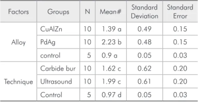
Table 2 presents a comparative analysis of the mean values for the principal factors versus the con-trol group. This analysis showed that, regarding the alloy factor (p = 0.025), PdAg post removal caused higher leakage values as compared to those of the control group. Regarding the technique factor, both
* Campos TN, Yamamoto E, Araki AT, Takahashi CU, Veiga JAL, Contin I. Temperature evaluation during the cast post removal with ultra-sonic vibration. Rev Odontol UNICID. In press 2006.
Table 1 - Descriptive analysis of leakage in the experimen-tal and control groups (mm).
Groups n Mean Standard
Deviation
Standard Error
Control 1 5 0.97 0.05 0.03
Carbide bur 2 CuAlZn 5 1.14 0.09 0.04
Carbide bur 3 PdAg 5 2.11 0.54 0.24
Ultrasound 4 CuAlZn 5 1.64 0.61 0.27
Ultrasound 5 PdAg 5 2.34 0.45 0.20
Table 2 - Mean leakage (mm) according to alloy and re-moval technique**.
Factors Groups N Mean# Standard
Deviation
Standard Error
Alloy
CuAlZn 10 1.39 a 0.49 0.15
PdAg 10 2.23 b 0.48 0.15
control 5 0.9 a 0.05 0.03
Technique
Carbide bur 10 1.62 c 0.62 0.20
Ultrasound 10 1.99 c 0.61 0.20
Control 5 0.97 d 0.05 0.03
techniques increased leakage as compared to the control group (p = 0.025).
Discussion
The apical seal of an endodontically treated tooth is an important factor for a successful restorative treatment. Concern with apical sealing has been the object of many researches investigating techniques and materials of endodontic illing, as well as the possibility of apical contamination during root prep-aration and with the use of a temporary restoration.
Coronal leakage is the main cause of failure of endodontic treatment.18 This leakage can progress and extend itself along the intraradicular post.20 The following are other reasons that may lead to the removal of an intraradicular retainer: presence of caries, inadequate design of the post (length and diameter) in relation to the dimensions of the canal, poor retention of the post or the core, badly execut-ed endodontic treatments, among others. The latter requires that an adequate illing be redone.
However, when the endodontic treatment is con-sidered clinically and radiographically acceptable, what should be the next step after removal of the intraradicular retainer?
Removal of the intraradicular retainer is a pro-cedure that requires care, since it can cause perfora-tions, cracks or root fractures. This procedure gen-erates heat and vibration that can disturb the apical seal without changing the radiographic image.
The results of the present research showed that statistically signiicant leakage occurred in all of the experimental groups. Comparisons between the ex-perimental groups and the control group were made to discard leakage due to procedures made before ce-mentation and removal of the intraradicular retain-ers. According to Table 1, there was higher leakage in Group 5 (PdAg-ultrasound), followed by Group 3 (PdAg-carbide bur), Group 4 (CuAlZn-ultrasound) and Group 2 (CuAlZn-carbide bur) consecutively.
In a previous research, removal of a CuAlZn intraradicular retainer with burs produced higher
temperatures than those produced by the removal of a PdAg one. The pressure on the handpiece was not standardized because the objective was to re-produce a clinical situation. The pressure developed was related to the hardness of the material. Cutting was intermittent but, because the CuAlZn alloy was softer, an extended length of the working time and more heat were produced. The PdAg alloy was more resistant to wear and required more pressure, so it was expected to generate higher temperatures than CuAlZn. However, the hardness made controlling of the handpiece more dificult and caused more dental wear, thus enlarging the canal and making refrigeration easier.4
In another study, removal of the same alloys fol-lowing the same method and with an ultrasound device generated a signiicant amount of heat, and the PdAg alloy showed much higher temperature values.*
The results of the previously mentioned articles did not present high temperatures on the internal surface of the canal maybe because the authors mea-sured it on the external surface of the teeth, and be-cause of refrigeration. It is to be supposed that the apical seal was disturbed mainly by vibration.
Ultrasound fractures the cement in the coronal area, leading to a migration of the fulcrum to the apical portion of the post. Ultrasonic vibration starts the moving of the post and generates stresses in the apical area, consequently moving the retainer.6
In the present research, two different alloys were selected with visibly different characteristics, mainly in relation to their hardness. Materials with higher hardness tend to conduct vibration better. Soft al-loys do not conduct ultrasound adequately.12,13 The dowel material thus inluences ultrasound action.15 Our results fortify these assertions as we observed higher leakage values with the harder alloy (PdAg) and with ultrasound.
Cardoso et al.5 (1995) stated that a root illing of at least 4 mm is necessary because, in their experi-ment, the post space preparations previously showed
a maximum leakage value of 3.23 mm.
In the present research, the external surface of the root was isolated to evaluate the inluence of post removal on the apical seal. Leakage was mea-sured in the cervico-apical direction, and the maxi-mum leakage mean value was 2.34 mm. Besides, an inverse leakage may be observed, as described by Habitante et al.11 (1989) who obtained smaller leak-age values with the apical preparation technique, varying from 0.83 to 1.85 mm.
Therefore, from a clinical standpoint, in relation to the removal of radicular posts, and based on the results obtained here, endodontic re-intervention is recommended especially when there is a root illing of less than or equal to 4 mm. It must also be con-sidered that, in most cases, the replaced posts had been in place for some time, and the age of the root illing is an important factor in terms of quality of the apical seal.
Conclusions
Based on the method used, it can be concluded that removal of the posts increased leakage through the apical seal both by means of the carbide burs and by means of ultrasound.
The degree of leakage was dependent on the type of alloy and on the removal technique, thus being directly related to the dificulty involved at the time of removal.
As a clinical guideline, it is suggested that end-odontic intervention should be repeated mainly in situations of dificult access.
Acknowledgment
The authors wish to thank the Foundation for the Scientiic and Technological Development of Den-tistry (FUNDECTO – Fundação para o Desenvolvi-mento Cientíico e Tecnológico da Odontologia) for a Scientiic Initiation Grant.
References
1. Alfredo E, Garrido AD, Souza-Filho CB, Correr-Sobrinho L, Sousa-Neto MD. In vitro evaluation of the effect of core diameter for removing radicular post with ultrasound. J Oral Rehabil. 2004;31(6):590-4.
2. Bando E, Kawashima T, Tiu IT, Nakano M. Removing dowels in difficult teeth. J Prosthet Dent. 1985;54(1):34-6.
3. Braga NM, Alfredo E, Vansan LP, Fonseca TS, Ferraz JA, Sousa-Neto MD. Efficacy of ultrasound in removal of in-traradicular posts using different techniques. J Oral Sci. 2005;47(3):117-21.
4. Campos TN, Yamamoto E, Mori M, Saito T. Evaluation of the temperature generated during the removal of posts with burs in high speed handpiece. Rev Odontol Univ São Paulo. 1998;12(3):253-6.
5. Cardoso RJA, Moura AAM, Antoniazzi JH. Análise compara-tiva in vitro da qualidade do selamento marginal pós-preparo para retentor intra-radicular realizado em tempos diversos, após obturação, frente a diferentes técnicas. RPG Rev Pós Grad. 1995;2(2):73-8.
6. Castrisos T, Abbott PV. A survey of methods used for post removal in specialist endodontic practice. Int Endod J. 2002;35(2):172-80.
7. de Deus G, Gurgel Filho ED, Ferreira CM, Coutinho Filho T. Intratubular penetration of root canal sealers. Pesqui Odontol Bras. 2002;16(4):332-6.
8. Garrido AD, Fonseca TS, Alfredo E, Silva-Sousa YT, Sousa-Neto MD. Influence of ultrasound, with and without water
spray cooling, on removal of posts cemented with resin or zinc phosphate cements. J Endod. 2004;30(3):173-6.
9. Gerstein H, Weine FS. Specially prepared burs to remove silver cones and fractured dowels. J Endod. 1977;3(11):408-10. 10. Gomes APM, Kubo CH, Santos DR, Padilha RQ. The
influ-ence of ultrasound on the retention of cast posts cemented with different agents. Int J Endod. 2001;34(2):93-9. 11. Habitante SM, Bombana AC, Pesce HF. Estudo comparativo da
influência do selamento marginal em canais radiculares obturados com e sem preparo apical. Rev Bras Odontol. 1989;46:18-22. 12. Hülsmann M. Methods for removing metal obstructions from
the root canal. Endod Dent Traumatol. 1993;9(6):223-37. 13. Imura N, Zuolo ML. Remoção de retentor intra-radicular com
aparelho de ultra-som. Rev Assoc Paul Cir Dent. 1997;51:262-7.
14. Leonardo MR, Salgado AA, da Silva LA, Tanomaru Filho M. Apical and periapical repair of dogs’ teeth with periapical lesions after endodontic treatment with different root canal sealers. Pesqui Odontol Bras. 2003;17(1):69-74.
15. Matsumura H, Salonga JP, Taira Y, Atsuta M. Effect of ul-trasonic instrumentation on bond strength of three dental cements bonded to nickel-chromium, alloy. J Prosthet Dent. 1996;75(3):309-13.
16. Paiva JG, Antoniazzi JH. Endodontia – Base para a prática clínica. 2a ed. São Paulo: Artes Médicas; 1993.
oxide-euge-nol-based sealers on the obturation of lateral canals. Pesqui Odontol Bras. 2002;16(2):127-30.
18. Saunders WP, Saunders EM. Coronal leakage as a cause of failure in root-canal therapy: a review. Endod Dent Traumatol. 1994;10(3):105-8.
19. Shemen BB, Cardash HS. A technique for removing cemented posts. J Prosthet Dent. 1985;54:200-1.
20. Tjan AH, Grant BE, Dunn JR. Microleakage of composite resin cores treated with various dentin bonding systems. J Prosthet Dent. 1991;66(1):24-9.
21. Usumez A, Cobankara FK, Ozturk N, Eskitascioglu G, Belli S. Microleakage of endodontically treated teeth with different dowel systems. J Prosthet Dent. 2004;92(2):163-9.
22. Williams VD, Bjorndal AM. The Masseran technique for the removal of fractured posts in endodontically treated teeth. J Prosthet Dent. 1983;49(1):46-8.