Reproducibility, reliability and validity
of measurements obtained from Cecile3
digital models
Abstract: The aim of this study was to determine the reproducibility, reliability and validity of measurements in digital models compared to plaster models. Fifteen pairs of plaster models were obtained from orth-odontic patients with permanent dentition before treatment. These were digitized to be evaluated with the program Cécile3 v2.554.2 beta. Two examiners measured three times the mesiodistal width of all the teeth present, intercanine, interpremolar and intermolar distances, overjet and overbite. The plaster models were measured using a digital vernier. The t-Student test for paired samples and interclass correlation coefi-cient (ICC) were used for statistical analysis. The ICC of the digital mod-els were 0.84 ± 0.15 (intra-examiner) and 0.80 ± 0.19 (inter-examiner). The average mean difference of the digital models was 0.23 ± 0.14 and 0.24 ± 0.11 for each examiner, respectively. When the two types of mea-surements were compared, the values obtained from the digital models were lower than those obtained from the plaster models (p < 0.05), al-though the differences were considered clinically insigniicant (differenc-es < 0.1 mm). The Cécile digital models are a clinically acceptable alter-native for use in Orthodontics.
Descriptors: Dental models; Reproducibility of results; Orthodontics.
Gustavo Adolfo Watanabe- Kanno(a)
Jorge Abrão(b)
Hiroshi Miasiro Junior(a) Alfonso Sánchez-Ayala(c) Manuel O. Lagravère(d)
(a) MSc Student, Graduate Program in
Orthodontics; (b)PhD, Associate Professor
– Department of Orthodontics, School of Dentistry, University of São Paulo, São Paulo, SP, Brazil.
(c) PhD Student, Graduate Program
in Prosthodontics, Department of Prosthodontics, Piracicaba Dental School, University of Campinas, Piracicaba, SP, Brazil.
(d)Research Associate, Graduate Program
in Orthodontics, Department of Dentistry, University of Alberta, Edmonton, Alberta, Canada.
Corresponding author:
Gustavo Adolfo Watanabe-Kanno Departamento de Ortodontia e Odontopediatria da FOUSP Av. Prof. Lineu Prestes, 2227, Cidade Universitária
São Paulo - SP - Brazil CEP: 05508-900 E-mail: [email protected]
Introduction
Digital technology is slowly inluencing differ-ent scidiffer-entiic areas and improving them. One of these areas is orthodontics, where diagnostic tools, digital photography and cephalometric software, among others, are improving this area.1 Plaster
models in orthodontics are necessary diagnostic tools but present several disadvantages such as the need for storage areas, high risk of breakage, loss of information and dificulty to send to other clini-cians in multidisciplinary cases.2,3 Some alternatives
to using plaster models have been suggested such as photocopies, photographs, holograms and digi-talization of points from the plaster cast.4,5 Digital
models now offer some advantages that include ease of storage and retrieval, ease of interofice trans-ferability, and possibly similar or better diagnostic ability.6 Some studies evaluated different methods
to calculate tooth size discrepancy and found no statistical signiicant differences in the measure-ments obtained from plaster and digital models, and suggested the need for further research to de-termine accuracy, reliability and reproducibility of digital models using new software versions.2,7 One
group of investigators found a signiicant difference between plaster and digital model linear measure-ments but concluded that the average difference did not appear to be clinically relevant and suggested that the software presented the advantage of being able to rotate and enlarge images on the screen.8,9
The accuracy and reliability of measurements made on computer-based models appeared to be as accu-rate and reliable as the ones obtained from plaster models.2 On the other hand, other investigators
found excellent reproducibility of digital models and signiicant differences in some measurements regarding reliability and validity, but these were not considered clinically signiicant.10,11
Cécile3, a digital modeling analysis software, was created in 2003 by Bibliocast, Montreuil. Some advantages of this software include the possibility of performing space analysis and creating virtual set-ups for treatment planning purposes. Nevertheless, there is a lack of literature with respect to its mea-surement validity, reliability and/or reproducibility. Digital models could be as reliable as plaster
models, and an additional tool for orthodontic di-agnosis. In this case, some fundamental factors such as spacing condition, teeth inclination, rota-tions, presence of interproximal contacts and other anatomical variations should also be considered.12-15
Because the need for evidence-based orthodontics is increasing, the degree of usefulness of different emerging methods ought to be evaluated. For these reasons, the objective of the present study is to de-termine the reproducibility, reliability, and valid-ity of measurements obtained from Cécile3 digital models.
Material and Methods
The study sample consisted of ifteen pairs of randomly selected initial model casts (upper and lower) from patients that presented for treatment at the orthodontic clinic, School of Dentistry, Universi-ty of São Paulo. Inclusion criteria was that all 12-18 year old patients had to have complete permanent dentition erupted from irst molar to irst molar, no caries lesions, no interproximal wear, no extractions nor previous orthodontic treatment.
Transver-sal distances were also measured from the occluTransver-sal view, and overjet and overbite were measured us-ing the analysis tools (Figure 2). The plaster models were measured using a vernier digital caliper (Mitu-toyo, model 500-144B, Suzano, SP, Brazil) with an accuracy of 0.01 mm. The measurements obtained are described in Table 1 and were measured by two different investigators, each repeating the measure-ment set three times.
Statistical analysis
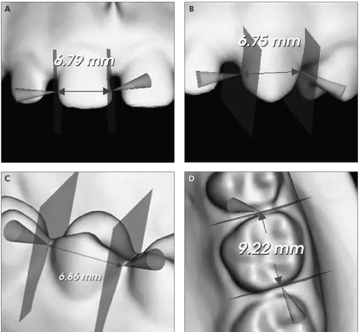
The data were analyzed using the statistical pro-gram SPSS for Windows version 16.0 (SPSS, Chica-go, IL, USA). Sample normality and homogeneity of variances were determined using the Shapiro-Wilks and Levene tests. The Intraclass correlation coefi-cient test (ICC) was used to determine the reproduc-ibility of the measures obtained. The Student t-Test was used to compare examiner reliability for both methods used and the validity between them. Figure 1 - Measurements of mesiodistal width of (A) incisor, (B) canine, (C) premolar and (D) molar using the Cécile3 tool, as shown from different views.
A B
Results
Intra- and inter-examiner reliability for both methods was generally high. The average intra-ex-aminer reliability was 0.852 ± 0.12 (range 0.706-0.940) and 0.824 ± 0.15 (range 0.663-0.927) for the plaster and Cécile3 digital models respectively. In-ter-examiner values were 0.818 ± 0.18 (range 0.575-0.931) and 0.782 ± 0.19 (range 0.506-0.917) for the plaster and Cécile3 digital models respectively. Overjet presented the highest intra-examiner reli-ability (0.996), while the mesiodistal width of the lower left canine presented the lowest value (0.537
for the plaster models and 0.367 for the Cécile3 dig-ital models). The maxillary interpremolar distance had the greatest inter-examiner reliability in plaster models (0.999) and the maxillary intercanine dis-tance had the greatest inter-examiner reliability in the Cécile3 digital models (0.998). The lowest inter-examiner value was found in the mesiodistal width of the lower left canine (0.318 in the plaster models and 0.158 in the Cécile3 digital models).
The mean measurement error for all measure-ments was obtained for the plaster and Cécile3 digi-tal models for both examiners. There were no sig-Figure 2 - Measurements of (A) intercanine, (B) interpremolar, (C) intermolar, (D) overjet and overbite using the Cécile3 tool.
A B
niicant differences in measurement errors in most measurements except for the lower right mandibular lateral incisor mesiodistal width, lower right irst premolar mesiodistal width, upper right lateral inci-sor mesiodistal width and lower interpremolar dis-tance (p < 0.05) for examiner 1; and except for the upper right canine mesiodistal width, lower inter-canine distance, lower interpremolar distance and overbite for examiner 2 (p < 0.05).
The second trial of measurements for each exam-iner was chosen to determine the validity of mea-surements (Table 2). All values obtained from the Cécile3 digital models were smaller than the values obtained from the plaster models with the exception of the upper interpremolar distance for examiner 2. The mean differences between plaster and Cécile3 digital models were 0.17 ± 0.06 mm for examiner 1. The lower right mandibular irst molar presented the highest variability (0.39 ± 0.28 mm). For examiner 2, the mean difference was 0.19 ± 0.06 mm, with the highest value obtained for overjet (0.31 ± 0.22 mm). The Student t-Test showed signiicant differences be-tween the measurements obtained from the Cécile3 and plaster models except for the irst molar, canine and central incisor of the lower left quadrant, second premolar and irst premolar from the upper right quadrant and upper left central incisor (p > 0.05)
for examiner 1. For examiner 2, the upper intermo-lar distance and lower intermointermo-lar distance did not present a signiicant difference (p > 0.05).
Discussion
The aim of this study was to determine if Cécile3 digital models are as reliable as plaster models. To achieve this, reproducibility, reliability and validity of the measurements obtained were evaluated.
Reproducibility of Cécile3 models
Reproducibility evaluates the agreement between two readings from the same sample (which in the present study would be for Cécile3 digital models and plaster models).16 While studying reproducibility
in orthodontics, Roberts, Richmond16 (1997) stated
that an ICC < 0.4 is considered low, between 0.4 and 0.75 is acceptable, and > 0.75 is good. The present study presented an ICC higher than 0.75 in the in-tra- and inter-examiner evaluations. Low values were only present for the mesiodistal width of the left man-dibular canine in plaster models (ICC-Intra-examin-er = 0.537 / ICC-Int(ICC-Intra-examin-er-examin(ICC-Intra-examin-er = 0.318) and Cécile3 models (ICC-Intra-examiner = 0.367 / ICC-Inter-ex-aminer = 0.158). These indings might be attributed to the fact that, in some cases, the interproximal area between the teeth is not clearly deined, which can alter the reproducibility of the measurements at the time of marking points. However, considering all the results, we can infer that the differences are clini-cally acceptable, and reproducibility is high. Stevens
et al.10 (2006), using the concordance correlation
co-eficient (CCC), showed that all 50 intra-examiner measurements had excellent reproducibility for both plaster and OrthoCad models with the exception of 7 measurements (4 plaster and 3 digital), which were considered good. Quimby et al.2 (2004) found a high
degree of reproducibility with an ICC > 0.90 for the measurements made on both plaster and computer-based models, measured by two examiners.
Reliability of Cécile3 digital models
Reliability was considered as the extent to which a measurement was repeatable under identical con-ditions, between Cécile3 digital models and plaster models.16 No statistical signiicant differences were
Table 1 - Measurement definitions.
Measurement Definition12,13,14,15
Mesiodistal Width
Greatest mesiodistal diameter from the anatomic mesial contact point to the anatomic distal contact point in each tooth, parallel to the occlusal surface.
Intercanine Distance
Straight distance between the crown tips of the canines.
Interpremolar Distance
Straight distance between the mesial fossae of the first premolars.
Intermolar Distance
Straight distance between the mesial fossae of the first molars.
Overjet
Distance between the incisal border of the more buccal upper central incisor and the buccal surface of the more lingual lower central incisor.
Overbite
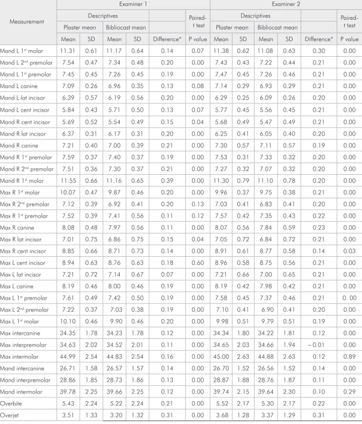
Table 2 - Measurement Means (mm)†.
Measurement
Examiner 1 Examiner 2 Descriptives
t test
Descriptives Paired-t Paired-tesPaired-t Plaster mean Bibliocast mean Plaster mean Bibliocast mean
Mean SD Mean SD Difference* P value Mean SD Mean SD Difference* P value Mand L 1st molar 11.31 0.61 11.17 0.64 0.14 0.07 11.38 0.62 11.08 0.63 0.30 0.00
Mand L 2nd premolar 7.54 0.47 7.34 0.48 0.20 0.00 7.43 0.43 7.22 0.44 0.21 0.00
Mand L 1st premolar 7.45 0.45 7.26 0.45 0.19 0.00 7.47 0.45 7.26 0.46 0.21 0.00
Mand L canine 7.09 0.26 6.96 0.35 0.13 0.08 7.14 0.29 6.93 0.29 0.21 0.00 Mand L lat incisor 6.39 0.57 6.19 0.56 0.20 0.00 6.29 0.25 6.09 0.26 0.20 0.00 Mand L cent incisor 5.84 0.43 5.71 0.50 0.13 0.07 5.77 0.45 5.56 0.45 0.21 0.00 Mand R cent incisor 5.69 0.52 5.54 0.49 0.15 0.04 5.68 0.49 5.47 0.49 0.21 0.00 Mand R lat incisor 6.37 0.31 6.17 0.31 0.20 0.00 6.25 0.41 6.05 0.40 0.20 0.00 Mand R canine 7.21 0.40 7.00 0.39 0.21 0.00 7.30 0.57 7.11 0.57 0.19 0.00 Mand R 1st premolar 7.59 0.37 7.40 0.37 0.19 0.00 7.53 0.31 7.33 0.32 0.20 0.00
Mand R 2nd premolar 7.51 0.36 7.30 0.37 0.21 0.00 7.27 0.32 7.07 0.32 0.20 0.00
Mand R 1st molar 11.55 0.66 11.16 0.65 0.39 0.00 11.30 0.79 11.10 0.78 0.20 0.00
Max R 1st molar 10.07 0.47 9.87 0.46 0.20 0.00 9.96 0.37 9.75 0.38 0.21 0.00
Max R 2nd premolar 7.12 0.39 6.92 0.41 0.20 0.13 7.03 0.41 6.83 0.41 0.20 0.00
Max R 1st premolar 7.52 0.39 7.41 0.56 0.11 0.12 7.57 0.42 7.35 0.43 0.22 0.00
Max R canine 8.08 0.48 7.97 0.56 0.11 0.00 8.07 0.56 7.84 0.59 0.23 0.00 Max R lat incisor 7.01 0.75 6.86 0.75 0.15 0.04 7.05 0.72 6.84 0.72 0.21 0.00 Max R cent incisor 8.85 0.66 8.71 0.73 0.14 0.00 8.91 0.61 8.77 0.58 0.14 0.03 Max L cent incisor 8.94 0.63 8.76 0.63 0.18 0.60 8.96 0.58 8.75 0.56 0.21 0.00 Max L lat incisor 7.21 0.72 7.14 0.67 0.07 0.00 7.21 0.66 7.00 0.65 0.21 0.00 Max L canine 8.19 0.46 8.00 0.46 0.19 0.00 8.19 0.42 7.98 0.42 0.21 0.00 Max L 1st premolar 7.61 0.49 7.42 0.50 0.19 0.00 7.58 0.45 7.37 0.46 0.21 0. 00
Max L 2nd premolar 7.22 0.37 7.03 0.38 0.19 0.00 7.10 0.41 6.90 0.41 0.20 0.00
Max L 1st molar 10.10 0.46 9.90 0.46 0.20 0.00 9.98 0.51 9.79 0.51 0.19 0.00
Max intercanine 34.35 1.78 34.23 1.78 0.12 0.00 34.34 1.80 34.22 1.81 0.12 0.00 Max interpremolar 34.63 2.02 34.52 2.01 0.11 0.00 34.65 2.03 34.66 1.94 - 0.01 0.00 Max intermolar 44.99 2.54 44.83 2.54 0.16 0.00 45.00 2.63 44.88 2.63 0.12 0.89 Mand intercanine 26.71 1.58 26.57 1.57 0.14 0.00 26.70 1.52 26.56 1.52 0.14 0.00 Mand interpremolar 28.86 1.85 28.73 1.86 0.13 0.00 28.87 1.88 28.76 1.87 0.11 0.00 Mand intermolar 39.78 2.25 39.66 2.25 0.12 0.00 39.74 2.15 39.64 2.30 0.10 0.29 Overbite 5.43 2.24 5.22 2.24 0.21 0.00 5.52 2.17 5.30 2.17 0.22 0.00 Overjet 3.51 1.33 3.20 1.32 0.31 0.00 3.68 1.28 3.37 1.29 0.31 0.00
Significant at P < .05; Max: maxillary, Mand: mandibular, L: left; R: right. †Time trial no. 2 of each examiner randomly selected for comparison. * + value
found in most variables measured for both examers. The measurement differences were clinically in-signiicant for examiner 1 (range 0.00-0.12 mm) and examiner 2 (range 0.00-0.09 mm). Similar results were obtained by Stevens et al.10 (2006), observing
a good reliability for the measurements obtained in OrthoCad and plaster models. Quimby et al.2 (2004)
demonstrated statistical differences between the plaster and digital models in all their measurements with the exception of the mandibular intercanine width. The measurements made on computer-based models showed greater variation in all categories ex-cept overbite and overjet. Most measurements dif-fered by less than 1 mm. These indings are similar to those of the present study where computer-based models appeared to be a clinically acceptable alter-native to conventional plaster models.
Validity of Cécile3 digital models
Validity was considered as the extent to which the Cécile3 digital models measured against the plaster models (gold standard).16 Plaster and Cécile3
digital models presented differences in mesiodistal tooth width measurements, intercanine distance, interpremolar distance, intermolar distance, overjet and overbite. The mean differences between the plas-ter and Cécile3 digital models for examiner 1 had a range between 0.07 mm and 0.39 mm; for examiner 2, the range was between 0.01 mm and 0.31 mm. Most of the obtained values were statistically dif-ferent (Table 2). Santoro et al.17 (2003) found
simi-lar results, where the mean differences ranged from 0.16 mm to 0.49 mm and were all statistically sig-niicant with the exception of overbite. Garino, Ga-rino8 (2002) and Rheude et al.6 (2005) found that
the measurements made from digital models were clinically acceptable, with reasonable reliability and reproducibility and adequate clinical informa-tion for diagnosis and treatment planning, thus eliminating the need for plaster models. Oliveira et al.1 (2007) did not ind signiicant differences with
the exception of the mesiodistal width of the lower right second premolar (p < 0.05). Zilberman et al.9
(2003) and Quimby et al.2 (2004) stated that even
when no signiicant differences were found, these appeared to be clinically acceptable. In the present
study, all values obtained from the Cécile3 digital models were smaller than those obtained from the plaster models. Similar results were found by Mul-len et al.11 (2007), who observed that the
measure-ments on the ball-bearing mounted models were on average 0.067 mm greater in the e-model software than the direct measurements obtained on the casts (range: 0 to –0.16 mm, p < .0045).
Limitations of Cécile3 digital models
Differences can be explained by the dificulty in locating the points, especially at the level of the in-terproximal contacts, which is affected by the oper-ator’s experience in handling a digital model. One disadvantage of digital models is that they have to be static in order to locate or mark the points need-ed to obtain a measurement.1 A digital model can be
blown up in the computer screen, which gives a sig-niicant advantage on locating landmarks because a 3-dimensional structure is viewed as a 2-dimen-sional image.14 In this study, the same dificulties
and advantages were experienced by the examiners. Prior training is required to use the software, since those more familiar with the computer resources are more capable of achieving more precise measure-ments. An extra dificulty observed was the presence of shadows (especially in crowded areas) in Cécile3 digital models resulting from the digitalization pro-cess. Also, occlusal anatomy and wear facets in Cécile3 digital models did not present a high dei-nition. With respect to overbite and overjet, it was shown that these measurements were inluenced by how the digital models were mounted in maximum intercuspidation on the computer. Finally, it was ob-served that Cécile3 digital models took considerably less time than plaster models to measure. This rep-resents a more eficient way of performing diagno-sis, as mentioned by Zilberman et al.9 (2003).
Conclusions
References
1. Oliveira DD, Ruellas ACO, Drummond MEL, Pantuzo MCG, Lanna ÂMQ. Confiabilidade do uso de modelos digitais tri-dimensionais como exame auxiliar ao diagnóstico ortodônti-co: um estudo piloto. Rev Dent Press Ortodon Ortop Facial. 2007;12(1):84-93.
2. Quimby ML, Vig KWL, Rashid RG, Firestone AR. The accu-racy and reliability of measurements made on computer-based digital models. Angle Orthod. 2004;74(3):298-303. 3. Martensson B, Ryden H. The holodent system, a new
tech-nique for measurement and storage of dental casts. Am J Or-thod Dentofacial Orthop. 1992;102(2):113-9.
4. Schirmer UR, Wilshire WA. Manual and computer aided space analysis: a comparative study. Am J Orthod Dentofacial Orthop. 1997;112(6):676-80.
5. Champagne M. Reliability of measurements from photocopies of study models. J Clin Orthod. 1992;26(10):648-50. 6. Rheude B, Sadowsky L, Ferriera A, Jacobson A. An evaluation
of the use of digital study models in Orthodontic Diagnosis and Treatment Planning. Angle Orthod. 2005;75(3):300-4. 7. Tomassetti JJ, Taloumis LJ, Denny JM, Fischer JR. A
com-parison of 3 computerized Bolton tooth-size analyses with a commonly used method. Angle Orthod. 2001;71(5):351-7. 8. Garino F, Garino GB. Comparison of dental arch
measure-ments between stone and digital casts. World J Orthod. 2002;3(3):250-4.
9. Zilberman O, Huggare JAV, Parikakis KA. Evaluation of the validity of tooth size and arch width measurements using con-ventional and three-dimensional virtual orthodontic models. Angle Orthod. 2003;73(3):301-6.
10. Stevens D, Flores-Mir C, Nebbe B, Raboud DW, Heo G, Ma-jor PW. Validity, reliability, and reproducibility of plaster vs
digital study models: Comparison of peer assessment rating and Bolton analysis and their constituent measurements. Am J Orthod Dentofacial Orthop. 2006;129(6):794-803. 11. Mullen SR, Martin CA, Ngan P, Gladwin M. Accuracy of
space analysis with emodels and plaster models. Am J Orthod Dentofacial Orthop. 2007;132(3):346-52.
12. Moorees CFA, Reed RB. Biometrics of crowding and spac-ing of the teeth in the mandible. Am J Phys Anthropol. 1954;12(1):77-88.
13. Keene HJ, Engel G. The mandibular dental arch, part IV: prediction and prevention of lower anterior relapse. Angle Orthod. 1979;49(30):173-80.
14. Bushang PH, Dermirjian A, Cadotte L. Permanent mesio-distal tooth size of French-Canadians. J Can Dent Assoc. 1988;54(6):441-4.
15. Santoro M. Mesiodistal dimensions and tooth size discrepancy of the permanent dentition of Dominican americans. Angle Orthod. 2000;70(4):303-7.
16. Roberts CT, Richmond S. The design and analysis of reliabil-ity studies for the use of epidemiological and audit indices in orthodontics. Br J Orthod. 1997;24(2):139-47.