Compared leaf anatomy of Nymphaea (Nymphaeaceae) species from Brazilian flood plain
Texto
Imagem
Documentos relacionados
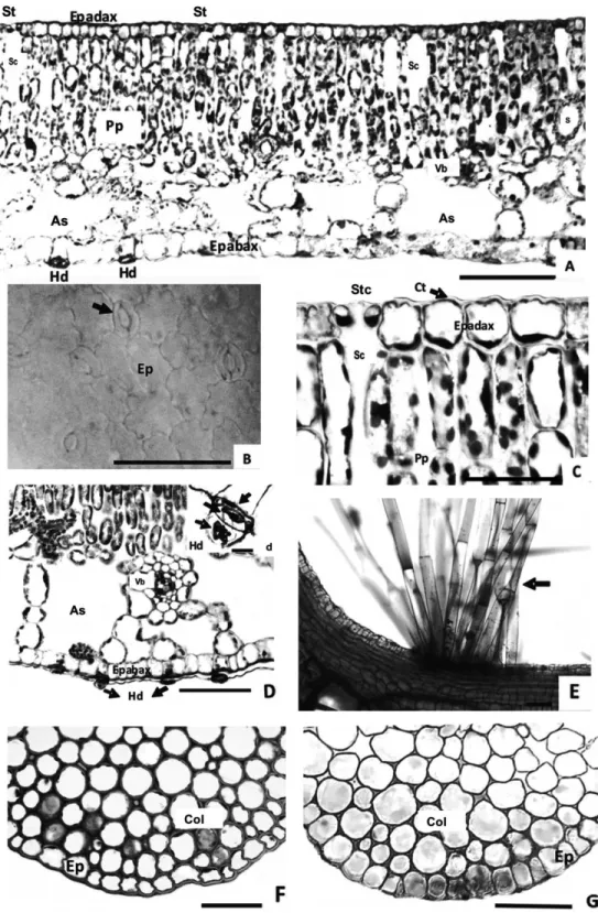
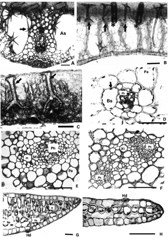
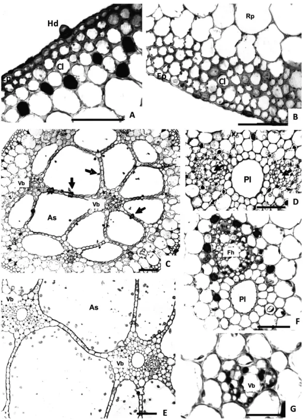
Anatomic features observed in the three studied species such as: midrib with complex vascular system, mesophyll consisting of tabular lobed chlorophyllous elements and fusoid
We studied communities of leaf litter ants from the Brazilian Atlantic Forest, compared richness estimates for genera and species, and built species accumulation curves with
The characters that contributed to the formation of the groups were a petiole with a contour that was circular or plano-convex, epicuticular wax in plates on the abaxial surface
A: apex of the culm with flowering branches; B: apex of the leaf-sheath and base of leaf blade; C: apex of the leaf-sheath, ligular region and base of leaf blade; D: detail of
The tepals (Figs. 2A, C) presented uniseriate epidermis with cuticle flange, and periclinal thick- walled cells in the abaxial face and thin-walled cells in the
The species examined here demonstrated ovarian struc- tures typical of the Eupatorieae, including an uniseriate epidermis with trichomes, outer mesophyll with closely juxtaposed
In the species studied here, stegmata were observed in the petiole epidermis, close to lignified cells, and also above the vascular bundles in the leaf blade.. They may therefore act
In most of the studied species, the epidermis, in adaxial surface is composed of ordinary epidermal cells, with sinuous anticlinal walls (Fig. 1 a-f), and thick, notably in
