Minor segmental wall motion
abnormalities detected in patients
with Chagas’ disease have adverse
prognostic implications
Divisão de Cardiologia, Departamento de Clínica Médica,
Faculdade de Medicina de Ribeirão Preto, Universidade de São Paulo, Ribeirão Preto, SP, Brasil
A. Pazin-Filho, M.M.D. Romano, O.C. Almeida-Filho, M.S. Furuta, L.F. Viviani, A. Schmidt, J.A. Marin-Neto and B.C. Maciel
Abstract
Recent data from our laboratory have shown that patients with the indeterminate form of Chagas’ disease can have impairment of left ventricular contractility, as evaluated by the slope of the left ventricle end-systolic pressure-dimension relationship. We also showed that Chagas’ disease patients with minimal baseline wall motion abnor-malities detected by two-dimensional echocardiography have more intense contractility impairment when compared to patients with the indeterminate form of the disease without this abnormality. The prognostic implications of these findings have not been established. We evaluated 59 patients (37-76 years, mean = 55 years) with differ-ent clinical forms of Chagas’ disease, who had normal left vdiffer-entricular global systolic function at baseline (57.6 ± 6.9%) and who had at least one additional echo during clinical follow-up (0.4-17.6; mean 4.6 years). Group 1 consisted of 14 patients with minor baseline left ventricle wall motion abnormalities and group 2 consisted of 45 patients without these abnormalities. During follow-up, global left ventricle systolic function deterioration was observed in 10 group 1 patients (71.4%) and in only 10 group 2 patients (22.2%; P < 0.005). Age and duration of follow-up were not independent determinants of left ventricular function deterioration in these patients. The present data indicate that mild segmental left ventricular wall motion abnor-malities are associated with worsening of systolic function in Chagas’ disease patients who have normal baseline global systolic perfor-mance.
Correspondence B.C. Maciel Divisão de Cardiologia Departamento de Clínica Médica FMRP, USP
Av. Bandeirantes, 3900 14048-900 Ribeirão Preto, SP Brasil
Fax: +55-16-3633-0869 E-mail: [email protected]
Publication supported by FAPESP.
Received May 18, 2005 Accepted January 2, 2006
Key words
•Echocardiography •Chagas’ disease •Left ventricular function •Indeterminate form of
Chagas’ disease
Chagas’ disease remains a significant public health issue in many countries and is a major cause of morbidity and mortality in Latin America. The social impact of this disease can be evaluated by the estimated human infection prevalence of 16 million
form (2,3). In the natural history of the dis-ease, the progression of cardiac involvement is heterogeneous and tends to be slow: about 70% of cardiac patients who have mild car-diac involvement, without heart failure, re-main clinically stable for periods ranging from 12 to 27 years of follow-up (4).
The majority of patients with Chagas’ disease remain in the indeterminate form of the disease for 10-30 years or even more. They have positive serology, no symptoms or signs of Chagas’ disease, and normal ECG and heart, esophagus and colon X-rays (5). Different diagnostic techniques (vector-cardiography, echo(vector-cardiography, radionu-clide ventriculography, exercise stress test, Holter monitoring, endomyocardial biopsy) have shown some degree of cardiac abnor-malities in a substantial number of these patients (5). Although their prognostic im-plications are not completely clear, these findings do not appear to significantly influ-ence the progression of the disease to symp-tomatic clinical forms, and patients with the indeterminate form usually have an excel-lent long-term prognosis. Despite the dis-crepant observations regarding ventricular systolic dysfunction in patients with the in-determinate form of Chagas’ disease, it should be pointed out that a substantial pro-portion of these patients may have minor impairment of segmental ventricular func-tion. We have previously shown (6) that patients with the indeterminate form of Chagas’ disease can have impairment of left ventricular contractility, as evaluated by the slope of the left ventricle end-systolic pres-sure-dimension relationship. Our results also showed that, independent of the clinical clas-sification, patients who had minor left ven-tricular segmental wall motion abnormali-ties (LVWMA) on baseline two-dimensional (2-D) echocardiogram presented a more de-pressed myocardial contractility when com-pared to patients who had only isolated con-duction abnormalities (6) on ECG. These data were consistent with a more extensive
and diffuse myocardial involvement in these patients. Although the clinical classification of patients with Chagas’ disease may vary from country to country (7), in Brazil, mild isolated regional wall motion abnormalities documented in Chagas’ disease patients are not unanimously considered to be a hall-mark for classifying a patient as presenting the cardiac form of the disease.
University Hospital Research Ethics Com-mittee.
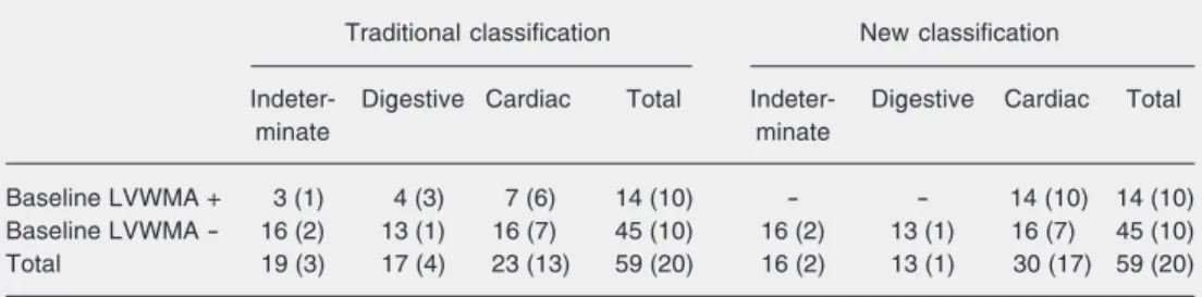
Fifty-nine patients (mean age 55.1 years; range: 37-76 years) who fulfilled the inclu-sion criteria were selected based on the tra-ditional clinical classification, 23 had the cardiac form (38%), 17 the digestive form (30%) and 19 the undetermined form (32%); using the new classification 30 had the car-diac form (51%), 13 the digestive form (22%) and 16 had the undetermined form (27%). The mean average follow-up was 4.6 years (range: 0.4-17.6 years). The patients were divided into two groups: 1) with LVWMA (N = 14) and 2) without LVWMA, at base-line 2-D echocardiography (N = 45). The two groups were analyzed to identify any worsening of left ventricular systolic func-tion (extension of LVWMA as documented by worsening of wall motion in at least two left ventricular segments based on semi-quantitative grading and/or reduction of at least 5% in left ventricular ejection fraction) during follow-up. 2-D left ventricular ejec-tion fracejec-tion was obtained off-line using the Simpson’s method, incorporated into the ul-trasound system.
Data are reported as mean ± SD. The chi-square test was used to compare proportions of patients with or without left ventricular deterioration during follow-up. A P value < 0.05 was considered to be statistically sig-nificant.
Baseline mean ejection fraction was 57.6
± 6.9%. Among patients presenting minor LVWMA, 71.4% showed worsening of left ventricular systolic function during follow-up while only 22.2% of those without LVWMA had left ventricular systolic func-tion deteriorafunc-tion (P = 0.004; chi-square test; Table 1). The number of patients with differ-ent clinical forms of Chagas’ disease who had worsening of left ventricular wall mo-tion on 2-D echocardiography during fol-low-up is shown in Table 2. The deteriora-tion of left ventricular systolic funcdeteriora-tion was notdependent on patient age or duration of follow-up (chi-square test).
The clinical manifestations of the chronic cardiac phase of Chagas’ disease are quite variable: patients can be asymptomatic or may present different types of arrhythmias (including potentially lethal arrhythmias), atrioventricular block, conduction
abnormali-Table 1. Deterioration of left ventricular systolic function during follow-up as a function of baseline left ventricular wall motion abnormalities (LVWMA).
Deterioration of Total
left ventricle systolic function during follow-up
Yes No
Baseline LVWMA
Present 10 4 14
Absent 10 35 45
Total 20 39 59
Data are reported as number of patients.
Table 2. Deterioration of left ventricular systolic function during follow-up of the different clinical forms of Chagas’ disease according to the traditional and new classification systems.
Traditional classification New classification
Indeter- Digestive Cardiac Total Indeter- Digestive Cardiac Total
minate minate
Baseline LVWMA + 3 (1) 4 (3) 7 (6) 14 (10) - - 14 (10) 14 (10)
Baseline LVWMA - 16 (2) 13 (1) 16 (7) 45 (10) 16 (2) 13 (1) 16 (7) 45 (10)
Total 19 (3) 17 (4) 23 (13) 59 (20) 16 (2) 13 (1) 30 (17) 59 (20)
ties on electrocardiogram, arterial or pulmo-nary embolism, or congestive heart failure (4). Left ventricular dysfunction, left ven-tricular aneurysm and congestive heart fail-ure are recognized as important predictors of mortality in these patients. The magnitude of changes in left ventricular systolic function in Chagas’ disease is quite variable depend-ing on the degree of myocardial involve-ment of each patient. Seginvolve-mental LVWMA based on echocardiography and radionuclide or contrast ventriculography has shown a number of abnormalities in chronic cardiac chagasic patients: apical dyskinesia was documented in 50-65% of symptomatic car-diac patients, while akinesia or hypokinesia was observed in the same region in 20% of patients; on the other hand, wall motion abnormalities involving the postero-inferior wall are reported in 15-20% of patients and are rarely detected in the anterolateral wall; finally, diffuse hypokinesia was observed in 15-40% of patients (8). A significant pro-portion of these patients have segmental wall motion abnormalities documented in more than one left ventricular region. It is espe-cially relevant to mention that this variable pattern of segmental involvement of the left ventricle is not related to obstructive lesions in epicardial coronary arteries, which are usually normal or even dilated in chagasic patients (4).
Evaluation of left ventricular systolic function in patients with the indeterminate form of Chagas’ disease has shown discrep-ant results ranging from absence of any ven-tricular function abnormality to the occur-rence of apical dyskinesia or akinesia in 10-68% of patients (8). Despite these heteroge-neous observations regarding the degree of
myocardial involvement in the indetermi-nate form of the disease, and even consider-ing the limitation related to inadequate clini-cal characterization of the indeterminate form in several investigations (5), it should be recognized that a substantial proportion of these patients have minor impairment of segmental ventricular function (9).
The present findings indicate that minor LVWMA documented at baseline 2-D echo-cardiography in Chagas’ disease patients who have normal global systolic function is a predictor of deterioration of ventricular func-tion during follow-up. In addifunc-tion, the pres-ent results suggest that patipres-ents who fulfill current criteria for the indeterminate form of Chagas’ disease (no symptoms or physical signs, specific serological positivity, normal ECG and heart, esophagus and colon X-rays) but have minor LVWMA in 2-D echo-cardiography should be considered to pres-ent the cardiac form of Chagas’ disease. The present findings also support the proposal that an objective assessment of left ventricle function should always be carried out in Chagas’ disease patients presenting the in-determinate form of the disease (8). We believe that this preliminary observation should be confirmed in a larger prospective study.
References
1. Wanderley DMV & Corrêa FMA (1995). Epidemiology of Chagas’ heart disease. São Paulo Medical Journal, 113: 742-749.
2. Coura JR (1988). Determinantes epidemiológicos da doença de Chagas no Brasil: a infecção, a doença e sua morbi-mortalidade.
Memórias do Instituto Oswaldo Cruz, 83 (Suppl 1): 392-402. 3. Dias JCP (1985). História natural. In: Cançado JR & Chuster M
(Editores), Cardiopatia Chagásica. Fundação Carlos Chagas, Belo Horizonte, MG, Brazil.
4. Marin-Neto JA, Simões MV & Maciel BC (1998). Other cardiomyo-pathies. In: Yusif S, Cairns JA, Camin AJ et al. (Editors), Evidence Based Cardiology. BMJ Books, London, UK.
5. Pereira-Barreto AC & Ianni BM (1995). The undetermined form of Chagas’ heart disease: concept and forensic implications. São Paulo Medical Journal, 113: 797-801.
6. Almeida-Filho OC, Maciel BC, Schmidt A et al. (2002). Minor
seg-mental dyssynergy reflects extensive myocardial damage and glo-bal left ventricle dysfunction in chronic Chagas disease. Journal of the American Society of Echocardiography, 15: 610-616.
7. Hagar JM & Rahimtoola SH (1995). Chagas’ heart disease. Current Problems in Cardiology, 20: 825-924.
8. Maciel BC, Almeida-Filho OC, Schmidt A et al. (1995). Ventricular function in Chagas’ heart disease. São Paulo Medical Journal, 113: 814-820.
9. Marin-Neto JA, Pazin-Filho A, Schmidt A et al. (2002). Forma inde-terminada da moléstia de Chagas. Proposta de novos critérios de caracterização e perspectivas de tratamento precoce da cardiomio-patia. Arquivos Brasileiros de Cardiologia, 79: 623-627.