Respiration, oxidative phosphorylation,
and uncoupling protein in
Candida
albicans
Departamento de Patologia Clínica, Faculdade de Ciências Médicas, Universidade Estadual de Campinas, Campinas, SP, Brasil
R.A. Cavalheiro, F. Fortes, J. Borecký, V.C. Faustinoni, A.Z. Schreiber and A.E. Vercesi
Abstract
The respiration, membrane potential (∆ψ), and oxidative phosphory-lation of mitochondria in situ were determined in spheroplasts ob-tained from Candida albicans control strain ATCC 90028 by lyticase treatment. Mitochondria in situ were able to phosphorylate externally added ADP (200 µM) in the presence of 0.05% BSA. Mitochondria in situ generated and sustained stable mitochondrial ∆ψ respiring on 5
mM NAD-linked substrates, 5 mM succinate, or 100 µM N,N,N’,N’-tetramethyl-p-phenylenediamine dihydrochloride plus 1 mM ascor-bate. Rotenone (4 µM) inhibited respiration by 30% and 2 µM antimycin A or myxothiazole and 1 mM cyanide inhibited it by 85%. Cyanide-insensitive respiration was partially blocked by 2 mM benzo-hydroxamic acid, suggesting the presence of an alternative oxidase. Candida albicans mitochondria in situ presented a carboxyatractylo-side-insensitive increase of ∆ψ induced by 5 mM ATP and 0.5% BSA,
and ∆ψ decrease induced by 10 µM linoleic acid, both suggesting the
existence of an uncoupling protein. The presence of this protein was subsequently confirmed by immunodetection and respiration experi-ments with isolated mitochondria. In conclusion, Candida albicans ATCC 90028 possesses an alternative electron transfer chain and alternative oxidase, both absent in animal cells. These pathways can be exceptional targets for the design of new chemotherapeutic agents. Blockage of these respiratory pathways together with inhibition of the uncoupling protein (another potential target for drug design) could lead to increased production of reactive oxygen species, dysfunction of Candida mitochondria, and possibly to oxidative cell death.
Correspondence
A.E. Vercesi
Departamento de Patologia Clínica Faculdade de Ciências Médicas UNICAMP
Caixa Postal 6111 13083-970 Campinas, SP Brasil
Fax: +55-19-3788-1118 E-mail: [email protected]
Research supported by CNPq/ PRONEX (No. 66.1192/1997-2), FAPESP (Nos. 98/13012-5 and 00/08019-2), and CAPES. R.A. Cavalheiro and V.C. Faustinoni are supported by CAPES. F. Fortes is the recipient of a doctoral fellowship from CNPq, and J. Borecký is the recipient of Visiting Professor from CNPq (No. 305381/02-4 NV).
Received November 18, 2003 Accepted June 8, 2004
Key words
•Candida albicans
spheroplasts
•Mitochondria •Respiratory chain •Mitochondrial membrane
potential
•Uncoupling protein
Introduction
Candidiases are common infections of the skin, oral cavity, esophagus, gastrointes-tinal tract, vagina, and vascular system, and have become a major cause of mortality in immunocompromised patients, including
(57%), C. tropicalis (13%), C. glabrata (10%), C. parapsilosis (6%), and C. krusei (3.4%), and 10.6% belonged to other yeast genera (5). The chemotherapy currently used against these fungi has a number of deficien-cies such as low specificity, toxicity, and drug resistance. Most antifungal drugs react with ergosterol or inhibit its production (6); however, there is an alarming increase in resistance to antifungal agents (3,7). There-fore, it is very important to search for bio-logical targets that could be exploited for the rational development of improved therapies. Mitochondria are the major source of cellular aerobic energy generated by oxida-tive phosphorylation. C. parapsilosis mito-chondria possess three types of respiratory pathways (8): i) the classical respiratory chain, ii) a secondary parallel respiratory chain (PAR) consisting of alternative ubi-quinone, cytochromes (cytbPAR and cytcPAR),
and a terminal oxidase (oxcPAR) insensitive
to antimycin A but inhibited by amytal (9) and by high concentrations of salicylhydrox-amic acid (SHAM), myxothiazol (10), and KCN (11), and finally iii) the constitutive alternative oxidase (12,13). The high resis-tance of C. parapsilosis to drugs (14) has been attributed to the presence of these two alternative electron transfer systems (8). Recently, Helmerhorst et al. (13) reported that C. albicans also contains a variety of respiratory systems whose expression de-pends on growth conditions. The authors reported that the C. albicans ability to regu-late the expression of individual complexes and the partitioning of electrons between both respiratory chains may be the reason why the cells can survive under conditions of oxidative stress.
Another mechanism of cell protection against oxidative stress may involve the re-cently described isoforms of uncoupling pro-tein (UCP, now UCP1; 15). The demonstra-tion of various UCP homologues in plants (16-18) and animals (17-18) raised the ques-tion of their true physiological role (18-21)
that should be more general than that of UCP1 - the production of heat in brown adipose tissue of hibernating mammals (15). Evidence has been presented that the new UCPs are involved in cell defense against oxidative stress (22-27). They are able to induce mild uncoupling, thus increasing the rate of respiration and decreasing the pro-duction of reactive oxygen species (ROS) (24-27).
A UCP was also detected recently in C. parapsilosis (28). Therefore, it is of great interest to study C. albicans UCP in order to understand its role in the ability of these cells to resist stress. In the present study, we dem-onstrated that spheroplasts obtained from cultures of C. albicans in the middle of their exponential phase of growth possess intact mitochondria able to phosphorylate ADP and an uncoupling protein (CaUCP) homolo-gous to the previously described C. parapsi-losis UCP (CpUCP) (28).
Material and Methods
Candida albicans
C. albicans ATCC 90028 was obtained from the American Type Culture Collection. It is a quality control strain for antifungal susceptibility testing and was maintained in sterile water and Sabouraud Dextrose Agar (BD, Difco, Franklin Lakes, NJ, USA). The cell cultures were grown in YEPG medium (29) at 37ºC and 200 rpm. To determine the inflexion point of exponential growth, samples were collected at 1-h intervals and culture density was determined spectropho-tometrically at 530 nm. Experimental points were fitted using a logistic dose-response model and the maximum growth rate during the exponential phase was calculated as its power parameter.
Spheroplast preparation
centrifugation for the standard preparation (29) and cells were washed once with cold water and once with buffer A (1 M sorbitol,
10 mM MgCl2, and 50 mM Tris-HCl, pH
7.8). Cells were resuspended in buffer A (3 ml/g of cells) supplemented with 30 mM dithiothreitol. After 15 min at room temper-ature with shaking, cells were harvested, resuspended in buffer A containing lyticase (1 mg/g of cells) and 1 mM DTT, and incu-bated at 30ºC until about 90% of cells were converted to spheroplasts (~60 min). The reaction was stopped with an equal volume of ice-cold buffer A and spheroplasts were washed twice with buffer A. The protein concentration of the final suspension was determined by the biuret method (30) in the presence of 0.2% deoxycholate.
Isolation of mitochondria
The spheroplasts were resuspended in buffer B1 (0.6 M mannitol, 1 mM EDTA,
0.5% BSA, 1 mM PMSF, and 10 mM Tris-HCl, pH 7.4). Spheroplasts were broken mechanically using a Dounce homogenizer with a maximum of 10 strokes and cell de-bris were removed by centrifugation at 1,000 g for 10 min. Mitochondria were pelleted by 10-min centrifugation at 10,500 g and washed with buffer B2 (0.6 M mannitol, 1 mM EDTA,
1% BSA, 10 mM Tris-HCl, pH 7.0). The last wash was made in buffer B2 medium without
BSA and EDTA (28) and mitochondrial pro-tein concentration was determined by the biuret method (30).
Mitochondrial membrane potential
The mitochondrial membrane potential (∆ψ) of permeabilized cells was monitored by measuring the fluorescence spectrum of safranine O with a Hitachi F4500 fluores-cence spectrophotometer (Hitachi, Tokyo, Japan) at excitation-emission wavelength of 495-586 nm (31). All experiments were per-formed at 28ºC in 2 ml of standard
incuba-tion medium containing 125 mM sucrose, 65 mM KCl, 10 mM HEPES, pH 7.2, 2.5 mM KH2PO4, and 1 mM MgCl2, plus 1 mg
mito-chondrial protein.
Oxygen uptake
Oxygen uptake was measured polaro-graphically at 28ºC using a Clark-type elec-trode (Yellow Springs Instruments, Yellow Springs, OH, USA) in 1.3 ml of standard incubation medium containing 1 mg mito-chondrial protein (28).
Immunodetection of Candida albicans UCP
Fifty micrograms of total proteins extract-ed from spheroplasts preparextract-ed from cultures of C. albicans and C. parapsilosis (positive control) was separated on 10% polyacryl-amide gel by standard SDS-PAGE. Protein bands were transferred to a nylon membrane (Hybond N) with a semi-dry blotting appara-tus (Amersham Biosciences AB (Pharma-cia) Uppsala, Sweden). The membrane was blocked overnight in 20 mM Tris-HCl, pH 7.4, 137 mM NaCl, 0.1% (v/v) Tween 20, and 10% (w/v) non-fat dry milk and incu-bated with the anti-AtPUMP1 polyclonal antibody (1:1000 dilution). After incubation with an anti-rabbit IgG alkaline phosphatase conjugate (1:5000 dilution), the membrane was developed in the dark in 100 mM Tris-HCl, pH 9.5, 100 mM NaCl, and 12.5 µM CSPD for 5 min. The bands were visualized by autoradiography and scanned with the Eagle-Eye photo documentation system (Eagle Eye Photo, Homer, AK, USA) (32).
Chemicals
viously applied to C. parapsilosis (29). In order to search for the presence of UCP and to examine its activity in these mitochondria we characterized the classical respiratory chain and its ability to sustain ∆ψ by oxidizing different respiratory substrates. Figure 1 shows that spheroplasts prepared from cell cultures grown to the middle expo-nential phase were permeable to mitochon-drial substrates and inhibitors without af-fecting the functional integrity of the sphero-plast mitochondria. These spherosphero-plast mito-chondria suspended in situ in reaction medi-um containing a cocktail of NAD-linked sub-strates generated and sustained a stable mem-brane potential and respiration rate of 43 nmol O2 min-1 mg protein-1 (Figure 1). The
subsequent inhibition of complex I by 4 µM rotenone resulted in a decrease of about 30% in the respiration rate (29 nmol O2 min-1 mg
protein-1) and
∆ψ. This suggests a significant contribution of this complex to ∆ψ genera-tion, in agreement with the results obtained by Helmerhorst et al. (13). They found the presence of a rotenone-sensitive and a pro-ton pumping NADH-Q oxidoreductase simi-lar to complex I in C. albicans ATCC 10231. The addition of 5 mM succinate restored ∆ψ but total oxygen consumption level was un-changed (29 nmol O2 min-1 mg protein-1).
The increase in ∆ψ by succinate addition was certainly the consequence of an increased electron flux through the classic respiratory pathway (coupled respiration - the only one that increases ∆µH+) associated with some
alteration in the partitioning of electrons among the three respiratory pathways (clas-sic, parallel, and via alternative oxidase) pres-ent in Candida yeast (13), resulting in a decrease in electron flux through the un-coupled pathways. Therefore, the total res-piration rate was not modified by the addi-tion of succinate. Blockage of complex III by 2 µM antimycin A or 2 µM myxothiazol promoted collapse of ∆ψ and a reduction of more than 90% in respiration (4 nmol O2
min-1 mg protein-1). Addition of 100 µM
Fluorescence
50 nmol O
2
495-586 nm
Rotenone Succinate
2 min
Cells TMPD
AA/Mix
KCN BHAM
29 29 43
4 28
6 0
Figure 1. Effect of substrates and inhibitors on Candida albi-cans spheroplast oxygen con-sumption (A) and mitochondrial membrane potential (B). C. albi-cans spheroplasts (1 mg/ml) were incubated in reaction me-dium containing 125 mM su-crose, 10 mM HEPES, pH 7.2, 2 mM Pi, 1 mM MgCl2, 65 mM
KCl, and 5 mM cocktail (malate, glutamate, pyruvate, and α -ke-toglutarate). The additions of the substrates 5 mM succinate and 100 µM TMPD/ascorbate (TMPD), and the inhibitors 4 µM rotenone, 2 µM antimycin A (AA) and/or 2 µM myxothiazol (Mix), 1 mM
KCN, and 2 mM BHAM, are indicated by the arrows. Numbers on the trace indicate O2
consumption rates in nmol O2 min-1 mg protein-1. TMPD =
tetramethyl-p-phenylenedia-mine dihydrochloride; BHAM = benzohydroxamic acid.
A
B
kDa
49
29
Cp Ca
Figure 2. Immunodetection of uncoupling proteins in a total protein extract of Candida albi-cans (2 bands in lane Ca) with polyclonal antibody raised against Arabidopsis PUMP1. A C. parapsilosis cell protein ex-tract containing CpUCP (single band in lane Cp) was used as a positive control. The positions of molecular mass standards are indicated by the arrows.
(TMPD), and lyticase were purchased from Sigma (St. Louis, MO, USA). Disodium 3- (4-methoxyspiro{1,2-dioethane-3,2'-(5'-chloro) tricyclo [3.3.1.13,7) decane}-4-yl]
phenylphosphate (CSPD) was from Tropix (Applied Biosystems (Tropix), Foster City, CA, USA). All other reagents were of ana-lytical grade.
Results and Discussion
pre-TMPD/1 mM ascorbate restored the respira-tion rate (28 nmol O2 min-1 mg protein-1).
Finally, inhibition of complex IV by 1 mM KCN was accompanied by a complete loss of ∆ψ and by a decrease of about 85% in respiration rate (6 nmol O2 min-1 mg protein-1).
Drastic changes in ∆ψ after inhibition of complexes III and IV suggest the essential role of these complexes in energy conserva-tion by C. albicans mitochondria. The CN-
-insensitive respiration was inhibited by 2 mM benzohydroxamic acid, suggesting the presence of alternative oxidases, as previ-ously described (8,13). In contrast, Helmer-horst et al. (13) reported that respiration by C. albicans ATCC 10231 cells was almost insensitive to 5 mM SHAM when grown in the absence of antimycin A. On the other hand, respiration by cells grown in the pres-ence of 10 µM antimycin A was completely inhibited by 5 mM SHAM, indicating that the alternative pathway was the only path-way utilized by C. albicans mitochondria when the classical cytochrome pathway was inhibited at the cytochrome c oxidase level. An uncoupling protein, another energy-dissipating system that can also modulate ∆ψ and the respiration rate, was recently identi-fied in the fungus kingdom as CpUCP from C. parapsilosis (28). Interestingly, UCP is absent in Saccharomyces cerevisiae (33). In the pres-ent study, the existence of a CpUCP homo-logue (CaUCP) was also detected in C. albi-cans mitochondria by immunoblot analysis using anti-AtPUMP1 antibody (32) and CpUCP (line Cp) as a positive control. The immunoblot revealed 2 protein bands of about 32 kDa (Figure 2, lane Ca) in a sample of total protein extract from C. albicans cells. The existence of two bands in C. albicans suggests the presence of more than one UCP, as ob-served in mammals and plants (17,18,34,35). Uncoupling proteins are known to be activated by free fatty acids (FFAs) and in-hibited by ATP (36-39). Accordingly, in situ ∆ψ measurements in C. albicans mitochon-dria demonstrated an increase of ∆ψ after
Fluorescence
Fluorescence
495-586 nm
A
B
495-586 nm
Cells
2 min
LA
ATP BSA
FCCP
BSA
Cells 2 min
c b
a
e
d FCCP
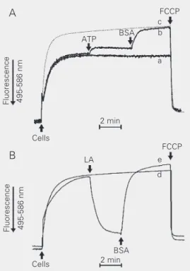
Figure 3. Effect of ATP and BSA (A) or LA (B) on mitochondrial ∆ψ of Candida albicans sphero-plasts in the presence of 1 µM CAT. Spheroplasts (1 mg/ml) were added to standard medi-um containing 5 mM substrate cocktail (malate, glutamate, pyruvate, and α-ketoglutarate) and 5 µM safranine O. Additions of 5 mM ATP (line b), 0.5% BSA (lines b and e), 10 µM LA (line e), and 1 µM FCCP are indicated by the arrows. Lines a and d are controls without additions and line c is a control with ATP and BSA present from the beginning of the experiment. BSA = bo-vine serum albumin; CAT = car-boxyatractyloside; ∆ψ = mem-brane potential; FCCP = carbo-nyl cyanide p-trifluoromethox-yphenylhydrazone; LA = linoleic acid.
consecutive additions of 5 mM ATP and 0.5% BSA, which binds non-covalently FFAs (Figure 3A, trace b) as compared to control experiments without additions (trace a). The dotted line (c) shows the ∆ψ generated by C. albicans mitochondria in the presence of ATP and BSA from the beginning of the experiment. In contrast, the addition of lino-leic acid caused a decrease in ∆ψ that was reversed by BSA (Figure 3B, trace e), reach-ing levels higher than control at the end (trace d), probably because BSA also binds the endogenous FFAs. These experiments were performed in the presence of 1 µM CAT to prevent the contribution of the ADP/ ATP carrier to the FFA-induced H+
re-up-take through FFA anion translocation (23). Indeed, it is known that the ADP/ATP car-rier in the absence of its substrates ADP and ATP can translocate FFA (23). In addition, the presence of glutamate, pyruvate, and malate/α-ketoglutarate prevented fatty acid anion transport through the corresponding carriers (23).
Figure 4. Effect of linoleic acid (LA) on oxidative phosphoryla-tion. Candida albicans mitochon-dria (1 mg/ml) were added (as indicated by the arrow at “Mito”) to 1.3 ml standard incu-bation medium (28ºC) with 5 mM substrate cocktail (malate, glutamate, pyruvate, and α -ke-toglutarate) and 200 µM ADP in the absence or in the presence of 2 µM LA (+LA). The numbers on the traces indicate O2
con-sumption rates in nmol O2 min-1
mg protein-1. RC = respiratory
control; ADP/O = ADP/O ratio.
observed uncoupling of oxidase phosphory-lation caused by linoleic acid can be attrib-uted to CaUCP. Figure 5 shows that, in the presence of ATP and BSA, C. albicans mito-chondria can efficiently phosphorylate ADP in situ, as indicated by the transient ∆ψ decrease induced by the addition of ADP.
The presence of uncoupling protein in Candida ssp implies a role of this protein in yeast mitochondrial energy metabolism and raises the possibility of its involvement in cell protection against ROS overproduction. This protective role against ROS has been described for UCP2 (22) and UCP3 (25). Mitochondria from underexpressing mice had significantly higher levels of oxidative dam-age than wild-type controls (26). In plants, leaf discs of transgenic tobacco plants over-expressing AtPUMP1 showed an increase in the tolerance to oxidative stress promoted by exogenous hydrogen peroxide compared to wild-type control plants (24).
The presence in C. albicans of an tive electron transfer chain (13) and alterna-tive oxidase (39) absent in animal cells of-fers exceptional targets for the design of new chemotherapeutic agents. Blockage of these respiratory pathways and/or inhibition of the uncoupling protein (another target for drug design) could lead to mitochondrial dys-function, increased production of ROS, and possibly to cell death.
Acknowledgments
The authors thank Prof. Dr. R. Docampo, University of Illinois at Urbana-Champaign, Urbana, IL, USA, for a critical reading of the manuscript.
20 nmol O
2
1 min +LA
Mito 22
23
10
17 ADP/O = 2.4 RC = 2.2
ADP/O = 1.1 RC = 1.4
Fluorescence 495-586 nm
Cells
2 min
FCCP
ADP Figure 5. ADP phosphorylation
by Candida albicans spheroplast mitochondria. Spheroplasts (1.0 mg/ml) were incubated in reac-tion medium containing 5 mM substrate cocktail (malate, glu-tamate, pyruvate, and α -ketoglu-tarate), 0.05% BSA, and 5 µM safranine O. Spheroplasts (cells), 200 µM ADP, and 1 µM FCCP were added where indicated by the arrows. Dotted line repre-sents a control experiment with-out ADP addition. FCCP = carbo-nyl cyanide p-trifluoromethoxy-phenylhydrazone.
ratio decreased from 2.4 to 1.1 in the pres-ence of 2 µM linoleic acid, whereas state 4 respiration, observed after ADP phosphory-lation, was stimulated by about 75% (Figure 4). These results mean that the linoleic acid-induced H+ recycling can efficiently divert
energy from oxidative phosphorylation in state 3 respiration even if the state 3 respira-tion rate is not modified, in agreement with Jarmuszkiewicz et al. (28). Therefore, the
References
1. Georgopapadakou NH & Tkacz JS (1995). The fungal cell wall as a drug target. Trends in Microbiology, 3: 98-104.
2. Rex JH, Pfaller MA, Rinaldi G, Polak A & Galgiani JN (1993). Antifun-gal susceptibility testing. Clinical Microbiology, 6: 367-381. 3. Sanglard D & Odds FC (2002). Resistance of Candida species to
antifungal agents: molecular mechanisms and clinical conse-quences. Lancet Infectious Diseases, 2: 73-85.
4. Calderone RA & Fonzi WA (2001). Virulence factors of Candida albicans. Trends in Microbiology, 9: 327-335.
Patologia Clínica, Hospital da Universidade de Campinas, Campi-nas, SP, Brazil.
6. Diamond RD (1991). The growing problem of mycoses in patients infected with the human immunodeficiency virus. Reviews of Infec-tious Diseases, 13: 480-486.
7. Rex JH, Rinaldi G & Pfaller MA (1995). Resistance of Candida species to fluconazole. Antimicrobial Agents and Chemotherapy, 39: 1-8.
8. Milani G, Jarmuszkiewicz W, Sluse-Goffart CM, Schreiber AZ, Vercesi AE & Sluse FE (2001). Respiratory chain network in mito-chondria of Candida parapsilosis: ADP/O appraisal of the multiple electron pathways. FEBS Letters, 508: 231-235.
9. Camougrand N, Cheyrou A, Henry MF & Guérin M (1988). The alternative respiratory pathway of the yeast Candida parapsilosis: oxidation of exogenous NAD(P)H. Journal of General Microbiology, 134: 3195-3204.
10. Camougrand N, Zniber S & Guérin M (1991). The antimycin-A-insensitive respiratory pathway of Candida parapsilosis: evidence for a second quinone involved specifically in its functioning. Bio-chimica et Biophysica Acta, 1057: 124-130.
11. Guérin M & Camougrand N (1986). The alternative oxidase of Can-dida parapsilosis. European Journal of Biochemistry, 159: 519-524. 12. Wagner AM (1995). A role for active oxygen species as second messengers in the induction of alternative oxidase gene expression in Petunia hybrida cells. FEBS Letters, 368: 339-342.
13. Helmerhorst EJ, Murphy MP, Troxler RF & Oppenheim FG (2002). Characterization of the mitochondrial respiratory pathways in Can-dida albicans. Biochimica et Biophysica Acta, 1556: 73-80. 14. Camougrand N, Velours G & Guérin M (1986). Resistance of
Can-dida parapsilosis to drugs. Biology of the Cell, 58: 71-78.
15. Ricquier D & Kader JC (1976). Mitochondrial protein alteration in active brown fat: a sodium dodecyl sulfate-polyacrylamide gel elec-trophoretic study. Biochemical and Biophysical Research Commu-nications, 73: 577-583.
16. Vercesi AE, Martins IS, Silva MAF, Leite HMF, Cuccovia IM & Chaimovich H (1995). PUMPing plants. Nature, 375: 24.
17. Bouillaud F, Couplan E, Pecqueur C & Ricquier D (2001). Homo-logues of the uncoupling protein from brown adipose tissue (UCP1): UCP2, UCP3, BMCP1 and UCP4. Biochimica et Biophysica Acta, 1504: 107-119.
18. Borecký J, Maia IG & Arruda P (2001). Mitochondrial uncoupling proteins in mammals and plants. Bioscience Reports, 21: 201-211. 19. Sluse FE & Jarmuszkiewicz W (2000). Activity and functional inter-action of alternative oxidase and uncoupling protein in mitochondria from tomato fruit. Brazilian Journal of Medical and Biological Re-search, 33: 259-268.
20. Hanák P & Jeñek P (2001). Mitochondrial uncoupling proteins and phylogenesis - UCP4 as the ancestral uncoupling protein. FEBS Letters, 495: 137-141.
21. Jeñek P, Costa AD & Vercesi AE (2000). Important amino acid residues of potato plant uncoupling protein (StUCP). Brazilian Jour-nal of Medical and Biological Research, 33: 1413-1420.
22. Mattiasson G, Shamloo M, Gido G et al. (2003). Uncoupling protein-2 prevents neuronal death and diminishes brain dysfunction after stroke and brain trauma. Nature Medicine, 8: 1062-1068.
23. Skulachev VP (1998). Uncoupling: new approaches to an old prob-lem of bioenergetics. Biochimica et Biophysica Acta, 1363: 100-124.
24. Brandalise M, Maia IG, Borecký J, Vercesi AE & Arruda P (2003). Overexpression of plant uncoupling mitochondrial protein in trans-genic tobacco increases tolerance to oxidative stress. Journal of Bioenergetics and Biomembranes, 35: 203-209.
25. Vidal-Puig AJ, Grujic D, Zhang CY et al. (2000). Energy metabolism in uncoupling protein 3 gene knockout mice. Journal of Biological Chemistry, 265: 16258-16266.
26. Brand MD, Pamplona R, Portero-Otin M, Requena JR, Roebuck SJ, Buckingham JA, Clapham JC & Cadenas S (2002). Oxidative dam-age and phospholipid fatty acyl composition in skeletal muscle mitochondria from mice underexpressing or overexpressing uncou-pling protein 3. Biochemical Journal, 368: 597-603.
27. Camougrand N, Velours G & Guérin M (1987). The energetic growth yields of the yeast Candida parapsilosis. Biology of the Cell, 61: 171-175.
28. Jarmuszkiewicz W, Milani G, Fortes F, Schreiber AZ, Sluse FE & Vercesi AE (2000). First evidence and characterization of an uncou-pling protein in fungi kingdom: CpUCP of Candida parapsilosis. FEBS Letters, 467: 145-149.
29. Milani G, Schreiber AZ & Vercesi AE (2001). Ca2+ transport into an
intracellular acidic compartment of Candida parapsilosis. FEBS Let-ters, 500: 80-84.
30. Gornall AG, Bardawill CJ & David MJ (1949). Determination of serum proteins by means of the biuret reaction. Biological Chemis-try, 177: 751-757.
31. Fortes F, Castilho RF, Catisti R, Carnieri EGS & Vercesi AE (2001). Ca2+ induces a cyclosporin A-insensitive permeability transition
pore in isolated potato tuber mitochondria mediated by reactive oxygen species. Journal of Bioenergetics and Biomembranes, 33: 43-51.
32. Borecký J, Maia IG, Costa ADT, Jeñek P, Chaimovich H, Andrade PMB, Vercesi AE & Arruda P (2001). Functional reconstitution of Arabidopsis thaliana plant uncoupling mitocondrial protein (AtPUMP1) expressed in Escherichia coli. FEBS Letters, 505: 240-244.
33. ElMoualij B, Duyckaerts C, Lamotte-Brasseur J & Sluse FE (1997). Phylogenetic classification of the mitochondrial carrier family of Saccharomyces cerevisiae. Yeast, 13: 573-581.
34. Hourton-Cabassa C, Matos AR, Zachowski A & Moreau F (2004). The plant uncoupling protein homologues: a new family of energy-dissipating proteins in plant mitochondria. Plant Physiology and Biochemistry, 42: 283-290.
35. ðá…ková M & Jeñek P (2002). Reconstitution of novel mitochondrial
uncoupling proteins UCP2 and UCP3. Bioscience Reports, 22: 33-47.
36. Jeñek P, Engstová H, ða…hová M, Vercesi AE, Costa ADT, Arruda P & Garlid KD (1998). Fatty acid cycling mechanism and mitochondrial uncoupling proteins. Biochimica et Biophysica Acta, 365: 319-327. 37. Jabçrek M, VaÍecha M, Gimeno RE, Dembski M, Jeñek P, Zhang M,
Burn P, Tartaglia LA & Garlid KD (1999). Transport function and regulation of mitochondrial uncoupling proteins 2 and 3. Journal of Biological Chemistry, 264: 26003-26007.
38. Jeñek P, Costa ADT & Vercesi AE (1996). Evidence for anion-translocating plant uncoupling mitochondrial protein in potato mito-chondria. Journal of Biological Chemistry, 261: 32743-32748. 39. Jarmuszkiewicz W, Sluse-Goffart CM, Vercesi AE & Sluse FE (2001).