The le ave s o f gre e n plants as we ll as a
cyano bacte rium , a re d alga, and fungi
co ntain insulin-like antige ns
1Laboratório de Q uímica e Função de Proteínas e Peptídeos,
Centro de Biociências e Biotecnologia, Universidade Estadual do Norte Fluminense, Campos dos Goytacazes , RJ, Brasil
2Laboratório de Tecnologia de DNA, Universidade Federal do Amazonas,
Manaus, AM, Brasil L.B. Silva1, S.S.S. Santos1,
C.R. Azevedo1, M.A.L. Cruz1,
T.M. Venâncio1,
C.P. Cavalcante2, A.F. Uchôa1,
S. Astolfi Filho2,
A.E.A. O liveira1,
K.V.S. Fernandes1 and
J. Xavier-Filho1
Abstract
We report the detection of insulin-like antigens in a large range of species utilizing a modified ELISA plate assay and Western blotting. We tested the leaves or aerial parts of species of Rhodophyta (red alga), Bryophyta (mosses), Psilophyta (whisk ferns), Lycopodophyta (club mosses), Sphenopsida (horsetails), gymnosperms, and an-giosperms, including monocots and dicots. We also studied species of fungi and a cyanobacterium, Spirulina maxima. The wide distribution of insulin-like antigens, which in some cases present the same electro-phoretic mobility as bovine insulin, together with results recently published by us on the amino acid sequence of an insulin isolated from the seed coat of jack bean (Canavalia ensiformis) and from the developing fruits of cowpea (Vigna unguiculata), suggests that path-ways depending on this hormone have been conserved through evolu-tion.
Co rre spo nde nce
J. Xavier-Filho
Laboratório de Q uímica e Função de Proteínas e Peptídeos Centro de Biociências e Biotecnologia, UENF
28015-620 Campos dos Goytacazes, RJ Brasil
Fax: + 55-22-2726-1520 E-mail: xavier@ uenf.br
Research supported by CAPES, CNPq, PRO NEX and FINEP and the Universidade Estadual do Norte Fluminense supporting body, FENO RTE.
Received December 6, 2001 Accepted January 31, 2002
Ke y wo rds
·Insulin
·Insulin-like
·Antigens
·Evolution
·Plants
·Cyanobacteria ·Algae ·Fungi
Intro ductio n
The peptide hormone insulin was discov-ered in 1921 by Banting and Best (1). Imme-diately after its discovery and isolation from dogs, two reports on the positive results ob-tained with similar purified preparations from several plants on the lowering of glucose levels in diabetic animals were published by Collip (2) and Best (3), investigators in-volved in the original discovery. One of them, (Collip) named the isolated protein gluco-kinin because he did not want to give the same name, insulin, to a protein originating
molecular weights, chromatographic prop-erties, immunological characteristics and bio-logical activities identical to those of verte-brate insulins. No further structural informa-tion was given for these molecules at the time.
Insulin is part of the signaling pathways involved in the internalization of glucose into several types of vertebrate cells (9,10). Other actions of insulin refer to its effects on protein synthesis and gene transcription (11). Besides pancreatic tissues in vertebrates, insulin has been detected in different tissues of members of several other phyla, suggesting that path-ways in which it is involved have been evolu-tionarily conserved (12-15). In plants, apart from the many reports on the lowering of blood sugar levels in diabetic animals by ex-tracts of plant parts (4,16,17), no place for insulin as a member of signaling pathways has been suggested by researchers in the fields of glucose mobilization or transport (18-21). Nevertheless, it has been shown that added insulin increases germination of some seeds (22; Oliveira AEA and Xavier-Filho J, unpub-lished results), accelerates synthesis of riboso-mal proteins in germinating maize embryos (23,24), and increases the activity of glyoxysomal enzymes involved in the conver-sion of fat to carbohydrate in maize (22).
The objective of the present study was to show that insulin-like antigens are present in the leaves of a multiplicity of green plant species, a red alga, a cyanobacterium, and fungi. In many cases these insulin-like anti-gens show characteristic molecular weights of the vertebrate insulins.
Mate rial and Me tho ds
Plant m ate rial
Leaves of the plants (Table 1) utilized were collected at the Rio de Janeiro Botani-cal Garden, loBotani-cally in Campos dos Goytaca-zes, Manaus (Amazon), or acquired com-mercially. Arabidopsis thaliana leaves were
kindly provided by Larissa Nogueira S. Menezes from the Departamento de Genética, Universidade Federal do Rio de Janeiro. After collection, leaves were washed in tap water and dried at 40ºC in a circulating air oven. After drying, leaves were powdered in a mortar and kept in sealed bottles for further protein extraction. The cyanobacterium Spirulina maxima was bought from Her-barium Laboratório Botânico Ltda., Curitiba, PR, Brazil. The red alga (Rhodophyta) Gracilariopsis sp was collected by André Touil of the Laboratório de Ciências Ambi-entais, CBB, Universidade Estadual do Norte Fluminense, Campos dos Goytacazes, in the Lagoa do Açu (Rio de Janeiro, RJ, Brazil). The yeast Saccharomyces cerevisiae and the fungus Shiitake (Lentinus edodes) were com-mercial products.
Antibo die s
A highly purified antibody (Cat. No. GGG7303/971577) against human insulin raised in guinea pigs was purchased from Peninsula Laboratories (San Carlos, CA, USA). A peroxidase-conjugated guinea pig anti-IgG antibody (A5545) raised in goats was bought from Sigma (St. Louis, MO, USA).
Pro te in e xtractio n
shak-ing. The suspensions were centrifuged at 4,000 g and the supernatants utilized for the assay (see below).
Po lyacrylam ide ge l e le ctro pho re sis and
We ste rn blo tting
Protein extracts (50 µl) and commercial bovine insulin (10 µg) were submitted to SDS-PAGE by the method of Laemmli (25). Electrophoresis was run for 4 h at 15 mA. Gels were stained with 0.25% (w/v) Coo-massie brilliant blue R in methanol/acetic acid/water (5/1/4) and destained in the same solution. Western blotting (26) to nitrocellu-lose membranes was performed using a semi-dry device and a buffer consisting of 25 mM Tris, 192 mM glycine and 20% methanol. Transfer was done at 1 mA/cm2 for 4 h. After
transfer and blocking with non-fat milk, pro-teins were identified using the guinea pig anti-insulin (human) antibody (Peninsula Laboratories) as primary antibody and an IgG peroxidase complex. A positive reaction was detected by chemiluminescence.
Insulin de te ctio n
The presence of insulin-like antigens in the bacteria, fungi and plant extracts studied was determined in an assay developed for 96-well plates (Nunc-Maxisorp). The con-trol insulin solution (bovine insulin, 1 µg/ 100 µl) and the different extracts, all pre-pared in 0.05 M carbonate/bicarbonate buffer, pH 9.6, were pipetted into the wells of a plate (100 µl of each solution) and left to stand for 16 h at 4°C. The solutions were then discarded and the wells were washed for 1 h (changing solutions every 10 min) with 0.05% Tween 20 in 0.1 M phosphate, 0.5 M NaCl, pH 7.6 (buffer A). After wash-ing, 300 µl of a blocking buffer (1% gelatin, 0.05% Tween 20 in 0.01 M phosphate buffer, 0.5 M NaCl, pH 7.6) was added to the wells for 1 h. The plates were again washed for 1 h as described above. After the washings, 50
µl of the anti-human insulin antibody solu-tion (1:5000 dilusolu-tion) prepared with block-ing buffer was added to the wells and left to stand at room temperature for 45 min. The plates were then washed with buffer A for 30 min, with changes every 5 min, and incu-bated with 50 µl of peroxidase-conjugated anti-IgG (1:2000) in blocking buffer at room temperature for 45 min. The plates were then washed four times with buffer A for 5 min each time. The substrate for peroxidase (10 mg ortho-phenylenediamine in 25 ml of 26 mM citrate/52 mM phosphate buffer, pH 5.0, with 10 µl hydrogen peroxide) was then added and the plates were left to stand for 10 min in the dark at room temperature. The reaction was stopped by the addition of 50 µl 3 N sulfuric acid and absorbance was read with an ELISA reader at 490 nm (27). A sample was considered positive when the reading at 490 nm was above that of the control (buffer without insulin).
Re sults
D e te ctio n o f insulin-like antige ns in so lutio n
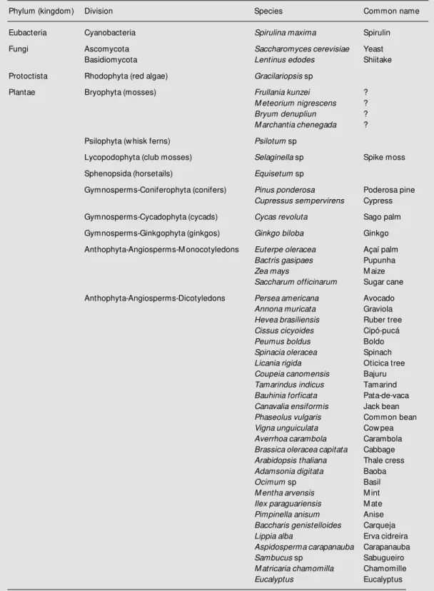
Table 1. List of phyla (green plants, cyanobacterium, red alga, and fungi) and species w hich tested positive for insulin-like antigens w ith an antibody to human insulin.
Phylum (kingdom) Division Species Common name
Eubacteria Cyanobacteria Spirulina maxima Spirulin
Fungi Ascomycota Saccharomyces cerevisiae Yeast
Basidiomycota Lentinus edodes Shiitake
Protoctista Rhodophyta (red algae) Gracilariopsis sp
Plantae Bryophyta (mosses) Frullania kunzei ?
M eteorium nigrescens ?
Bryum denupliun ?
M archantia chenegada ?
Psilophyta (w hisk ferns) Psilotum sp
Lycopodophyta (club mosses) Selaginella sp Spike moss
Sphenopsida (horsetails) Equisetum sp
Gymnosperms-Coniferophyta (conifers) Pinus ponderosa Poderosa pine
Cupressus sempervirens Cypress
Gymnosperms-Cycadophyta (cycads) Cycas revoluta Sago palm
Gymnosperms-Ginkgophyta (ginkgos) Ginkgo biloba Ginkgo
Anthophyta-Angiosperms-M onocotyledons Euterpe oleracea Açaí palm
Bactris gasipaes Pupunha
Zea mays M aize
Saccharum officinarum Sugar cane
Anthophyta-Angiosperms-Dicotyledons Persea americana Avocado
Annona muricata Graviola
Hevea brasiliensis Ruber tree
Cissus cicyoides Cipó-pucá
Peumus boldus Boldo
Spinacia oleracea Spinach
Licania rigida Oticica tree
Coupeia canomensis Bajuru
Tamarindus indicus Tamarind
Bauhinia forficata Pata-de-vaca
Canavalia ensiformis Jack bean
Phaseolus vulgaris Common bean
Vigna unguiculata Cow pea
Averrhoa carambola Carambola
Brassica oleracea capitata Cabbage
Arabidopsis thaliana Thale cress
Adamsonia digitata Baoba
Ocimum sp Basil
M entha arvensis M int
Ilex paraguariensis M ate
Pimpinella anisum Anise
Baccharis genistelloides Carqueja
Lippia alba Erva cidreira
Aspidosperma carapanauba Carapanauba
Sambucus sp Sabugueiro
M atricaria chamomilla Chamomille
Eucalyptus Eucalyptus
We ste rn blo tting
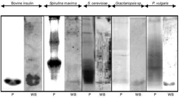
Some of the plant materials that tested positive for insulin-like antigens in the plate assay utilized and listed in Table 1 were further analyzed by Western blotting after SDS-PAGE using the same anti-human in-sulin antibody as for the plate assay. Some of the results obtained are presented in Figure 1. Extracts from S. maxima, S. cerevisiae, Gracilariopsis sp and Phaseolus vulgaris all showed bands that stained positive by West-ern blotting. In addition to a positive signal confirming the findings for the plate assay (above) the position of the bands suggests that the stained materials have the same mo-lecular weight as bovine insulin.
D iscussio n
A considerable number of reports on the presence of insulin-like molecules in ex-tracts from many different species ranging from unicellular prokaryotes and eukaryotes to multicellular invertebrates and vertebrates have been published. These molecules have always been detected by employing antibod-ies raised against vertebrate insulins and dif-ferent chromatographic techniques. Many of these have proved to be functional in
bio-logical insulin assays (12,13,29,30). The presence of insulin-like antigens in plants was previously reported by Collier et al. (8) in a study on the flowering plants spinach, rye and Lemna gibba.These investigators also showed that the isolated molecules behave like vertebrate (porcine) insulins in reversed-phase HPLC, bind to insulin receptors on lymphocytes and stimulate glucose oxidation and lipogenesis in isolated rat adipocytes.
In addition, we recently reported that some leguminous plants (dicotyledons) con-tain insulin-like antigens. In fact these anti-gens isolated from seed coats, leaves and developing fruits were shown to be proteins with molecular weights similar to those of vertebrate insulins and to possess the same amino acid sequence as bovine insulin (31; Venâncio TM, Oliveira AEA, Machado OLT, Fernandes KVS and Xavier-Filho J, unpub-lished results).
We report here on the screening of sev-eral green plants, algae, fungi, and a cyano-bacterium for the presence of insulin-like antigens (Table 1). Particularly interesting was the finding of insulin-like antigens in Arabidopsis thaliana, the model plant for genetic and biochemical studies (32,33). However, we could not find any sequences related to vertebrate insulins or plant
insu-Figure 1. SDS-polyacrylamide gel elect rophoresis and W est ern blotting of extracts from leaves and cells of Phaseolus vulgaris, Gracilariopsis sp, Saccharomy-ces cerevisiae, Spirulina maxi-ma, and bovine insulin. Western blot w as done using an anti-hu-man insulin antibody and the re-action w as visualized by chemi-luminescence. P, protein (Coo-massie brilliant blue staining); WB, Western blotting. Spirulina maxima S. cerevisiae Gracilariopsis sp
Bovine insulin P. vulgaris
lins like those from jack bean (Canavalia ensiformis) (31) and cowpea (Vigna ungui-culata) (Venâncio TM, Oliveira AEA, Ma-chado OLT, Fernandes KVS and Xavier-Filho J, unpublished results) in the published genome sequences of this plant (34). Ex-tracts of P. vulgaris leaves, the dicot A. thaliana and the legume V. unguiculata tested positive for insulin-like antigens in the plate assay (Table 1) and gave a positive reaction in Western blots (Figure 1).
A suggestion that algae may contain or depend on insulin for metabolic processes was previously made by Legros et al. (35) who showed the presence of insulin recep-tors in the plasma membrane of the unicellu-lar green alga Acetabularia mediterranea. In the present study we show that extracts of the red alga Gracilariopsis sp tested positive for insulin-like antigens in the plate assay (Table 1) and presented a band at roughly the same position as bovine insulin in Western blots (Figure 1).
The presence of insulin in fungi would seem to be undisputed since a pseudogene for preproinsulin was cloned from Neuros-pora crassa (ascomycete) (36) and insulin-like proteins were purified from S. cerevi-siae, also an ascomycete, by Best et al. in 1924 (37). More recently, insulin-like anti-gens were isolated from the fungus Aspergil-lus fumigatus, also an ascomycete (29). In the present study we detected insulin-like antigens in the edible fungus Shiitake (Lentinus edodes), a basidiomycete, and in S. cerevisiae. In yeast extracts we showed the presence of a band in Western blots at the same position as bovine insulin (Figure 1).
We also included in our study the cyano-bacterium Spirulina maxima, widely culti-vated for food and dietary supplements (38). It is worth noting here that the cyanobacteria are photosynthesizing organisms accepted to have given origin to the chloroplasts of green plants through endosymbiontic pro-cesses occurring millions of years ago. Ex-tracts of S. maxima have tested positive for
insulin-like antigens (Table 1) and a band was detected in Western blots after SDS-PAGE (Figure 1).
The presence of insulin in extra-pancre-atic tissues from vertebrates, invertebrates, microorganisms or plants is still denied by most researchers. The grounds for this are many, including the fact that the insulins found in non-pancreatic tissues would not be true insulins but rather what is called insulin-like or insulin-related molecules with simi-lar albeit different structures which are not involved in metabolic processes (glucose metabolism). These molecules include the insulin-like growth factors from vertebrates, bombyxins from silkworm, molluscan insu-lin-like peptides, and others (14). In spite of the many reports on the effects of extracts from plant parts on the reduction of blood sugar levels of diabetic animals (4,16,17), no function for insulin has been suggested by workers in the fields of glucose mobiliza-tion or transport (18-21). Nevertheless, it has been shown recently that insulin increases seed germination (22; Oliveira AEA and Xavier-Filho J, unpublished results), accel-erates synthesis of ribosomal proteins in ger-minating maize embryos (23,24), and in-creases the activity of glyoxysomal enzymes in maize (22).
Re fe re nce s
1. Banting FG & Best CH (1922). The inter-nal secretion of the pancreas. Journal of Laboratory and Clinical M edicine, 7: 465-480.
2. Collip JB (1923). Glucokinin. A new hor-mone present in plant tissue. Preliminary paper. Journal of Biological Chemistry, 56: 513-543.
3. Best CH (1924). Recent w ork on insulin. Endocrinology, 8: 617-629.
4. Gray AM & Flatt PR (1997). Nature’s ow n pharmacy: The diabetes perspective. Pro-ceedings of the Nutrition Society, 56: 507-517.
5. Khanna P, Nag TN, Jain SC & M ohan S (1974). Extraction of insulin from a plant source. 3rd International Congress on Plant Tissue and Cell Cultures, July 21-26, Leicester, UK.
6. Khanna P, Nag TN, Chandrajaia S & M ohan S (1976). Process for isolation of insulin from plant source. GrantedUnited States Patent, 3: 945-988.
7. Khanna P, Jain SC, Panagariya A & Dixit VP (1981). Hypoglycemic activity of poly-peptide-P from a plant source. Journal of Natural Products, 44: 648-655.
8. Collier E, Watkinson A, Cleland CF & Roth J (1987). Partial purification and character-ization of an insulin-like material from spin-ach and Lemna gibba G3. Journal of Bio-logical Chemistry, 262: 6238-6247. 9. Baum ann CA, Ribon V, Kanzaki M ,
Thurmond DC, M ora S, Shigematsu S, Bickel PE, Pessin JE & Saltiel AR (2000). CAP defines a second signalling pathw ay required for insulin-stimulated glucose transport. Nature, 407: 202-207. 10. Brüning JC, Gaut m am D, Burks DJ,
Gillette J, Schubert M , Orban PC, Klein R, Krone W, M üller-Wieland D & Kahn CR (2000). Role of brain insulin receptor in control of body w eight and reproduction. Science, 289: 2122-2125.
11. Alper J (2000). New insights into type 2 diabetes. Science, 289: 37-39.
12. LeRoith D, Shiloach J, Heffron R, Rubino-vitz C, Tanenbaum R & Roth J (1985). Insulin-related material in microbes: simi-larities and differences from mammalian insulins. Canadian Journal of Biochemis-try and Cell Biology, 63: 839-849. 13. LeRoith D, Delahunty G, Wilson GL,
Rob-erts Jr CT, Shemer J, Hart C, Lesniak M A, Shiloach J & Roth J (1986). Evolutionary aspects of the endocrine and nervous sys-tems. Recent Progress in Hormone Re-search, 42: 549-587.
14. Chan SJ & Steiner DJ (2000). Insulin through the ages: phylogeny of a grow th promoting and metabolic regulatory hor-mone. American Zoologist, 40: 213-222. 15. Sow er SA, Suzuki K & Reed KL (2000).
Perspective: Research activity of entero-pancreatic and brain/central nervous sys-tem hormones across invertebrates and vertebrates. American Zoologist, 40: 165-178.
16. Platel K & Srinivasan K (1997). Plant foods in the management of diabetes mellitus: vegetables as potential hypoglycaemic agents. Nahrung, 41: 68-74.
17. Ernst E (1997). Plants w ith hypoglycemic activity in humans. Phytomedicine, 4: 73-78.
18. Ward JM , Kühn C, Tegeder M & Frommer WB (1998). Sucrose transport in higher plants. International Review of Cytology, 178: 41-71.
19. Wobus U & Weber H (1999). Sugars as signal molecules in plant seed develop-ment. Biological Chemistry, 380: 937-944. 20. Williams LE, Lemoine R & Sauer N (2000). Sugar transporters in higher plants - a di-versity of roles and complex regulation. Trends in Plant Sciences, 5: 283-290. 21. Lemoine R (2000). Sucrose transporters
in plants: update on function and struc-ture. Biochimica et Biophysica Acta, 1465: 246-262.
22. Goodman DBP & Davis WL (1992). Insulin accelerates the post germinative devel-opment of several fat storing seeds. Bio-chemical and Biophysical Research Com-munications, 190: 440-446.
23. Sánchez de Jiménez E, Beltrán-Penã E & Ortíz-López A (1999). Insulin-stimulated ribosomal protein synthesis in maize em-bryonic axis during germination. Physiolo-gia Plantarum, 105: 148-154.
24. Dinkova TD, Aguilar R & Sánchez de Jiménez E (2000). Expression of maize eukaryotic initiation factor (eIF) iso4E is regulated at the translational level. Bio-chemical Journal, 351: 825-831. 25. Laemmli UK (1970). Cleavage of
struc-tural proteins during the assembly of the head of bacteriophage T4. Nature, 227: 680-685.
26. Tow bin H, Staehelin NT & Gordon J (1979). Electrophoretic transfer of pro-teins from polyacrylamide gels to nitrocel-lulose sheets; procedures and some ap-plications. Proceedings of the National Academy of Sciences, USA, 176: 4350-4354.
27. Gebara VCB, Petricevich VL, Raw I & Da Silva WD (1995). Effect of saponin from Quillaja saponaria (M olina) on antibody, tumour necrosis factor and interferon-g production. Biotechnology and Applied Biochemistry, 22: 31-37.
28. Raven PH, Evert RF & Eichhorn SE (1992). Biology of Plants. 5th edn. Worth Publish-ers, New York, NY, USA.
29. LeRoith D, Shiloach J, Roth J & Lesniak M A (1980). Evolutionary origins of verte-brate hormones: Substances similar to mammalian insulins are native to unicellu-lar eukaryotes. Proceedings of the Na-tional Academy of Sciences, USA, 77: 6184-6188.
30. LeRoith D, Shiloach J, Roth J & Lesniak M A (1981). Insulin or a closely related molecule is native to Escherichia coli. Journal of Biological Chem istry, 256: 6533-6536.
31. Oliveira AEA, M achado OLT, Gomes VM , Xavier Neto J, Pereira AC, Vieira JGH, Fernandes KVS & Xavier-Filho J (1999). Jack bean seed coat contains a protein w ith complete sequence homology to bo-vine insulin. Protein and Peptide Letters, 6: 15-21.
32. Negrutiu I, Beeftink F & Jacobs M (1975). Arabidopsis thaliana as a model system in somatic cell genetics. 1. Cell and tissue culture. Plant Science Letters, 5: 293-304. 33. M einke DW, Cherry J, Dean C, Rounsley SD & Koornneef M (1998). Arabidopsis thaliana: A model plant for genome analy-sis. Science, 282: 662-682.
34. The Arabidopsis Information Resource (2001). Available at : ht t p://w w w . arabidopsis.org. Accessed November 20, 2001.
35. Legros F, Uyt denhoef P, Dum ont I, Hanson B, Jeanmart J, M assant B & Conard V (1975). Specific binding of insu-lin to the unicellular alga Acetabularia mediterranea. Protoplasma, 86: 119-134. 36. M uthukumar G & Lenard J (1991). A preproinsulin-like pseudogene from Neu-rospora crassa. M olecular and Cellular En-docrinology, 82: 275-283.
37. Best CH, Smith RG & Scott DA (1924). An insulin-like material in various tissues of the normal and diabetic animal. American Journal of Physiology, 68: 161-182. 38. Kay RA (1991). M icroalgae as food and