Abstract
Submitted: August 11, 2016 Modiication: October 26, 2016 Accepted: November 4, 2016
Chondroblastic osteosarcoma
mimicking periapical abscess
Lesions of non-endodontic origin may mimic periapical abscess. Osteosarcoma is a rare malignant lesion. Case report: The present report describes a case of chondroblastic osteosarcoma in the periapical region of teeth #29, #30, and #31 of an 18-year-old male. Clinical history showed self-reported discomfort in the right posterior gingiva for over a month. Physical examination showed a small expansion and redness of the right mandibular buccal and lingual cortical plates, but no signs of pain or inlammation were observed. All the teeth responded positively to pulp sensibility. Periapical and panoramic radiographs showed slight periapical radiolucency in the roots of teeth #29 and #30, clear periodontal ligament space widening, and evident loss of lamina dura. Incisional biopsy was performed, and based on microscopic indings the diagnosis of chondroblastic osteosarcoma was conirmed. Conclusions: Non-endodontic diseases associated with tooth root apex, such as chondroblastic osteosarcoma, should be included in differential diagnosis of jaw lesions that resemble periapical abscess.
Keywords: Osteosarcoma. Periapical abscess. Apical periodontitis. Differential diagnosis. Endodontics.
Fernanda Paula YAMAMOTO-SILVA1
Brunno Santos de Freitas SILVA2
Aline Carvalho BATISTA1
Elismauro Francisco de MENDONÇA1
Décio dos Santos PINTO-JÚNIOR3
Carlos ESTRELA1
http://dx.doi.org/10.1590/1678-7757-2016-0424
1Universidade Federal de Goiás, Departamento de Ciências Estomatológicas, Goînia, GO, Brasil. 2Centro Universitário de Anápolis, Departamento de Medicina Oral, Anápolis, GO, Brasil.
3Universidade de São Paulo, Faculdade de Odontologia, Departmento de Estomatologia, São Paulo,
SP, Brasil.
Corresponding address: Carlos Estrela Departamento de Ciências Estomatológicas -
Introduction
A periapical radiolucency associated with a vital tooth constitutes a diagnostic challenge2. Periapical
lesions can be of endodontic or non-endodontic origin. Therefore, periapical radiolucency associated with root apices showed by radiographic examinations may be or not a consequence of infection of the root canal system4,12,13, which may involve progressive changes in periapical structures with subsequent bone resorption12. Conventional radiographic images
are frequently used to detect apical periodontitis.
The diagnosis of a periapical radiolucency requires careful and correct management of information obtained from patient history, clinical examination, pulp vitality testing, and radiography analysis2,4,13.
The establishment of diagnostic procedures, such as examination of signs and symptoms, as well as complementary examinations, is indispensable to obtain differential diagnosis. Lesions of non-endodontic origin may be associated with the periapical area of the tooth3,5,8,16,18.
Osteosarcoma is a rare malignant neoplasm (incidence of 0.7 per million)20 of mesenchymal origin
characterized by the production of immature bone13. It
represents 5% to 13% of total osteosarcomas6,7,9,10,13-15
and occurs in the maxilla and mandible with approximately equal frequency13.
In the present report we describe a case of chondroblastic osteosarcoma resembling a periapical abscess in an 18-year-old male.
Case report
An 18-year-old male patient was referred to the dental clinic of the Federal University of Goiás (Goiânia, GO, Brazil) with a chief complaint of a “discomfort on the right posterior gingiva” for over a month. At physical examination, a small expansion and redness were found in the buccal and lingual cortical plates of the right mandible in the region of teeth #29, #30,
and #31, but no signs of pain or inlammation were
observed (Figure 1A). The overlying mucosa appeared intact.
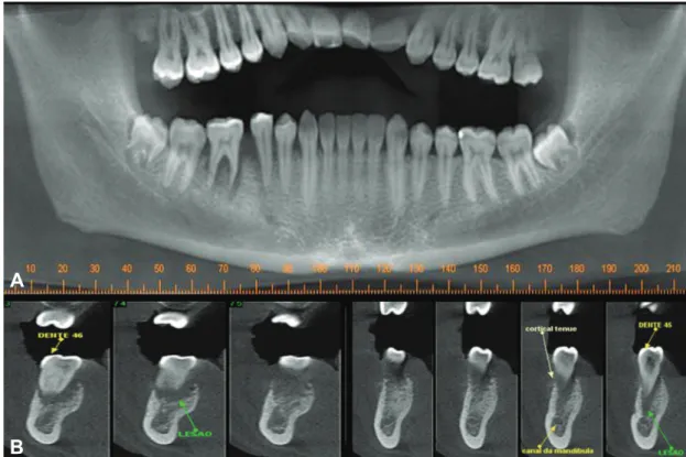
Periapical and panoramic radiographs showed periapical radiolucency ranging from 4 to 5 mm in the mesial and distal roots of teeth #29 and #30, a slight rarefaction of the inter-dental alveolar bone, a clear
periodontal ligament space widening, and an evident loss of lamina dura (Figure 1 B-C).
The patient reported no history of dental trauma. Neither cracks on the crowns of teeth #29 and #30, particularly on the mesial and distal marginal ridges, nor previous restorative treatments, were found. Pulp
vitality testing using tetraluoroethane spray (Endo-Ice; Hygenic Corp, Akron, OH) conirmed positive
response in all teeth associated with radiolucent lesions, and therefore root canals were not treated.
Cone beam computed tomography (CBCT) images were acquired. The imaging examinations showed
undeined periapical osteolytic lesion, represented
by a hypodense lesion with hyperdense areas inside, and thinning of the lingual cortical plate, associated with the roots of teeth #29 and #30. Alterations in trabecular bone were also seen (Figures 2 and 3).
Based on location of the lesion, its radiographic
indings and the fact that periapical inlammatory
lesions are the most frequent injuries in the affected region, the initial diagnosis included periapical abscess. However, given that the lesion had no relationship with pulp necrosis associated with the previous mentioned clinical and radiographic characteristics, intraosseous malignancies were considered, and an incisional biopsy was carried out. Microscopic evaluation showed a proliferation of round to spindle-shaped cells, with occasional cellular pleomorphism and variable osteoid production (Figure 4 A–B). Additionally, a focal area presented proliferation of atypical chondroblastic cells. Immunohistochemical reaction with Ki-67 marker showed evident cellular activity in the specimen (Figure
4C). Based on microscopic indings, a inal diagnosis of chondroblastic osteosarcoma was conirmed.
The patient returned to the dental clinic 10 days after the incisional biopsy presenting with a considerable enlargement of the region. At this time, the patient was referred to Araújo Jorge Cancer Hospital (Goiânia, GO, Brazil). The treatment consisted of hemimandibulectomy with wide surgical margins and adjuvant chemotherapy.
After surgical treatment, the patient underwent
reconstruction of the right mandible with a ibular
graft (Figure 5A). At the follow-up one year later, a bone scintigraphy was performed and no signs of bone metastases were seen (Figure 5B).
Figure 1- Clinical aspect (A) showing expansion of buccal and lingual cortical plates of the right mandible in the region of teeth #29, #30, and #31, with some redness and apparently intact overlying mucosa. Periapical and panoramic radiographs (B-C) showing slight apical radiolucency in the roots of teeth #29 and #30 with loss of lamina dura and periodontal ligament space widening
Figure 2- Cone beam computed tomography (A–B) showing a well-deined osteolytic lesion associated with the roots of teeth #29 and #30. Cross-sectional image (B) allowed observing alterations in the trabecular bone
A
Discussion
Osteosarcoma (osteogenic sarcoma) is a primary malignant tumor of the bone in which the neoplastic cells produce osteoid or bone matrix20. Excluding
nonhematopoietic lesions, osteosarcomas are the most common primary malignant bone tumors13, which
occur in the maxilla and mandible with approximately equal frequency13,20, predominantly in adolescents and
young adults, between the ages of 10 and 20 years,
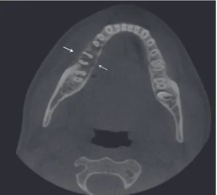
Figure 3- Axial plane cone beam computed tomography showing a thinning of the buccal and lingual cortical plates (arrows).
Figure 4- Histopathologic examination demonstrated round to spindle-shaped cells with cellular pleomorphism (A) and osteoid production
(B). A focal area presenting proliferation of atypical chondroblastic cells was observed (arrows) (hematoxylin-eosin, original magniication
100X). Immunohistochemical reaction with Ki-67 (C) marker showed evident cellular activity in the specimen
and is slightly more common in males20.
An increase in volume in the affected area and pain are the most common symptoms of osteosarcoma13, and when it occurs in the maxilla, nasal obstruction
may be present13,17. The etiology of osteosarcoma
are still unknown, however it has been associated with preexisting conditions, including prior radiation,
fibrous dysplasia, Paget’s disease, and chronic osteomyelitis19,20,21.
In the case reported here, clinical findings were in accordance with previous descriptions of
osteosarcoma6,9,10,13-15,17,19,21,22, since the patient was an
18-year-old man who complained of pain, the lesion was in the posterior region of the mandible, and clinical
examinations showed a swelling in the affected area. The clinical features of the lesion associated with presence of pain, swelling, and radiographic image of increased periodontal space mimicked a clinical condition of periapical abscess. Thus, based on these aspects, the initial differential diagnosis included periapical abscess. However, the positive response to pulp vitality testing suggested absence of root canal infection, which led to the recommendation of complementary examinations such as CBCT and an incisional biopsy. Due to the presence of an ill-deined radiolucency and periodontal space widening related with a vital pulp, intraosseous malignancies were considered in the differential diagnosis.
Common radiographic indings of osteosarcoma usually range from dense sclerosis to a mixed sclerotic radiolucent lesion and to a completely radiolucent process. The limits are undeined, making it dificult to determine tumor size and extension. The classic sunburst appearance is caused by osteophytic bone production on the surface of the lesion, especially in occlusal radiographs. An early radiographic change consists of a widening of the periodontal ligament space caused by tumor iniltration13. Considering the dificulty
to determine tumor extension using conventional radiography, in the case reported here CBCT was used to determine the degree of bone destructioncaused by the tumor, as well as its location and extension. A hypodense lesion with hyperdense areas inside,
thinning of the lingual cortical plate, and an undeined
periapical osteolytic lesion associated with the roots of teeth #29 and #30 were found.
The macroscopic essential criterion to characterize an osteosarcoma is the direct production of osteoid by malignant mesenchymal cells13. In addition to osteoid
formation, tumor cells may produce chondroid material and ibrous connective tissue. Depending on the relative amounts of osteoid, cartilage, or collagen ibers, this type of tumor can be subdivided in osteoblastic, chondroblastic, and ibroblastic1,13. Osteosarcomas
of the jaws are generally better differentiated than the extragnathic ones, and they commonly exhibit chondroblastic differentiation, characterized by lobules of atypical-appearing chondrocytes in lacunae20.
In the present report, the microscopic analysis showed a proliferation of round to spindle-shaped cells, with occasional cellular pleomorphism and variable osteoid production, as well as a focal area presenting proliferation of atypical chondroblastic cells. Cellular
activity was confirmed by immunohistochemical reaction with Ki-67 marker. The histopathological
examination conirmed the diagnosis of chondroblastic
osteosarcoma.
Takahama, et al.22 (2003)analyzed the clinic
pathological features and immunohistochemical expression of p53, MDM2, CDK4, PCNA, and Ki67 proteins in 25 head and neck osteosarcomas. The immunohistochemical analysis displayed positivity in 88% of the cases for Ki-67. Paparella, et al.15 (2013)
analyzed 74 cases of osteosarcoma of the jaws and found a predominant chondroblastic pattern, which leads to the conclusion that these lesions may be associated with a worse prognosis. Bennet, et al.1
(2001) conducted a 30-year retrospective review of osteosarcoma of the jaws and compared the clinical behavior of the tumors. Their goals were to assess how they differ from the reported characteristics of tumors of other sites and to report observations of clinical
and diagnostic signiicance. They reported that most
osteosarcomas had areas of chondroid formation in addition to neoplastic osteoid, the main complication was local recurrence, and metastasis was rare and occurred as a solitary process or in late stages of the disease. This was in contrast to lesions metastatic to the jaws, which were higher grade in appearance and had metastasized widely, early in the disease process. Primary osteosarcoma occurring in patients with a history of radiotherapy was typically more aggressive.
The recommended treatment of osteosarcoma has historically been the surgical resection of the lesion with safe margins associated with chemotherapy11,13,20.
In the present report, the treatment consisted of a hemimandibulectomy with wide surgical margins and adjuvant chemotherapy, due to the highly malignant features of the tumor, with a worse prognosis and a
high risk of local recurrence. The patient underwent
reconstruction of the right mandible, and clinical and
radiographic follow-up one year later conirmed tumor remission. Bone scintigraphy was conducted and showed no signs of bone metastases.
In summary, osteosarcomas could present similar
features of some inlammatory periapical lesions,
such as periapical abscess, since it also present pain, swelling and variable radiographic changes3.
Exceptional care should be paid to endodontic
diagnoses based on clinical and radiographic indings.
Since periapical lesions of non-endodontic origin may mimic periapical abscess and apical periodontitis, they
should be considered before root canal treatment.
Conclusions
Non-endodontic diseases associated with tooth root apex, such as chondroblastic osteosarcoma, should be included in the differential diagnosis of jaw lesions that resemble periapical abscess. Periapical lesions may be misdiagnosed at their early stages if malignant tumors are not suspected.
Acknowledgments
This study was partially supported by grants from
CNPq – National Council for Scientiic and Technological
Development (grant 457536/2014 to F.P.Y.S.) and PNPD/CAPES – Post-Doctoral National Program, Coordination of Higher Education and Graduate Training (process 02/2014 to B.S.F.S.). The authors also thank Dr. Alexandre Belotti for his technical
assistance. The authors deny any conlicts of interest
related to this study.
Reference
1- Bennett JH, Thomas G, Evans AW, Speight PM. Osteosarcoma of the jaws: a 30-year retrospective review. Oral Surg Oral Med Oral Pathol
Oral Radiol Endod. 2000;90:323-32.
2- Bregni RC, Contreras E, Hiraki KR, Vargas PA, León JE, Almeida OP.
Epithelioid osteosarcoma of the mandible: a rare case with unusual
immunoproile. Oral Surg Oral Med Oral Pathol Oral Radiol Endod.
2008;105:e47-52.
3- Bueno MR, Carvalhosa AA, Castro PH, Pereira KC, Borges FT, Estrela
C. Mesenchymal chondrosarcoma mimicking apical periodontitis. J Endod. 2008;34:1415-9.
4- Carvalhosa AA, Araújo Estrela CR, Borges AH, Guedes OA, Estrela C. 10-year follow-up of calcifying odontogenic cyst in the periapical
region of vital maxillary central incisor. J Endod. 2014;40:1695-7. 5- Carvalhosa AA, Zandonade RM, Souza Castro PH, Araújo Estrela
CR, Borges AH, Estrela C. 8-year follow-up of central giant cell lesion mimicking apical periodontitis. J Endod. 2014;40:1708-12.
6- Chaudhary M Chaudhary SD. Osteosarcoma of jaws. J Oral Maxillofac Pathol. 2012;16:233-8.
7- Chittaranjan B, Tejasvi MA, Babu BB, Geetha P. Intramedullary osteosarcoma of the mandible: a clinicoradiologic perspective. J Clin
Imaging Sci. 2014;31:4-6.
8- Faitaroni LA, Bueno MR, Carvalhosa AA, Bruehmueller Ale KA,
Estrela C. Ameloblastoma suggesting large apical periodontitis. J Endod. 2008;34:216-9.
9- Fernandes R, Nikitakis NG, Pazoki A, Ord RA. Osteogenic sarcoma of the jaw: a 10-year experience. J Oral Maxillofac Surg.
2007;65:1286-91.
10- Garrington GE, Scoield HH, Cornyn J, Hooker SP. Osteosarcoma
of the jaw. Analysis of 56 cases. Cancer. 1967;20:377-91.
11- Lukschal LF, Barbosa RM, Alvarenga RL, Horta MC. Osteosarcoma
in the maxila: case report. Rev Port Estomatol Med Dent Cir Maxilofac. 2013;54:48-52.
12- Nair PN, Sjögren U, Figdor D, Sundqvist G. Persistent periapical radiolucencies of root-illed human teeth, failed endodontic treatments,
and periapical scars. Oral Surg Oral Med Oral Pathol Oral Radiol Endod. 1999;87:617-27.
13- Neville BW, Damm DD, Allen CM, Bouquot JE. Oral maxillofacial pathology, 3rd ed. Philadelphia: Elsevier; 2009.
14- Nthumba PM. Osteosarcoma of the jaws: a review of literature
and a case report on synchronous multicentric osteosarcomas. World J Surg Oncol. 2012;10:240.
15- Paparella ML, Olvi LG, Brandizzi D, Keszler A, Santini-Araujo E, Cabrini RL. Osteosarcoma of the jaw: an analysis of a series of 74
cases. Histopathology. 2013;63:551-7.
16- Pontes FS, Fonseca FP, Jesus AS, Garcia-Alves AC, Araújo LM, Nascimento LS, et al. Nonendodontic lesions misdiagnosed as apical
periodontitis lesions: series of case reports and review of literature. J Endod. 2014;40:16-27.
17- Praveena NM, Maragathavalli G. Osteosarcoma of maxilla. J Indian Acad Oral Med Radiol. 2012;24:236-8.
18- Rodrigues CD, Villar-Neto MJ, Sobral AP, Silveira MM, Silva LB, Estrela C. Lymphangioma mimicking apical periodontitis. J Endod.
2011;37:91-6.
19- Saini R, Abd Razak NH, Ab Rahman SA, Samsudin AR.
Chondrosarcoma of the mandible: a case report. J Can Dent Assoc. 2007;73:175-8.
20- Saito K, Unni KK. Malignant tumours of bone and cartilage. In: Barnes L, Eveson JW, Reichart P, Sidransky D. World Health Organization
Classiication of Tumours. Pathology and genetics of head and neck
tumours. Lyon: IARC Press; 2005.
21- Soares RC, Soares AF, Souza LB, Santos ALV, Pinto LP. Osteosarcoma of mandible initially resembling lesion of dental periapex: case report.
Rev Bras Otorrinolaringol. 2005;71:242-5.
22- Takahama A Jr, Alves FA, Pinto CA, Carvalho AL, Kowalski LP, Lopes
MA. Clinicopathological and immunohistochemical analysis of