Videothoracoscopy for Isolated Atrial Fibrillation Ablation through
Bipolar Radiofrequency
Alexandre Siciliano Colafranceschi
1,2, Andrey José de Oliveira Monteiro
1,2, Eduardo Souza Leal Botelho
1,2, Leonardo
Secchin Canale
1, Arnaldo Rabischoffsky
2, Ieda Prata Costa
1,2, Fernando Eugênio dos Santos Cruz Filho
2, Roberto Luiz
Menssing da Silva Sá
1,2, Ana Luiza Boechat
2, Luis Alberto Oliveira Dallan
3Instituto Nacional de Cardiologia1, Rio de Janeiro, RJ; Hospital Pró-Cardíaco2, Rio de Janeiro, RJ; Instituto do Coração da Faculdade de Medicina da Universidade de São Paulo3; São Paulo, SP - Brazil
Summary
Background:The prevalence of atrial fibrillation, expenses with the healthcare system and the associated high morbidity and mortality have justified the search for new therapeutic approaches.
Objective: To evaluate the reproducibility of the surgical technique, its safety and the initial outcome of the video-assisted surgery for the isolated atrial fibrillation ablation with bipolar radiofrequency.
Methods: Ten patients (90% men) with symptomatic atrial fibrillation (50% paroxystic type) that was refractory to drug therapy, with no heart disease that required concomitant surgical treatment, were submitted to arrhythmia ablation guided by thoracoscopy from May 2007 to May 2008. Clinical, laboratory and image variables were prospectively collected before, during surgery and at the postoperative follow-up.
Results: The surgery was carried out as planned in all patients. There was no intra-thoracic structure iatrogenic lesion or deaths. At the mean 6-month follow-up, 80% of the patients were free of atrial fibrillation. There was a significant improvement in the symptoms of New York Heart Association Functional Class heart failure (2.4 ± 0.5 to 1.6 ± 0.7; p=0.011). There was no evidence of pulmonary vein stenosis at the angiotomography in this series.
Conclusion: The video-assisted surgery for the treatment of atrial fibrillation is reproducible and safe. There is a heart failure symptom evolution improvement after the surgery. (Arq Bras Cardiol 2009; 93(3) : 310-318)
Key words: Video-assisted Surgery; Catheter Ablation; Atrial Fibrillation.
Mailing address: Alexandre Siciliano Colafranceschi • Rua Dona Mariana,143, sala A 12 – Botafogo - 22.280-020 - Rio de Janeiro, RJ - Brazil
E-mail: [email protected]
Manuscript received June 16, 2008; revised manuscript received August 26, 2008; accepted September 11, 2008.
Introduction
The atrial fibrillation (AF) is the most common sustained cardiac arrhythmia in clinical practice and its prevalence is estimated between 0.4% and 1% of the general population1 and in 6% of those older than 65 years2.
The AF is an independent risk factor for mortality with a relative risk for all ages of 1.5 for men and 1.9 for women3.
In the last 20 years, hospital admissions due to AF increased 66%4. In Brazil, AF is the fifth major cause of hospital admission at the Sistema Unico de Saude (SUS – Brazilian Public Healthcare System) and affects almost 10% of the population treated by specialized cardiology services with a quaternary treatment level of complexity5. The high prevalence of this arrhythmia, the AF-related public healthcare costs and the elevated morbidity and
mortality associated to it, have justified the search for a better understanding of its physiopathological bases and new therapeutic approaches.
Minimally-invasive, curative, safer and more effective procedures than the long-term drug therapy can become first-choice treatments of AF6.
When analyzing its electrophysiological bases, it can be observed that the AF that occurs repeatedly is associated to triggers located around the orifices of the pulmonary veins in 90% of the patients7.
The transition zone between the endothelium of the pulmonary veins and the left atrial endocardium constitutes an anatomical substrate formed by two kinds of juxtaposed tissues. The fact that these tissues present distinct electrical properties can promote the development of triggering foci of AF episodes. The electrical isolation of the pulmonary veins can, therefore, terminate the arrhythmia.
Some authors still believe that the ablation of the peri-cardiac parasympathetic ganglia can increase the atrial fibrillation-free survival of patients submitted to electrical isolation of the pulmonary veins8.
of new devices, capable of generating localized cell damage and throughout the entire tissue thickness, as the bipolar radiofrequency, thus substituting the lesions created by the traditional surgery of cutting and suturing, the surgical procedure becomes faster and less bloody and equally effective as the traditional technique. Therefore, the cardiovascular surgeon has increased possibilities of action and a favorable scenario is created for the minimization of the surgical access and exclusion of the need for extracorporeal circulation in the clinical approach of patients with AF.
Wolf et al9 described a video-assisted surgical technique for the ablation of AF and reported their experience in little more than 20 patients. On the other hand, however, the reproducibility of this minimally-invasive technique and the possible risks associated to it and the fact that the lungs are kept in collapse for a period of time are still to be defined.
The objectives of this prospective analysis include: • To evaluate the reproducibility of the video-assisted, minimally-invasive surgical treatment of AF refractory to drug therapy.
• To evaluate its safety.
• To evaluate the impact of the surgery on the postoperative follow-up.
Methods
This protocol was approved by the National Ethical Committee in Research (CONEP) in 2004 and registered u n d e r # 1 1 1 4 1 , d e c i s i o n # 2 5 5 4 / 2 0 0 4 , p r o c e s s 25000.164698/2004-53. The Ethical Committee in Research of Instituto Nacional de Cardiologia (National Institute of Cardiology), Rio de Janeiro, corroborated the approval by CONEP.
Studied population
From May 2007 to May 2008, ten (10) patients were submitted to the surgical procedure at the Instituto Nacional de Cardiologia, in Rio de Janeiro/RJ, Brazil.
All patients presented heart failure classified according to the New York Heart Association (NYHA) functional class between II and III (Table 1).
The inclusion and exclusion criteria were defined as following:
Inclusion criteria
• Men or women aged 18 to 80 years;
• Minimum of three months of history of AF (intermittent or continuous) symptomatic and refractory to clinical treatment;
• Ejection fraction >35%;
• Patients able to understand and sign the free and informed consent form;
• Patients committed to clinical follow-up at return visits;
• Minimum postoperative follow-up of three months.
Exclusion criteria:
• Functional class IV (NYHA);
• Previous cardiac or thoracic surgery;
• Indication for concomitant cardiac or thoracic surgery;
• Cerebrovascular accident (CVA) or transient ischemic attack (TIA) in the last six months;
• Presence of atrial and/or ventricular thrombus; • Comorbidities that increase the surgical risk, depending on the evaluation of the main investigator;
Table 2 summarizes the preoperative characteristics of the patients. Some patients presented concomitant structural heart disease with no functional consequences and with no indication for surgical treatment or another intervention.
The mean time of the AF in this population was 5.2 ± 3.2 years (3 months to 12 years). The history related to the AF is shown in Table 2.
Tissue ablation system by bipolar radiofrequency
The components of the tissue ablation system by bipolar radiofrequency (RF) (Atricure Inc, Cincinnati – OH) used in this clinical study, include the disposable RF bipolar tweezers, the ablation and sensing unit (ASU) and a dissector specifically developed for this purpose.
The use of the RF is performed only in the atrial tissue,
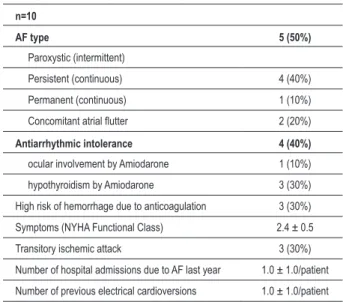
Table 1 - History of atrial fibrillation
n=10
AF type 5 (50%)
Paroxystic (intermittent)
Persistent (continuous) 4 (40%)
Permanent (continuous) 1 (10%)
Concomitant atrial flutter 2 (20%)
Antiarrhythmic intolerance 4 (40%)
ocular involvement by Amiodarone 1 (10%)
hypothyroidism by Amiodarone 3 (30%)
High risk of hemorrhage due to anticoagulation 3 (30%)
Symptoms (NYHA Functional Class) 2.4 ± 0.5
Transitory ischemic attack 3 (30%)
Number of hospital admissions due to AF last year 1.0 ± 1.0/patient
Number of previous electrical cardioversions 1.0 ± 1.0/patient
N - total number of patients; AF - atrial fibrillation; NYHA - New York Heart
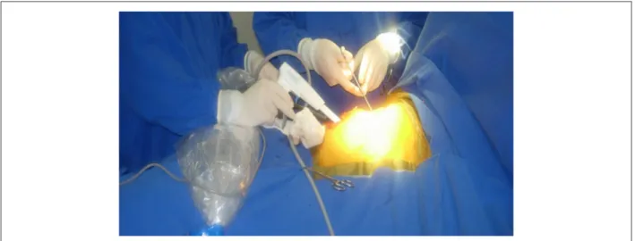
Figure 1 -Patient in left lateral decubitus position. Image at the end of the procedure on the right. Infra-axillary incision and chest drain are shown.
Figure 2 -Introduction of the bipolar tweezers guided by thoracoscopy.
avoiding structures such as the coronary arteries and the pulmonary veins.
Preoperative period
All patients were evaluated at the preoperative period, with a clinical history and physical examination. The risks, benefits and alternatives to the proposed procedure were discussed at this initial assessment and all patients signed the Free and Informed Consent Form.
Table 2 - Preoperative characteristics
n=10
Age 51.1 ± 11.1 yrs (38-71 yrs)
Male sex 9 (90%)
Weight 77.1 ± 10.9 kg (58-100 kg)
Height 1.69 ± 0.79 m (1.59-1.85 m)
Body surface (BS) 1.87 ± 0.16 m2
SAH 4 (40%)
DM 2 (20%)
COPD 1 (10%)
Smoking 2 (20%)
Dyslipidemia 5 (50%)
CAD 1 (10%)
Orovalvular disease 1 (10%)
Cardiomyopathy 2 (20%)
Dilated 1 (10%)
Hypertrophic 1 (10%)
Previous AF ablation 0
N - total number of patients, SAH - systemic arterial hypertension, DM - diabetes
mellitus, COPD - chronic obstructive pulmonary disease, DAC - coronary artery
disease, AF - atrial fibrillation, Kg - kilograms, m - meters, m2 - square meters.
The patients that accepted to participate in the study were asked to undergo the following preoperative examinations:
• Blood (Complete blood count, with platelet count, Coagulogram blood biochemistry);
• 12-derivation Electrocardiogram; • Chest teleradiography;
• 24-hour Holter.
Three-channel 24-hour Holter monitoring was used to assess the presence or absence of atrial fibrillation. AF was considered when any rhythm with intervals between the irregular R waves and the absence and absence of P waves (atrial electrical activity) was present for at least 30 seconds.
• Other examinations
disease underwent, at least once, a non-invasive risk stratification (myocardial scintigraphy, ergometric stress test or stress echocardiogram) to rule out concomitant asymptomatic obstructive coronary disease.
Surgical technique
The surgical procedure involves instrumentation on both sides of the thorax and it was carried out as previously detailed9. Briefly, the right side is initially performed: the patient is initially positioned on left lateral decubitus with the right arm abducted over the head (Figure 1). A 12-mm trocar is positioned under the sixth or seventh rib in order to allow the introduction of the 10-mm and 30-degree videothoracoscopy optical instrument inside the chest. This trocar is placed medially 2 cm to the mid-axillary line (Figure 2).
With the help of the optical instrument, the ribs are counted. A 4-6 cm incision is performed under the third rib, from lateral border of the nipple toward to mid-axillary line (Figure 1) The fat tissue in the mid-axillary hollow is placed posteriorly. The ribs are carefully retracted and the pericardium is directly observed. The pericardium is opened 2 to 4 cm anteriorly and parallel to the phrenic nerve, exposing the heart 3 cm above the vena cava and right atrium junction. The opening of the pericardium, inferiorly, must be carried out as distally as possible. The pericardium is repaired with separate 2-0 cotton stitches. The dissection of the tissue between the superior vena cava and the right upper pulmonary vein is carried out by blunt dissection, separating the tissues with the tip of the aspirator. The same maneuver is used to access the oblique sinus between the inferior vena cava and the right lower pulmonary vein.
A second incision is made for the positioning of a tissue dissector especially developed for this purpose. This dissector is directed to the oblique sinus, just above the inferior vena cava, behind the right pulmonary veins and is advanced superiorly to the pericardial reflection in the upper margin of the superior pulmonary vein. A rubber catheter is adapted at the tip of the dissector and goes behind the pulmonary veins with the removal of the dissector. The same maneuver is performed by exchanging the rubber catheter by the lower paddle of the bipolar tweezers.
The RF energy is administered and the occurrence of cell damage throughout the entire tissue thickness is, in theory, achieved after 10-15 seconds, when a beeper sound is emitted by the system.
A test of electrical conduction from the pulmonary veins to the left atrium is then used. This test is only possible to be performed in patients that are presenting a non-fibrillatory rhythm at the moment of the procedure.
Using a conventional pacemaker generator, the pulmonary veins are stimulated with a heart rate 20% > the basal rate and the atrial capture is verified, confirming or not the passage of the stimulus from the pulmonary veins to the left atrium. After the electrical isolation of the pulmonary vein, the non-capture of the pacemaker stimulus
by the left atrium confirms the electrical disconnection of the pulmonary veins. In case of left atrial capture, the bipolar RF must be administered again. At the end of the procedure, a tubular chest drain number 20 or a Blake-type silicone drain is positioned through one of the incisions (Figure 1).
The patients must be repositioned on the operating table in right lateral decubitus and the left side of the procedure is carried out similarly to what was performed on the right. The amount of tissue to be dissected, however, is smaller on the left when only the Marshall’s ligament dissection is performed, to allow the access along the upper border of the left upper pulmonary vein. The access to the oblique sinus inferiorly does not require additional dissection.
Intravenous heparin is not administered to the patients. The tweezers are kept occluding the pulmonary veins for a maximum of 20 seconds.
The endotracheal tube is removed while the patient is still in the operating room, whenever possible. The computed variables of the perioperative period include the time of right and left pulmonary collapse to perform the procedure, the need for electrical cardioversion after the isolation of the pulmonary veins and the confirmation of the electrical isolation through the electrical conduction test from the pulmonary veins to the left atrium. We also evaluated the need for conversion to thoracotomy and the occurrence of lesions in the intra-thoracic structures, such as the phrenic nerve, heart and lungs.
Postoperative follow-up
• D r u g p r e s c r i p t i o n s a n d c a r d i a c r h y t h m management:
The patients have their preoperative medications re-introduced in the morning after the surgical procedure and these are maintained during the postoperative follow-up. All patients were advised to use an oral anticoagulant for at least 6 months.
The desired strategy was for patients to attain hospital discharge in sinus rhythm. In order to achieve that, the use of intravenous antiarrhythmic drugs or electrical reversion was used as necessary, during the intrahospital postoperative period.
• Anatomic evaluation of the pulmonary veins: To evaluate the pulmonary veins, a computed angiotomography of the thorax was performed on the third month of postoperative follow-up. The tomographic study was carried out through the volumetric acquisition of data with 1 mm of collimation during the administration of nonionic contrast. The acquired images were processed by obtaining axial images and multiplane and three-dimensional reconstructions. Pulmonary vein stenosis was considered when there was any 50% decrease in its diameter.
• Other variables analyzed in the postoperative period.
Table 5 – Drugs
n=10 pre-operative post-operative
Beta-Blocker 5 (50%) 6 (60%)
Digitalis 2 (20%) 0
Amiodarone 3 (30%) 3 (30%)
Propafenone 2 (20%) 3 (30%)
Ca+2 channel blockers 2 (20%) 2 (20%)
Warfarin 8 (80%) 8 (80%)
ASA 2 (20%) 2 (20%)
ACEI 5 (50%) 5 (50%)
N - number of patients; ASA - Aspirin; ACEI - angiotensin-converting enzyme inhibitor.
of permanence of the chest drain and the complications that occurred during the intra-hospital period.
Statistical analysis
The McNemar test was used to compare proportions of paired groups and the Chi-square test to compare two non-paired proportions.
The Student’s t test was used for paired values and the Student’s t test for independent samples for the analysis of numerical variables.
To evaluate the occurrence of associations between the numerical variables, Pearson’s coefficient of correlation was used. Spearman’s coefficient was used for the correlation between ordinal variables.
Statistical significance was set at p <0.05.
Results
The surgical procedure was carried out in both sides of the thorax of the 10 patients, as previously planned. There was no need for conversion or enlargement of the
Table 3 - Perioperative variables
n=10
Right alveolar closing time (mean ± SD) 64.5 ± 24.2 minutes
Electrical isolation of right pulmonary veins
(confirmation) 10 (100%)
Left alveolar closing time (mean ± SD) 54.5 ± 15.2 minutes
Electrical isolation of left pulmonary veins
(confirmation) 9 (90%)
Electrical cardioversion in the OR 5 (50%)
Sinus rhythm at the end of the procedure 9 (90%)
Extubation at the end of the procedure 10 (100%)
SD - standard deviation of the mean; Confirmation - electrical conduction test of pulmonary veins confirming electrical isolation.
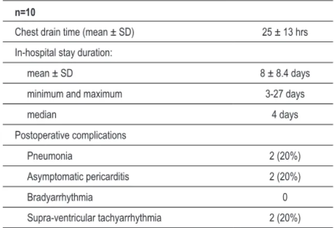
Table 4 - Intra-hospital postoperative variables
n=10
Chest drain time (mean ± SD) 25 ± 13 hrs
In-hospital stay duration:
mean ± SD 8 ± 8.4 days
minimum and maximum 3-27 days
median 4 days
Postoperative complications
Pneumonia 2 (20%)
Asymptomatic pericarditis 2 (20%)
Bradyarrhythmia 0
Supra-ventricular tachyarrhythmia 2 (20%)
N - total number of patients; SD - standard deviation of the mean.
thoracotomy in any of the cases as well as no operative mortality in this series.
There was no surgical injury of the cardiac chambers, of basal vessels or the phrenic nerve. None of the patients needed a transfusion of blood components during the hospital stay. The data related to the surgical intervention were grouped in Table 3 and the postoperative data in Table 4.
The mean postoperative follow-up of the patients was 6 months (3 to 12 months). There was no significant difference regarding the drugs used in the pre- and postoperative periods (Table 5).
Five patients underwent surgery in 2007 and the other five in 2008. All the complications described in Table 4 occurred in patients submitted to surgery in 2007 (p=0.048).
The mean hospital stay duration in 2007 was 12.4 days, whereas in 2008 it was 3.6 days (p=0.098). The time of right and left pulmonary collapse was significantly reduced with the accumulation of experience (Chart 1).
There was a decrease in the NYHA functional class in the preoperative period when compared to that assessed in the postoperative period: 2.4 ± 0.5 to 1.6 ± 0.7 (p=0.011).
Eight of the 10 patients (80%) presented sinus rhythm at the final assessment of the postoperative follow-up. The analysis of the angiotomography of the pulmonary veins did not identify pulmonary vein stenosis in any of the 10 patients.
Discussion
Surgery X atrial fibrillation
To define the best treatment for atrial fibrillation has been a challenge. The maintenance of the sinus rhythm can result in fewer symptoms, lower risk of cerebrovascular accident, discontinuation of anti-arrhythmic and oral anticoagulant drugs, improved exercise capacity, better quality of life and lower mortality10.
Chart 1 - Time of right and left pulmonary collapse (in minutes) in relation to the dates of surgeries.
treatments. The surgical treatment of the Maze (also known as “Maze III”), in addition to being a well-established surgical technique, is also extremely effective for the treatment of atrial fibrillation.
Dr. James Cox reports success rates > 95% for patients submitted to this procedure11. In our country, Kalil et al12 have reported more than 80% of AF-free patients after 14 months of mean postoperative follow-up. However, the complexity and morbidity associated to the long time needed to carry out the surgical procedure, limit its use by the surgical community in general.
Efforts to simplify the surgery have used several alternative sources of energy to create lesions that block the bidirectional electrical conduction in the atrial tissue, including the unipolar radiofrequency13. The endocardial use of the unipolar RF (in contrast to the bipolar RF) does not allow the easy identification of lesion occurrence throughout the tissue thickness (transmural), possibly producing incomplete and ineffective lesions14. Additionally, lesions that are not limited to the target-tissue can result in tissue damage of neighboring structures, particularly the esophagus and the pulmonary veins15. Gaynor et al16 initially reported the use of the tissue ablation system by bipolar radiofrequency to create linear and transmural lesions during heart surgery. The bipolar tweezers liberates energy between two electrodes, through tissues
fixed between the arms, which essentially eliminates the risk of lesion in adjacent tissues.
Surgery x catheter ablation
The development of less invasive procedures for the treatment of atrial fibrillation significantly increased after the identification of the importance of the pulmonary veins in the pathogenesis of this arrhythmia14. After that, the techniques of catheter ablation started to be broadly used for the treatment of AF. With the disseminated use, varied and severe complications occurred and ended up promoting the learning and encouraging the industry so that the evolution of technique and the technology used could result in more effective and safer procedures.
However, the catheter ablation of the atrial fibrillation by applying unipolar RF to the endocardial tissue still presents variable rates of success, frequently needing re-interventions and occasionally, resulting in severe complications such as fistula formation between the left atrium and the esophagus, which are potentially fatal15. Furthermore, the use of irrigated RF catheters, in an effort to reach deeper lesions, increases the risk of cardiac eruption and tissue perforation16.
uses bipolar RF for tissue ablation without reaching the electrical isolation of the pulmonary veins. This device is also capable of perceiving the tissue electrical conductance and defining the occurrence of lesions along the entire tissue thickness17.
An additional benefit of the bipolar RF is that the energy released is confined to the tissue between the electrodes. This limits the lateral spread of heat, reducing the possibility of lesion in adjacent tissues. The use of the bipolar tweezers allows, potentially, a higher uniformity of results independently of the surgeon, a fact that does not occur with the catheter ablation by unipolar RF18.
The video-assisted surgery tries to minimize the concerns related to the aggressiveness of the classic “Maze II” surgical intervention. This procedure prevents the sternotomy. The epicardial approach also allows the procedure to be carried out without extracorporeal circulation (ECC), with the heart beating and without the need for heparin use7. It is important to emphasize, however, that the surgical procedure proposed here aims at promoting only the electrical isolation of the pulmonary veins and connecting lesions are not contemplated.
Results x physiopathology of the AF
There is a concern that the isolation of the pulmonary veins without performing the connecting lesions is not so effective19 and that, potentially, it will allow the manifestation of atrial flutter-type supra-ventricular tachycardia.
Two of the 10 patients in this series presented atrial flutter after the surgery, one of whom already presented this arrhythmia in the preoperative period. The atrial tachyarrhythmias (including atrial flutter), after the AF ablation, are primarily related to incomplete ablation lines and that did not reach the entire tissue thickness20,21. The use of bipolar RF technology reduces this risk by conceptually guaranteeing the “transmurality ” of the lesions. In addition to the electrical conductance calculated by the device during the use of the bipolar energy, the electrical isolation of the pulmonary veins could be tested in all patients in non-fibrillatory rhythm. In one patient, however, the electrical isolation of the left pulmonary veins could not be demonstrated, although the bipolar RF was applied four times. This patient presents the four pulmonary veins connected to the left atrium in anatomical form at the angiotomography. Perhaps the methodology of the conductance test favored the electrical conduction of the pacemaker stimulus from the pulmonary vein to left atrium through the electrodes of the bipolar tweezers, which were, until then, maintained open inside the cavity until the conductance test was conclusive.
From this case on, the bipolar tweezers started to be removed from the cavity to perform the conductance test of the pulmonary veins. This patient remains in sinus rhythm.
The video -assisted surgery, however, does not contemplate the ablation of right atrial flutter circuit which can co-exist with the atrial fibrillation22.
There is a debate on the efficacy of the isolation of the pulmonary veins in situations of continuous (persistent or permanent) atrial fibrillation, which comprehends 50% of this sample19. This surgical approach previews a broad isolation of the pulmonary veins and left atrial antrum, resulting in the isolation of a large left atrial volume. Although the extension of left atrial tissue that must be isolated for the procedure to be successful is debatable, there is evidence that the longer the extension of the left trial ablation, the higher the success rates of the procedure23.
Reports by Wolf et al9, who used a technique that was similar to the one described here, demonstrated that 100% of the patients with continuous (persistent or permanent) atrial fibrillation are arrhythmia-free after six months. However, this approach does not contemplate the pathogenesis of the permanent atrial fibrillation, when there is an electrical and mechanical remodeling of the atrial tissue.
In our series, the only patient that did not leave the operating room in sinus rhythm was a patient refractory to attempts of cardiac rhythm reversion (permanent AF) and since then, this model of patient has been discouraged from undergoing the procedure. A second patient, with persistent AF and high ventricular response was submitted to the procedure successfully, but had early AF recurrence. This patient presents non-obstructive hypertrophic myocardiopathy and several restrictions (severe adverse events) to the use of antiarrhythmic drugs. He kept the sinus rhythm for two weeks after the procedure, but it was not possible to submit him to electrical cardioversion due to the presence of atrial thrombus.
Considering the current knowledge, the occurrence of episodes of tachyarrhythmias for up to four weeks is frequent, and the early recurrence of AF does not mean treatment failure24.
Surgery x complications: the effect of the learning curve
Although minimally invasive (reduced direct tissue trauma), this procedure requires general anesthesia, orotracheal intubation and independent pulmonary ventilation with pulmonary collapse. All these aspects present potential deleterious effects. Both thorax sides are manipulated and all patients receive bilateral chest drainage. The complications that occur in the postoperative period, despite not being fatal, result in a long ICU and hospital stay duration. The operative complications, time of hospital stay during the postoperative period and time of pulmonary collapse for carrying out the surgery were dependent on the date when it was performed.
References
1. Go AS, Hylek EM, Phillips KA, Chang Y, Henault LE, Selby JV, et al. Prevalence of diagnosed atrial fibrillation in adults: national implications for rhythm management and stroke prevention: the AnTicoagulation and Risk Factors in Atrial Fibrillation (ATRIA) Study. JAMA. 2001; 285: 2370-5.
2. Feinberg WM, Blackshear JL, Laupacis A, Kronmal R, Hart RG. Prevalence, age distribution, and gender of patients with atrial fibrillation: analysis and implications. Arch Intern Med. 1995; 155: 469-73.
3. Benjamin EJ, Wolf PA, D’Agostino RB, Silbershatz H, Kannel WB, Levy D. Impact of atrial fibrillation on the risk of death: the Framingham Heart Study. Circulation. 1998; 98: 946-52.
4. Friberg J, Buch P, Scharling H, Gadsbphioll N, Jensen GB. Rising rates of hospital admissions for atrial fibrillation. Epidemiology. 2003; 14: 666-72.
5. Fornari LS, Calderaro D, Nassar IB, Lauretti C, Nakamura L, Bagnatori R, et al. Misuse of antithrombotic therapy in atrial fibrillation patients: frequent, pervasive and persistent. J Thromb Thrombolysis. 2007; 23 (1): 65-71.
6. Lip GYH, Tse HF. Management of atrial fibrillation. Lancet. 2007; 370: 604-18.
7. Haissaguerre M, Jais P, Shah DC, Takahashi A, Hocini M, Quiniou G, et al. Spontaneous initiation of atrial fibrillation by ectopic beats originating in the pulmonary veins. N Engl J Med. 1998; 339: 659-66.
8. Scanavacca M, Pisani CF, Hachul D, Lara S, Hardy C, Darrieux F, et al. Selective atrial vagal denervation guided by evoked vagal reflex to treat patients with paroxysmal atrial fibrillation. Circulation. 2006; 114 (9): 876-85.
9. Wolf RK, Schneeberger EW, Osterday R, Miller D, Merril W, Rege JB, et al. Video-assisted bilateral pulmonary vein isolation and left atrial appendage exclusion for atrial fibrillation. J Thorac Cardiovasc Surg. 2005; 130:
797-802.
10. Friedman PA, Hammill SC. Atrial fibrillation therapies-rate or rhythm control? Business Briefing: US Cardiol. 2004. [Accessed 2009 Feb 19]. Available from: http://www.touchcardiology.com./articles/atrial-fibrillation-therapies-rate.
11. Cox JL, Schuessler RB, Boineau JP. The development of the Maze Procedure for the treatment of atrial fibrillation. Semin Thorac Cardiovasc Surg. 2000; 12 (1): 2-14.
12. Kalil RAK, Albrecht A, Lima GG, Vasconcellos D, Cunha B, Hatem D, et al. Resultados do tratamento cirúrgico da fibrilação atrial crônica. Arq Bras Cardiol. 1999; 73 (2): 144-8.
13. Damiano RJ. Alternative energy sources for atrial ablation: judging the new technology. Ann Thorac Surg. 2003; 75: 329-30.
14. Ouyang F, Antz M, Ernset S, Hachiya H, Mavrakis H, Deger FT, et al. Recovered pulmonary vein conduction as a dominant factor for recurrent atrial tachyarrhythmias after complete circular isolation of the pulmonary veins. Circulation. 2005; 111: 127-35.
15. Gillinov AM, Petterson G, Rice TW. Esophageal injury during radiofrequency ablation for atrial fibrillation. J Thorac Cardiovasc Surg. 2001; 122: 1239-40.
16. Gaynor SL, Diodato MD, Prasad SM, Ishii Y, Schuessler RB, Bailey MS, et al. A prospective, singlecenter clinical trial of a modified Cox maze procedure with bipolar radiofrequency ablation. J Thorac Cardiovasc Surg. 2004; 124: 535-42.
17. Prasad SM, Maniar HS, Schuessler RB, Damiano RJ. Chronic transmural atrial ablation by using bipolar radiofrequency energy on the beating heart. J Thorac Cardiovasc Surg. 2002; 124: 708-13.
have been related to the occurrence of the described respiratory complications, which occurred in the beginning of the experience and resulted in a longer hospital stay duration.
A change in the anesthetic conduct and in the objective assessment of the neuromuscular block status at the end of the procedure (with the use of equipment that test the residual level of neuromuscular blockers) allowed more safety in the extubation procedure and decreased to zero the respiratory complications after the procedure.
All patients received common analgesic medication (dipyrone and acetaminophen) or non-steroidal anti-inflammatory drugs during hospital stay. None of the patients needed opioids in the postoperative period. The chest pain, however, was not an objectively collected variable.
It becomes evident the importance of the learning curve when incorporating new procedures and technologies to the clinical practice.
Limitations
The small number of patients and the short postoperative follow-up period in this longitudinal, non-randomized study with no control group has limitations that are inherent to this design, especially regarding the inference of clinical outcomes. Patient selection biases were not controlled. The use of 24-hour Holter monitoring alone in the postoperative
period as the tool that defined the occurrence or not of atrial fibrillation in the postoperative period might have underestimated the recurrence of AF.
Conclusion
The video-assisted surgical technique for the treatment of atrial fibrillation is reproducible. The surgical procedure is safe; however, it requires the learning from the professionals involved in this new therapeutic approach modality.
The initial clinical results of heart failure symptom improvement and atrial-fibrillation-free survival are encouraging, as they provide a technical alternative to the invasive treatment of atrial fibrillation.
Potential Conflict of Interest
No potential conflict of interest relevant to this article was reported.
Sources of Funding
There were no external funding sources for this study.
Study Association
18. Scanavacca MI, Sosa E. Ablação por cateter da fibrilação atrial: técnicas e resultados. Arq Bras Cardiol. 2005; 85 (4): 295-301.
19. Oral H, Scharf C, Chugh A, Hall B, Cheung P, Good E, et al. Catheter ablation for paroxysmal atrial fibrillation: segmental pulmonary vein ostial ablation versus left atrial ablation. Circulation. 2003; 108: 2355-60.
20. Gaita F, Riccardi R, Caponi D, Shah S, Garberoglio L, Vivalda L, et al. Linear cryoablation of the left atrium versus pulmonary vein cryoisolation in patients with permanent atrial fibrillation and valvular heart disease. Circulation. 2005; 111: 136-42.
21. Kobza R, Hindricks G, Tanner H, Schirdewahn P, Dorszewski A, Piorkowski C, et al. Late recurrent arrhythmias after ablation of atrial fibrillation: incidence, mechanisms and treatment. Heart Rhythm. 2004; 1: 676-83.
22. Moreira W, Timmermans C, Wellens HJ, Mizusawa Y, Philippens S, Perez D,
et al. Can common-type atrial flutter be a sign of an arrhythmogenic substrate in paroxysmal atrial fibrillation? clinical and ablative consequences in patients with coexistent paroxysmal atrial fibrillation/atrial flutter. Circulation. 2007; 116 (24): 2786-92.
23. Marrouche NF, Dresing T, Cole C, Bash D, Saad E, Balaban K, et al. Circular mapping and ablation of the pulmonary vein for treatment of atrial fibrillation: impact of different catheter technologies. J Am Coll Cardiol. 2002; 40: 464-74.