DOI: 10.1590/0004-282X20150135
ARTICLE
Injecction of trigger points in the temporal
muscles of patients with miofascial syndrome
Infiltrações de pontos-gatilho na musculatura temporal em pacientes com fibromialgia e
cefaleia
Svetlana Sabatke1, Rosana Herminia Scola2, Eduardo S. Paiva3, Pedro André Kowacs4
Fibromyalgia (FM) is a non-articular rheumatic syndrome, characterized by chronic widespread pain with tender points
on palpation of speciic painful body sites, in the absence of other known organic disease. he classiication criteria were
described in 1990 by the American College of Rheumatology1.
It is considered the second most common rheumatic
dis-ease, afecting mainly women. Many other diseases may be
associated with FM, and most patients have other associat-ed comorbidities2,3,4,5. Myofascial pain syndrome (MPS) is a
common regional pain syndrome, considered to be the
com-plaint of pain most present in medical practice. he pain -ful symptoms are the result of hyperalgesia of small trigger
points, which radiate pain to distant sites6,7.MPS can afect
the orofacial region, and called masticatory myofascial pain syndrome (MMPS). In patients with FM, facial pain inten-sity is correlated with generalized muscle pain8.he occur
-rence of headaches in patients with FM is great, where their symptoms can be manifested by myofascial pain referral of trigger points9,10,11,12,13. Considering the temporal muscles
trig-ger points pain with their respective radiation areas are rep-resented in Figure 1. One way of treating MPS is the injection of anesthetic or saline at trigger points. Treatment is aimed at decreasing the intensity of facial pain, and may also decrease headache symptoms by modulating pain12,13,14,15,16,17,18,19,20. he
1Universidade Federal do Paraná, Hospital das Clínicas, Curitiba PR, Brazil;
2Universidade Federal do Paraná, Hospital das Clínicas, Divisão de Neuromuscular, Curitiba PR, Brazil; 3Universidade Federal do Paraná, Hospital das Clínicas, Disciplina de Reumatologia, Curitiba PR, Brazil;
4Universidade Federal do Paraná, Hospital das Clinicas, Departamento de Medicina Interna, Divisão de Neurologia, Seção de Dor de Cabeça, Curitiba
PR, Brazil.
Correspondence: Svetlana Sabatke; UFPR; Av Iguaçu, 2820 / sala 901; 80240-030 Curitiba PR, Brasil; E-mail: [email protected] Conflict of interest: There is no conflict of interest to declare.
Received 22 October 2014; Received in final form 23 May 2015; Accepted 11 June 2015. ABSTRACT
Objective: The aim was to examine the effect of blocking trigger points in the temporal muscles of patients with masticatory myofascial pain syndrome, fibromyalgia and headache. Method: Seventy patients with one trigger point were randomly divided into 3 groups: injection with saline or anesthetic and non-injected (control). Results: Pain was reduced in 87.71% patients injected with saline and 100% injected with anesthetic. Similar results were obtained for headache frequency. With regard to headache intensity, the injection groups differed from the control group, but not between themselves. Conclusion: Treatment with injection at trigger points decreased facial pain and frequency and intensity of headache. Considering the injected substance there was no difference.
Keywords: headache, fibromyalgia, infiltration, trigger point, myofascial pain syndrome.
RESUMO
Objetivo: Comparar o efeito terapêutico do bloqueio de pontos-gatilho na musculatura temporal com soro fisiológico e anestésico em pacientes com síndrome da dor miofascial mastigatória, fibromialgia e cefaleia, entre sí e com controles não-infiltrados. Método: Setenta pacientes que apresentaram pelo menos um ponto-gatilho na musculatura temporal foram aleatoriamente divididas em 3 grupos: infiltração com soro fisiológico, infiltração com anestésico e controle (não-infiltradas). Resultados: Houve redução na intensidade de dor na face em 87,71% dos pacientes infiltrados com soro fisiológico e em 100% dos pacientes infiltrados com anestésico, mas não no grupo controle. Houve similaridade dos resultados considerando a frequência da cefaléia. Quanto à intensidade da cefaléia, tanto a infiltração com soro fisiológico, quanto com anestésico foram efetivos e sem diferença significativa entre sí, ao contrário do grupo controle. Conclusões:
O tratamento com infiltração diminui a dor na face, bem com a frequência e a intensidade da cefaléia. Quando considerado a substância infiltrada não há diferenças no tratamento.
aims of the present study were to investigate the efective
-ness of blocking trigger points using diferent substances (sa -line versus anesthetic) in the temporal muscles of patients with MMPS, FM and headache.
METHOD
his study was approved by the Ethics Committee for Human Research of the Hospital das Clinicas of the Federal University
of Parana and consisted of a randomized, double-blind study
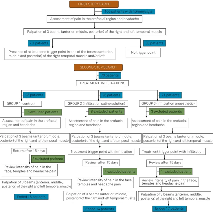
with control group. he research was divided into two stages, illustrated in the lowchart presented (Figure 2), and in each,
the patients read and signed an informed consent form. In the
irst stage of the study, we included patients with ibromyal
-gia, diagnosed according to the classiication criteria of the
American College of Rheumatology1; patients who were under
treatment in the period between January 2007 and June 2008,
in the ibromyalgia clinic of Hospital das Clinicas of the Federal University of Paraná. Exclusion criteria were: present for irst
visit and still not under treatment at the clinic, male patients, and inability to read and/or understand the informed consent
form. he initial sample consisted of 100 female patients with ibromyalgia, aged 23-70 years. Patients initially attended the selection visit (irst stage) and selected attended the visit of treatment, and the follow-up visit (second stage). In the irst
stage the patients were evaluated for the presence or no of pain in the region of the face and/or neck and headache without
classify them.Next, they were examined for trigger points in the temporal muscle (right and left). he presence or absence
of pain was scored using a numerical scale (0-no pain,
1-pres-ence of sensitivity, 2-until the nail bed of the examiner’s index inger turned whitish8.
In the second stage, trigger points were injected with
sa-line or anesthetic; the patients included here were those ex
-amined in the irst stage of the study who had at least one
trigger point in one of the temporal muscles (right or left) regardless of the palpation of these points cause or not a
headache. Exclusion criteria were: evidence of inlammatory
rheumatic disease, heart disease or uncontrolled hyperten-sion, uncontrolled diabetes mellitus, blood dyscrasias, local infection, systemic infection, local skin changes and history
of allergy to anti-inlammatory medication prescribed after
injection6. Of the 100 patients examined in the irst stage, 70
patients were included in this second stage (treatment with injection) and randomly divided into three groups (group with saline injection, group with anesthetic injection and control group). In the follow-up visit, the study subjects were
re-evaluated for efectiveness of intervention used, which
was compared to the status of the controls not subjected to therapeutic intervention.
Group 1 (control) consisted of 23 patients. Of these, 5 dropped out and 2 did not return for reassessment after 15 days, leaving 16 patients in this group. Group 2 (injection with saline 0.9%) 26 patients. Of these, 5 dropped out, 4 did not return for reassessment after 15 days, 2 were discarded due to the absence of a trigger point and 1 was ruled out
due to being allergic to anti-inlammatory medication pre -scribed after the injection procedure, leaving 14 patients in
this group. Group 3 (injection with anesthetic: 2% lidocaine
without vasoconstrictor) consisted of 21 patients. Of these 3 dropped out and 1 did not return for reassessment after
15 days, leaving 17 patients in this group. he characteris -tics of the saline, anesthetic and control groups are shown in Figure 1, as well as information about the discontinued pa-tients. In the saline and anesthetic groups, the procedure was performed in a double-blinded fashion and with the patient
lying down. he trigger points were located using manual
palpation of the skin disinfected with 70% alcohol, a freezing spray (-40°C) was applied to avoid pain during needle pen-etration20,21.In the procedure, we used a carpule syringe with
a short 30G needle6. An amount of 0.2 to 0.5 ml of anesthetic
or saline was injected at each trigger point8,20.he injection
procedure, massage and stretching the temporal muscles af-ter the procedure were performed as described by Simons
et al. and other studies as well. Eight subjects of saline and
anesthetic groups received nimesulide tablets 100 mg, to take twice a day for two days. While taking the medication, patients were instructed to apply warm, moist compresses three to four times a day for 10 to 15 minutes, or to soak the injection area in a warm bath22.
Statistical analysis
he 70 patients included in the second stage were random -ly divided by SigmaStat for Windows version 2.0 into three groups (group with saline injection, group with anesthetic
injection and control group). he Wilcoxon test was used for
comparison of intra-group results, and the Kruskal-Wallis test was used to compare the results between the groups. Two by two comparison of the groups in relation to the likelihood of improvement was carried out with logistic regression, control-ling for patient age and adopting the Wald test, and if not
pos-sible, we used the Fisher exact test with Bonferroni. Correction values p < 0.05 indicate signiicance statistical.
RESULTS
In the irst stage, all selected patients (100%) had some
type of pain in the region of the face and/or neck as well as
someone headache. Sensitivity in the temporal muscle oc-curred in 90% of patients. Despite that the patients had trig-ger points in all the temporal muscles, these points occurred less in the right posterior (27 patients) and left posterior (29 patients) and more in the left anterior (62 patients) and right
anterior (61 patients). Headache was produced in 93 to 98% of trigger points when palpated (Table 1). he results below per -tain to the records of the second stage, in which we analyzed the patients who were injected compared to the control group.
Facial pain intensity
Both saline and anesthetic treatments signiicantly re -duced the intensity of facial pain, unlike the control group
Figure 2. Flow chart of research stages.
FIRST STEP SEARCH
100 patients with fibromyalgia
Assessment of pain in the orofacial region and headache
Palpation of 3 beams (anterior, middle, posterior) of the right and left temporal muscle
70 patients 30 patients
Presence of at least one trigger point in one of the beams (anterior,
middle and posterior) of the right temporal muscle and/or left No trigger point
SECOND STEP SEARCH
70 patients
TREATMENT INFILTRATIONS
23 patients 26 patients 21 patients
GROUP 1 (control) GROUP 2 (infiltration saline solution) GROUP 3 (infiltration anaesthetic)
5 excluded patients 8 excluded patients 3 excluded patients
Assessment of pain in the orofacial region and headache
Assessment of pain in the orofacial region and headache
Assessment of pain in the orofacial region and headache
Palpation of 3 beams (anterior, middle, posterior) of the right and left temporal muscle
Palpation of 3 beams (anterior, middle, posterior) of the right and left temporal muscle
Palpation of 3 beams (anterior, middle, posterior) of the right and left temporal muscle
Return after 15 days Treatment trigger point with infiltration Treatment trigger point with infiltration
Review after 15 days Review after 15 days
2 excluded patients
4 excluded patients 1 excluded patient
Review intensity of pain in the face, temples and headache pain
Palpation of 3 beams (anterior, middle, posterior) of the right and left temporal muscle
Ended 16 patients
Review intensity of pain in the face, temples and headache pain
Review intensity of pain in the face, temples and headache pain
Palpation of 3 beams (anterior, middle, posterior) of the right and left temporal muscle
Palpation of 3 beams (anterior, middle, posterior) of the right and left temporal muscle
(p = 0.004 and p < 0.001; Table 2). In a two by two
compari-son of the groups, there was a statistically signiicant difer -ence when the comparison was carried out with the control
group, but no diference between the groups treated with
saline and anesthetic (p = 0.003 and p = 0.005; Table 3). In 85.71% of patients treated with saline, 100% treated with an-esthetic and 43.75% of the control, there was reduction in fa-cial pain intensity. In a two by two comparison of the groups in relation to reduction in facial pain intensity, a statistically
signiicant diference was found when the comparison was
between the anesthetic-treated group and control group (p < 0.001; Table 3).
Pain in the temples or above the ears
Only two individuals in the saline group, two in the
con-trol group and ive in the anesthetic group said that they
did not feel pain in the temples or above the ears 15 days
after treatment. he improvement occurred in 14.29% of
those treated with saline, in 29.41% of those treated with
anesthetic and 12.50% in the control group. Two by two
comparison of the groups showed no statistically signii
-cant diference (Table 3).
Weekly headache frequency
Regarding the weekly headache frequency, unlike the control group, both the saline and anesthetic groups showed
decreased frequency, demonstrated by statistically signii
-cant diferences (p = 0.037 and p = 0.002; Table 2). Two by two comparison of the groups showed a statistically signiicant diference when the comparison was carried out with the control group, but no diference between the groups treated
with saline and anesthetic (p = 0.003 and p = 0.002; Table 3).
Headache intensity
Regarding headache intensity, both saline and
anesthet-ic treatments were efective, difering from the control group (p = 0.008 and p = 0.001; Table 2). Headache intensity de -creased in patients treated with saline by 64.29% and with
Table 1. Result of palpation of each temporal muscle.
Musc. Temp. Pain on muscle palpation Presence of trigger points Palpation causes headache
0 1 2 3 n No Yes n No Yes n
post. lt. 11 (11%) 40 (40%) 44 (44%) 5 (5%) 100 71 (71%) 29 (29%) 100 2 (7%) 27 (93%) 29
med.lt. 5 (5%) 28 (28%) 56 (56%) 11 (11%) 100 52 (52%) 48 (48%) 100 3 (6%) 45 (94%) 48
ant. lt. 2 (2%) 13 (13%) 60 (60%) 25 (25%) 100 38 (38%) 62 (62%) 100 1 (2%) 61 (98%) 62
post. rt. 26 (26%) 43 (43%) 29 (29%) 2 (2%) 100 73 (73%) 27 (27%) 100 2 (7%) 25 (93%) 27
med.rt. 12 (12%) 32 (32%) 51 (51%) 5 (5%) 100 58 (58%) 42 (42%) 100 2 (5%) 40 (95%) 42
ant. rt. 3 (3%) 21 (21%) 49 (49%) 27 (27%) 100 39 (39%) 61 (61%) 100 2 (3%) 59 (97%) 61
musc. temp.: temporal muscle; post. lt.: left posterior; ant. lt.: left anterior; post. rt.: right posterior; med. rt.: right median; ant. rt.: right anterior; n: total patients; 0: absence of pain; 1: presence of sensitivity; 2: presence of pain; 3: escape response.
Table 2. Comparison between groups.
Group n Intensity of
facial pain p*
Weekly frequency of
headache p*
Intensity of
headache p*
Saline Pre-injection 14 7.8 ± 1.6 4.3 ± 2.4 8.6 ± 1.8
Post-15 days 14 2.8 ± 3.7 0.004 2.1 ± 2.7 0.037 5.1 ± 4.0 0.008
Decrease 14 5.0 ± 4.0 2.1 ± 3.3 3.5 ± 3.8
Anesthetic Pre-injection 17 6.5 ± 1.8 4.1 ± 1.9 7.8 ± 1.6
Post-15 days 17 1.6 ± 2.1 < 0.001 1.9 ± 1.7 0.002 4.4 ± 2.5 0.001
Decrease 17 4.9 ± 1.9 2.2 ± 2.2 3.4 ± 2.8
Control Pre-injection 16 7.0 ± 1.8 2.9 ± 2.5 7.9 ± 2.4
Post-15 days 16 5.8 ± 4.1 0.209 3.1 ± 2.3 0.919 6.6 ± 2.5 0.173
Decrease 16 1.3 ± 3.6 -0.1 ± 1.5 1.3± 3.2
(*) non-parametric wilcoxon test; p < 0.05.
Table 3. Two by two comparison of groups.
Groups compared Intensity of facial pain p*
Decrease in intensity of facial pain p**
Pain in temples or above ears p***
Weekly frequency of headache p*
Intensity of headache p***
Saline x Anesthetic 0.704 0.196 0.384 0.966 0.220
Saline x Control 0.003 0.026 0.801 0.003 0.756
Anesthetic x Control 0.005 < 0.001 0.204 0.002 0.112
anesthetic by 82.35%, compared to 56.25% in the control group. In a two by two comparison of the groups, no
statisti-cally signiicant diference was found (Table 3).
DISCUSSION
he study involved a sample of only female patients, be
-cause FM is a chronic pain syndrome that afects mainly wom -en2,3,4. here was agreement with the age group in which is the
disease occurs, 20 to 60 years17. he mean age was close to that
of most recent studies demonstrating that the prevalence in women peaks around 55 to 64 years, ages similar to that of
menopause, in which there is a signiicant hormonal decline, reinforcing the possible inluence of hormonal factors2.
It is believed that the high percentage (100%) of patients with pain in some region of the face and/or neck occurred
because of this study being speciically for the assessment of
pain in this region; pain in this region is often not observed
due to the lack of emphasis given by the examiner, mainly
when the pain is not just a localized problem but rather a condition with generalized muscle pain like FM10.In these
pa-tients, sensitivity in the temporal muscle was identiied by
the reporting of pain at the temples or above the ears,
con-irming a high pain sensitivity in the temporal muscle of pa -tients with MPS7.he strong presence of MMPS as well as
headache conirmed the association of FM with other condi
-tions. he high prevalence of both in this study reinforced the
need for individual evaluation of each patient, to identify the
comorbidity, inluencing the measurement of symptoms and
assisting in the treatment modality and thereby controlling the disease11,12.
Our percentage of 100% of patients with headache was higher than that reported by some authors, i.e., 53 to 82%, but in agreement with others observing more than 91%10,11. We
found at least one trigger point in the temporal muscle in 70%
of patients palpated in the irst stage of the study, conirming
the association with MPS, higher than other reports of 18%1,13.
In agreement with others noting 68 to 72%14,15.he high per
-centage of patients found with MMPS conirms the associa
-tion of temporomandibular disorders in patients with ibro -myalgia, and reinforces the hypothesis of these disorders as a possible cause of the facial pain9. he irradiation of pain from
these trigger points can cause headache and toothache11,22.
Headache may be a manifestation of muscle sensitivity9,16.
Pain irradiation from trigger points located in the temporal muscle can be manifested by headache11,15,16,17,22. his study
did not aim to classify the headache, but when the treatment
of myofascial pain (trigger points iniltration in the temporal
muscle), there were changes both in intensity and in
head-ache frequency. hus, these patients was possible to state
that there was a presence of a secondary headache
myofas-cial syndrome. he presence of trigger points in these areas is
consistent, because it is the most typical location of migraine
pain. Also, it is among the most common areas of radiating pain in patients with migraine, including tension-type chron-ic headache14,12,16.
Representing one of the forms of recommended treat-ment13,17, injection of trigger points in the temporal
mus-cle was used to test the efect of this type of treatment on
myofascial pain and headache. Although there are reports that the injection procedure is required in only 20 to 25%
of patients with MPS, it is considered most efective treat -ment18,23. he amount of anesthetic injected at each trigger
point was about 0.2 ml to 0.5 ml of 2% lidocaine without va-soconstrictor6,17. Lidocaine was the anesthetic of choice, as
in other studies. It is proven that this causes necrosis of the muscle tissue injected, but with rapid regeneration occur-ring in 16 days6,16,17,18,19,24,25.
As part of the treatment, the muscles were stretched after
the injection to enhance the efectiveness of treatment6. To
minimize soreness after injection, in addition to hemostasis
care, anti-inlammatory medication was used for 48 hours to -gether with warm, moist compresses, despite that there have been studies without stretching and heat treatment after in-jection20. In the control group, anti-inlammatory medication
was not used, because the patients were already receiving
treatment in the ibromyalgia clinic and had been undergo -ing medical treatment without results for alleviation of pain.
hese patients were continued use of medications. Since the purpose of administering an anti-inlammatory medication was only to relieve soreness after the iniltra -tion procedure, whether or anesthetic physiological serum, non-medicated control group. In addition, medication use was restricted for only two days and believe it has not
inter-fered in the evaluation that took place 15 days after the irst
visit for all groups. Failure to use warm, moist compresses in
the control group may have inluenced the results obtained. In this study, both local anesthetic and saline were efec -tive in the treatment of trigger points21. Both groups showed
a reduction in pain intensity compared with control, with no
statistically signiicant diference between them. he control
group also showed improvement, but this was not statistically
signiicant. However, there are studies showing greater eica -cy of anesthesia and without injection treatments, and there
are diferent comparisons with regard to the type of substance
injected, also testing dry needling13,18,22,24,25. But there is agree
-ment that the efectiveness of the treat-ment is not related to
the nature of the substance injected. Probably the relief occurs by needle contact with the trigger point, breaking its vicious cycle16,24. After treatment, there was a decrease in pain in all the
temporal muscles examined, a inding conirmed by palpation.
When asking about pain at the temples or above the ears, the patients said that there was no improvement. We suggest that
this negative inding could be related to treatment failure in
References
1. Wolfe F, Smythe HA, Yunus MB, Bennett RM, Bombardier C, Goldenberg DL et al. The American College of Rheumatology 1990 criteria for the classification of fibromyalgia. Arthritis Rheum.1990;33(2):160-72. doi:10.1002/art.1780330203
2. Moldofsky HK. Disordered sleep in fibromyalgia and related myofascial facial pain conditions. Dent Clin North Am. 2001;45(4):701-13.
3. Antonelli MA, Vawter, RL. Nonarticular pain syndromes: differentiating generalized, regional, and localized disorders. Postgrad Med.1992;91(2):95-8.
4. Okeson JP. Dores bucofaciais de Bell: tratamento clínico da dor bucofacial. São Paulo: Quintessence; 2006.
5. Fernández-de-las-Peñas C, Alonso-Blanco C, Cuadrado ML, Gerwin RD, Pareja JA et al. Myofascial trigger points and their relationship to headache clinical parameters in chronic tension-type headache. Headache. 2006;46(8):1264-72. doi:10.1111/j.1526-4610.2006.00440.x
6. Fernández-de-las-Peñas C, Ge HY, Arendt-Nielsen L, Cuadrado ML, Pareja JA et al. The local and referred pain from myofascial trigger points in the temporalis muscle contributes to pain profile in chronic tension-type headache. Clin J Pain. 2007;23(9):786-92. doi:10.1097/AJP.0b013e318153496a
7. Venâncio RA, Alencar FG, Zamperini C. Different substances and dry-needling infections in patients with myofascial pain and headaches. Cranio. 2008;26(2):96-103. doi:10.1179/crn.2008.014
8. Hong CZ,, Kuan TS, Chen JT, Chen SM. Referred pain elicited by palpation and by needling of myofascial trigger points: a comparison. Arch Phys Med Rehabil. 1997;78(9):957-60. doi:10.1016/S0003-9993(97)90057-5
9. Olson GB, Savage S, Olson J. The effects of collagen hydrolysat on symptoms of chronic fibromyalgia and temporomandibular joint pain. Cranio. 2000;18(2):135-41.
10. Cimino R, Michelotti A, Stradi R, Farinaro C. Comparison of clinical and psychologic features of fibromyalgia and masticatory myofascial pain. J Orofac Pain. 1998;12(1):35-41.
11. Boomershine CS, Crofford LJ. A symptom-based approach to pharmacologic management of fibromyalgia. Nat Rev Rheumatol. 2009;5(4):191-9. doi:10.1038/nrrheum.2009.25
12. Williams DA, Schilling S. Advances in the assessment of fibromyalgia. Rheum Dis Clin North Am. 2009;35(2):339-57. doi:10.1016/j.rdc.2009.05.007
13. Hameroff SR, Crago BR, Blitt CD, Womble J, Kanel J. Comparison of bupivacaine, etidocaine, and saline for trigger-point therapy. Anesth Analg. 1981;60(10):752-5. doi:10.1213/00000539-198110000-00011
14. Sollecito TP, Stoopler ET, DeRossi SS, Silverton S. Temporomandibular disorders and fibromyalgia: comorbid conditions? Gen Dent. 2003;51(2):184-7.
15. Bohr T. Problems with myofascial pain syndrome and fibromyalgia syndrome. Neurology. 1996;46(3):593-7. doi:10.1212/WNL.46.3.593
16. Fricton JR. Myofascial pain. Baillieres Clin Rheumatol 1994;8(4):857-80. doi:10.1016/S0950-3579(05)80052-4
17. Wheeler AH. Myofascial pain disorders: theory to therapy. Drugs. 2004;64(1):45-62. doi:10.2165/00003495-200464010-00004
18. 18. Han S, Harrison P. Myofascial pain syndrome and trigger-point management. Reg Anesth. 1997;22(1):89-101. doi:10.1016/S1098-7339(06)80062-3
19. Cummings TM, White AR. Needling therapies in the management of myofascial trigger point pain: a systematic review. Arch Phys Med Rehabil. 2001;82(7):986-91. doi:10.1053/apmr.2001.24023
20. Carlson CR, Okeson JP, Falace DA, Nitz AJ, Lindroth JE. Reduction of pain and EMG activity in the masseter region by trapezius trigger point injection. Pain. 1993;55(3):397-400. doi:10.1016/0304-3959(93)90018-K
21. Littlejohn GO. Balanced treatments for fibromyalgia. Arthritis Rheum. 2004;50(9):2725-9. doi:10.1002/art.20486
22. Simons DG, Travell JG, Simons, LS. Dor e disfunção miofascial: manual de pontos-gatilho. Porto Alegre: Artmed; 2005.
23. Jaeger B, Skootsky SA. Double-blind, controlled study of different myofascial trigger point injection techniques. Pain. 1987;30(suppl 1):S292. doi:10.1016/0304-3959(87)91645-9
24. Hong C. Lidocaine injection versus dry needling to myofascial trigger point. The importance of the local twitch response. Am J Phys Med Rehabil. 1994;73(4):256-63. doi:10.1097/00002060-199407000-00006
25. McMillan AS, Nolan A, Kelly PJ. The efficacy of dry needling and procaine in the treatment of myofascial pain in the jaw muscles. J Orofac Pain. 1997;11(4):307-14.
26. Goldenberg DL, Burckhardt C, Crofford L. Management of fibromyalgia syndrome. J Am Med Assoc. 2004;292(19):2388-95. doi:10.1001/jama.292.19.2388
he present study demonstrated that anesthetic and sa -line injection, compared to the control group, caused a reduc-tion in both the intensity and frequency of headache, with no
statistically signiicant diference between the two injection groups. he study reinforced the evidence that treatments
aimed at muscles decrease headache symptoms, through a central mechanism of pain modulation12,14,15,16,17,18,19,20.