Quim. Nova, Vol. 33, No. 1, 30-32, 2010
Artigo
† This paper is dedicated to our colleague and friend Clara M. A. Tanaka in memoriam
*e-mail: [email protected]
ABIETATRIENES DITERPENOIDS FROM Sagittaria montevidensis SSP Montevidensis
Clara M. A. Tanaka†, Vanessa S. C. O. Radke e Cleuza C. da Silva
Departamento de Química, Universidade Estadual de Maringá, Av. Colombo, 5790, 87020-900 Maringá – PR, Brasil Celso V. Nakamura
Departamento de Análises Clínicas, Universidade Estadual de Maringá, Av. Colombo, 5790, 87020-900 Maringá – PR, Brasil Pollyanna L. de Oliveira, Lucilia Kato e Cecília M. A. de Oliveira*
Instituto de Química, Universidade Federal de Goiás, Campus II, 74000-970 Goiânia – GO, Brasil
Recebido em 1/10/08; aceito em 14/7/09; publicado na web em 25/11/09
The antimicrobial properties of the hexane, hexane/EtOAc and methanol fractions of the fresh petioles of Sagittaria montevidensis ssp montevidensis (Alismataceae) were evaluated against fungi and Gram-negative and Gram-positive bacteria. A new abietatriene-type diterpenoid, 3β,7α–dihydroxi-abieta-8,11,13-triene and the known 3β-hydroxy-abieta-8,11,13-trien-7-one were isolated from the most active fraction tested and the structures of these compounds were elucidated by data including IR, EIMS, and 1D and 2D NMR spectra.
Keywords: Sagittaria; abietatrienes; antimicrobial activity.
INTRODUCTION
The genus Sagittaria (Alismataceae) comprises 25 species of herbaceous aquatic plants that are distributed throughout the Ame-ricas in tropical regions. No evidence has been found for the use of Sagittaria montevidensis ssp montevidensis in traditional medicine. However, others Sagittaria species are known to produce antibacterial compounds such as clerodane, pimarane, labdane and rosane-type diterpenoids.1,2 During of our survey of the active substances from Alistamaceae species,3-6 fractions of different polarity deriving from the crude methanolic extract of S. montevidensis ssp monteviden-sis were tested against a representative set of fungi and bacteria strains. Chromatographic treatment of the most active fraction led to the isolation of a new diterpenoid abietatriene derivative, 3β,7α– dihydroxi-abieta-8,11,13-triene (1), along with a known diterpenoid, 3β-hydroxy-abieta-8,11,13-trien-7-one (2). The structures of the compounds were determined by spectroscopic analysis including EIMS, IR, and 1D and 2D 1H, 13C NMR data and also by comparison of its NMR data with those of related compounds.
EXPERIMENTAL
General experimental procedures
1D and 2D 1H, 13C NMR spectra were acquired on a Varian Mercury plus BB spectrometer, operating at 300.059 MHz (1H) and 75.458 MHz (13C) for a CDCl
3 solution using TMS as an internal standard. Mass spectrometry was performed on a Shimadzu GC-MS QP 2000A, 70 eV.
Plant material
The plant material was collected in Curitiba, Paraná, Brazil and authenticated by Dr. M. do C. Amaral (IB-UNICAMP). A voucher specimen (# UEC 115194) was deposited in the Herbarium of the Ins-tituto de Biologia, Universidade Estadual de Campinas, Campinas-SP.
Extraction and isolation
Fresh petioles (86.0 g) of S. montevidensis ssp montevidensis were extracted with MeOH. After removal of the solvent by vacuum, the resi-due (32.0 g) was partitioned between EtOAc and H2O. The EtOAc (6.0 g) extract was subjected to column chromatography on a silica gel 60 to give n-hexane, n-hexane/EtOAc (95:5, 90:10, 80:20, 70:30, 60:40, 50:50) and MeOH fractions. These subfractions were named subfractions A, B, C, D, E, F, G and H, respectively. The fraction E eluted with n-hexane/ EtOAc 70:30 (602.0 mg) was further puriied by column chromatography (CC) on silica gel 60 using an n-hexane-EtOAc gradient solvent system to obtain compounds 1 (12.0 mg) and 2 (40.0 mg).
3β-hydroxy-abieta-8,11,13-trien-7-one (2)7,8 Yellowish oil: RMN- 1H δ (300 MHz, CDCl
3): 7.80 (d, 2.1 Hz, H-14), 7.40 (dd, 8.2; 2.1 Hz, H-12), 7.28 (d, 8.2 Hz, H-11), 3.36 (dd, 4.8, 10.8 Hz, H-3), 2.90 (sept, 6.0 Hz, H-15), 2.73 (d, 11.4 Hz, H-6β), 2.72 (d, 6.6 Hz, H-6α), 2.40 (m, H-1), 1.86 (dd, 11.4 and 6.6, H-5), 1.82-1.90 (m, H-2), 1.24 (d, 6.0 Hz, H-16), 1.24 (d, 6.0 Hz, H-17), 1.24 (s, H-20), 1.05 (s, H-19), 0.97 (s, H-18). NMR 13C (75 MHz; CDCl
3): 199.7 (C-7), 153.0 (C-9), 147.0 (C-13), 132.7 (C-12), 130.5 (C-8), 125.0 (C-14), 123.9 (C-11), 78.0 (C-3), 48.4 (C-5), 38.7 (C-4), 37.4 (C-10), 36.1 (C-1), 35.7 (C-6), 33.5 (C-15), 27.4 (C-2), 27.3 (C-19), 23.7 (C-20), 23.6 (C-17), 23.2 (C-16), 14.8 (C-18).
Microorganisms used and growth condition
The fractions above were assayed against positive and Gram-negative bacteria by a broth microdilution assay to determine the minimal inhibitory concentrations (MICs) as described below. The assays were performed with Escherichia coli ATCC 25922, Pseudomonas aeruginosa ATCC 15442, Bacillus subtilis ATCC 6623, and Staphylococcus aureus ATCC 25923 obtained from the American Type Culture Collection (ATCC, Rockville, MD). For the antifungal assay, a single clinical isolate of each species (Candida albicans, C. krusei, C. parapsilosis, and C. tropicalis), obtained from vaginal mucosa, was selected for testing. The bacteria were maintained in Tryptic Soy Agar (Difco). The yeast was maintained in Sabouraud Dextrose Agar (Difco).
Antifungal susceptibility testing
Abietatrienes diterpenoids from Sagittaria montevidensis ssp Montevidensis 31 Vol. 33, No. 1
were determined by microdilution techniques in RPMI 1640 (Sigma Chemical Co., St. Louis, MO) for yeast.9 Inoculates were prepared in the same medium at a density adjusted to a 0.5 McFarland turbidity standard (106 colony-forming units [CFU] m/L) and diluted to a 1:10 ratio for the broth microdilution procedure. Microtiter trays were in-cubated at 37º C, and the MICs were recorded after 24 h of incubation. Two susceptibility endpoints were recorded for each isolate. The MIC was deined as the lowest concentration of compounds at which the tested microorganism does not demonstrate visible growth. Nystatin (Sigma Chemical Co., St. Louis, MO, USA) was included in the test as a control. When the MIC was equal or smaller than 100 µg/mL, the antimicrobial activity was considered signiicant. If the fractions displayed a MIC from 100 to 500 µg/mL, the antimicrobial activity was considered moderate; from 500 to 1000 µg/mL, the antimicrobial activity was considered weak; over 1000 µg/mL, the fractions were considered inactive.10
Antibacterial susceptibility testing
The MICs of all the extracts and reference antibiotics (tetracycline, vancomycin, and penicillin - Sigma Chemical Co., St. Louis, MO, US) were determined by microdilution techniques in Mueller-Hinton broth (Difco) according to CLSI.9 Inoculates were prepared in the same medium at a density adjusted to a 0.5 McFarland turbidity standard (108 colony-forming units [CFU])/mL] and diluted 1:10 for the broth microdilution procedure. Microliter plates were incubated at 37 ºC and the MICs were recorded after 24 h of incubation. Two susceptibility endpoints were recorded for each isolate. The MIC was deined as the lowest concentration of compounds at which the tested microorganism did not demonstrate visible growth.
RESULT AND DISCUSSION
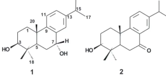
The results of the preliminary screening for antimicrobial activity of all fractions are summarized in Table 1, which shows that six among ten fractions demonstrated signiicant activity against at least one of the microorganisms tested. However, the most active fraction was hexane/ EtOAc (70:30, fraction E) with an MIC = 31.2 µg/mL against C. krusei. Therefore, it was puriied by sequential chromatographic techniques, with the metabolites 1 and 2 (Figure 1) as the major constituents. Compound 2 was identiied as 3β-hydroxy-abieta-8,11,13-trien-7-oneby comparison of the IR, 1H NMR, and 13C NMR spectra with those already published. We now report the characterization of 1.
Compound 1 [α]D = +17 (MeOH, c.0.011), was isolated as a yellowish oil. HRMS suggested a molecular formula of C20H30O2, m/z 302.2232 (calculated m/z 302.2238). The IR spectrum showed absorption bands at νmax 3400 and 1600 cm-1, which could be attribu-ted to hydroxyl groups and double bonds, respectively. The 1H NMR (Table 2) spectrum showed signals for a trisubstituted aromatic ring at δH 7.19 (1H, d, J=1.8 Hz), 7.18 (1H, d, J=8.2 Hz) and 7.12 (1H, dd, J=8.2 and 1.8 Hz), oxymethine at 4.85 (1H, t, J= 3.0 Hz) and 3.35 (1H, dd, J=10.8 and 5.4 Hz), methylenes at 2.30 (1H, m), 2.00 (2H, ddd, J=9.0, J=5.7 and 3.0 Hz), 1.70-1.80 (2H, m) and 1.56 (1H, dd, J=13.2 and 5.4 Hz), a septet at 2.87 (1H, J=6.9 Hz) and methyl groups at1.23 (6H, d, J=6.9 Hz), 1.13 (3H, s), 1.09 (3H, s) and 0.90 (3H, s). From the 13C NMR (Table 2) and DEPT 20 carbons were detected and assigned by HMQC to a aromatic ring (δC 146.9, 146.8, 136.1, 127.9, 126.9 and 124.7), two oxygenated methine (δC 78.9 and 68.7), three methylene groups (δC 36.8, 28.5 and 28.2), two methines (δC 43.8 and 33.7), two sp3 quaternary carbons (38.7 and 37.8) and inally four methyl signals at 28.1, 24.2, 24.1 (2x) and 15.7. Comparison of above data and those of 3β-hydroxy-abieta-8,11,13-trien-7-one2,7,8 showed a good agreement except for the presence of an additional oxymethine group in the compound 1 replacing the carbonyl group observed in 2. Analysis of the 1H -1H COSY experiment (Figure 2) showed correlations between the oxymethine at δH 4.85 (δC 68.7) and protons H-5, H-6 indicating that the extra hydroxyl group was possibly situated on C-7. This assumption was further conirmed by an HMBC experiment, where correlation of H-5 with the C-7 was observed. Additional HMBC correlations of C-3/H-18, H-19, H-1β and C-12/H-14, H-11 and a NOESY experiment, showing that the axial methyl protons at C-19 gave cross peaks with the 20-methyl protons and the H-2β, conirmed the structural similarities of 1 and 2. The presence and location of the isopropyl group linked to quater-nary aromatic carbon was supported by chemical shift and NOESY Figure 1. Structure of abietatrienes 1 and 2
Table 1. Minimum inhibitory concentrations MICs (µg/mL) of the fractions of S. montevidensis spp montevidensis
A B C D E F G H References
Gram-positive bacteria Staphylococcus aureus ATCC
25923 1000 125 1000 125 62.5 125 250 >1000 0.00975ª
Bacillus subtilis ATCC 6623 >1000 >1000 250 250 125 125 125 >1000 0.09b
Gram-negative bacteria
Escherichia coli ATCC 25922 >1000 >1000 >1000 >1000 1000 1000 >1000 >1000 0.78c Pseudomonas aeruginosa ATCC
15442 >1000 >1000 >1000 >1000 >1000 >1000 >1000 >1000 3.125
c
Fungi
Candida albicans >1000 >1000 >1000 500 500 250 500 1000 1.0d
Candida parapsilosis 500 500 500 500 125 250 250 500 8.0d
Candida tropicalis >1000 500 1000 250 125 250 250 >1000 8.0d
Candida krusei 500 62.5 250 62.5 31.2 62.5 62.5 500 4.0d
aPenicillin; bVancomycin; cTetracycline; dNystatin data from experiments in triplicate, MIC deined as the lowest concentration for which no growth was observed in
de Oliveira et al.
32 Quim. Nova
correlations of H-16 (H-17) with H-14. This assumption was further conirmed by a NOE experiment, where correlation of H-14 with the H-7 was observed.
The NOESY and NOE experiments as well as coupling constants established the relative coniguration of the compound 1. Proton H-3 (J3ax,2eq=5.4 and J3ax,2ax =10.8 Hz) had an α-disposition as showed by its coupling constants. It was further supported by NOESY correlations of H-3 with H-5 and H-3 with Me-19. No NOESY correlation was obser-ved between H-7 and H-5 or H-7 and H-19, but the coupling constant corresponding to H-7 (bt, J = 3.0 Hz) and the 13C NMR chemical shift values11 suggests an α-disposition of the hydroxyl group at the C-7. These conigurations (Figure 2) were supported by an NOE spectrum that exhibited effects between H-3 (δ 3.35) with H-5 (δ 1.65) and H-1α (δ 1.56); H-7 (δ 4.85) with H-6β (δ 2.00) and H-14 (δ 7.19). Therefore, compound 1 was assigned to be 3β,7α –hydroxi-abieta-8,11,13-triene.
Finally, a literature survey revealed that abietane-type diterpenoids are recognized to inhibit Gram-positive bacteria and some fungi species with appreciable MIC values.12Therefore, compounds 1 and 2 may merit further experiments to evaluated their antimicrobial activities.
Figure2. COSY, NOESY and NOE correlations observed for compound 1 Table 2.1H, 13C NMR data of the compound 1 (CDCl
3), COSY and HMBC correlations
13C 1H COSY HMBC
1 36.8 1β: 2.30 (dt, J=13.2 and 3.0); 1α:1.56 (ldd, J=5.4 and 13.2 Hz) H-20
2 28.2 1.70-1.80 (m) H-1β H-1β
3 78.9 3.35 (dd, J=10.8 and 5.4 Hz) H-2 H-18; H-19 and H-1β
4 38.7 - H-18 and H-19
5 43.8 1.65 (dd, J=9.0 and 5.7 Hz) H-6 H-20
6 28.5 2.00 (ddd, J= 9.0, 5.7 and 3.0 Hz) H-5
7 68.7 4.85 (bt, J =3.0 Hz) H-6 H-5
8 136.1
-9 146.8 - H-20
10 37.8 - H-20
11 124.7 7.18 (1H, d, J=8.2 Hz)
12 126.9 7.12 (1H, dd, J= 8.2 and 1.8 Hz) H-11 and H-14
13 146.9
-14 127.9 7.19 (1H, d, J= 1.8 Hz)
15 33.7 2.87 (1H, sep, J=6.9 Hz) H-16 and H-17 H-16 and H-17
16 24.1a 1.23 (3H, d, J=6.9 Hz)
17 24.2a 1.23 (3H, d, J=6.9 Hz)
18 15.7 0.90 (3 H, s) H-19
19 28.1 1.09 (3H, s) H-18
20 24.1 1.13 (3H, s)
a interchangeable signals
SUPPLEMENTARY MATERIAL
Available at http://quimicanova.sbq.org.br, in format .PDF, with access free.
ACKNOWLEDGEMENTS
The authors wish to thank the Conselho Nacional de Desenvolvi-mento Cientíico e Tecnológico (CNPq) for their inancial support and for Coordenação de Aperfeiçoamento de Pessoal de Ensino Superior (Capes) for the fellowship provided to P. L. de Oliveira.
REFERENCES
1. Liu, X.-T.; Pan Q.; Shi, Y.; Williams, I. D.; Sung, H. H.-Y.; Zhang, Q.; Liang, J.-Y.; Ip, N. Y.; Min, Z. D.; J. Nat. Prod. 2006, 69, 255.
2. Liu, X.-T.; Shi, Y.; Yu, B.; Williams, I. D.; Sung, H. H.-Y.; Zhang, Q.; Liang, J.-Y.; Ip, N. Y.; Min, Z. D.; Planta Medica 2007, 73, 84.
3. Tanaka, C. M. A.; Sarragiotto, M. H.; Spector, J. Z.; Marsaioli, A. J.; Phytochemistry 1997, 44, 1547.
4. Costa, M.; Tanaka, C. M. A.; Imamura, P. M.; Marsaioli, A. J.; Phytochemistry 1999, 50, 117.
5. Radke, V. S. C. O.; Tanaka, C. M. A.; Biochem. Syst. Ecol. 2004, 32, 529. 6. Radke, V. S. C. O.; Amaral, M. C. E.; Schuquel, I. T. A.; da Silva, C. C.; de
Oliveira, C. M. A.; Tanaka, C. M. A.; J. Braz. Chem. Soc. 2007, 18, 444. 7. Burnell, R. H.; Coté, C.; Théberge, N.; J. Nat. Prod. 1993, 9, 1459. 8. Seca, A. M. L.; Silva, A. M. S.; Bazzocchi, I. L.; Jimenez, I. A.;
Phytochem-istry 2008, 69, 498.
9. CLSI - Clinical and Laboratory Standards Institute; Approved standard M7-A6; Methods for dilution antimicrobial susceptibility tests for bacteria that grow aerobically, Pennsylvania, US., 2005a.
10. Rios, J. L.; Recio, M. C.; J. Ethnopharmacol. 2005, 100, 80.
11. Del Corral, J. M. M.; Gordaliza, M.; Salinero, M. A.; San Feliciano, A.; Magn. Reson. Chem. 1994, 32, 774.
Quim. Nova, Vol. 33, No. 1, S1-S4, 2010
Material Suplementar
†This paper is dedicated to our colleague and friend Clara M. A. Tanaka in
memoriam
*e-mail: [email protected]
ABIETATRIENES DITERPENOIDS FROM Sagittaria montevidensis SSPmontevidensis
Clara M. A. Tanaka†, Vanessa S. C. O. Radke e Cleuza C. da Silva
Departamento de Química, Universidade Estadual de Maringá, Av. Colombo, 5790, 87020-900 Maringá – PR, Brasil
Celso V. Nakamura
Departamento de Análises Clínicas, Universidade Estadual de Maringá, Av. Colombo, 5790, 87020-900 Maringá – PR, Brasil
Pollyanna L. de Oliveira, Lucilia Kato e Cecília M. A. de Oliveira*
Instituto de Química, Universidade Federal de Goiás, Campus II, 74000-970 Goiânia – GO, Brasil
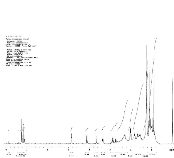
Figure 1S. 1H NMR Spectrum of the compound 1 at 300 MHz in CDCl
de Oliveira et al.
S2 Quim. Nova
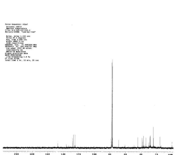
Figure 2S. 13C NMR Spectrum of the compound 1 at 75 MHz in CDCl
Abietatrienes diterpenoids from Sagittaria montevidensis ssp montevidensis S3
Vol. 33, No. 1
de Oliveira et al.
S4 Quim. Nova