A
rti
c
le
J. Braz. Chem. Soc., Vol. 19, No. 5, 831-835, 2008. Printed in Brazil - ©2008 Sociedade Brasileira de Química 0103 - 5053 $6.00+0.00
*e-mail: [email protected]
Hydroxylation of a Hederagenin Derived Saponin by a Xylareaceous Fungus Found
in Fruits of
Sapindus saponaria
Michael Murgu,
aLuiz F. Arruda Santos,
aGezimar D. de Souza,
aCristina Daolio,
aBernd Schneider,
bAntônio Gilberto Ferreira
aand Edson Rodrigues-Filho*
,aaDepartamento de Química, Universidade Federal de São Carlos, CP 676, 13565-905 São Carlos-SP, Brazil
bMax Planck Institute for Chemical Ecology, Beutenberg Campus, Hans-Knöll-Straße 8, D-07745 Jena, Germany
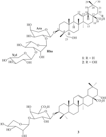
Durante nossos estudos visando a obtenção de microorganismos endofíticos associados a plantas tropicais, um fungo do grupo Xylareaceae foi isolado das partes internas dos frutos de Sapindus saponaria. Os frutos de S. saponaria acumulam grande quantidade de saponinas triterpênicas e sesquiterpênicas. A saponina 3-O-( -D-xilopiranosil)-(1 3)- -L-ramnopiranosil-(1 2)- -L-arabinopiranosil-hederagenina foi isolada por métodos cromatográfios após saponificação do extrato bruto obtido dos frutos de S. saponaria e admistrada junto ao meio de cultivo usado para crescimento do fungo. Após extração dos metabólitos obtidos, uma nova saponina foi purificada por CLAE em escala preparativa e caracterizada como um derivado 22 -hidroxilado. Essa saponina hidroxilada teve sua estrutura molecular elucidada através da interpretação de dados de EM/EM e RMN.
During our screening of tropical plants for endophyte microorganisms, a Xylareaceous fungus was found living on the internal part of Sapindus saponaria fruits. The fruits of S. saponaria
accumulate great amounts of triterpenoidal and sesquiterpenoidal saponins. The saponin 3O( -D-xylopyranosyl)-(1 3)- -L-rhamnopyranosyl-(1 2)- -L-arabinopyranosyl-hederagenin was isolated using chromatographic methods, after alkaline hydrolysis of the crude extract obtained fromS. saponaria fruits and added to the culture medium used to grows the fungus. A new saponin was isolated from this experiment by preparative scale HPLC and characterized as a 22 -hydroxy derivative. The structure of this hydroxylated saponin was elucidated based on interpretation of MS/MS data and NMR spectra.
Keywords: Sapindus saponaria, saponin, Xylaria, biotransformation, hydroxylation
Introduction
As part of our investigations on the chemical aspects of tropical plants and microorganisms interactions, we have been studying the plant Sapindus saponaria (Sapindaceae). Following many other Sapindaceas, S. saponaria produces annually great amounts of small fruits where a sap is accumulated.1 Studying the glycoside composition of
this soap by means of mass spectrometry coupled with liquid chromatography (LC-MS), it was found that those fruits accumulate on their pericarp huge amounts of monodesmosidic triterpene (SAPs) and bisdesmosidic sesquiterpene (acyclic sesquiterpene oligoglycosides, ASOGs) acetylated saponins.2 Saponins are usually referred
as antifungal and antibacterial compounds, being the monodesmosidic the most active ones.3,4 These compounds
form a group of chemical weapons used by plants in the fight against fungi, and the protection against pathogenic microbes is probably their natural role in plants.4-6 Despite
this status of saponins as antimicrobial compounds, during our chemical studies on S. saponaria fruits it was observed that a fungus grows from a hairy structure attached to the seeds. The seeds are in contact with the pericarps where saponins are accumulated. In our previous studies this fungus was isolated and tentatively classified as a member of Xylareaceae, based on morphological characteristics and secondary metabolite profile, and we have also reported that this fungus is able to metabolize the glycosides present in
S. saponaria fruits.7 Thus, using LC-MS it was shown that
Hydroxylation of a Hederagenin Derived Saponin by a Xylareaceous Fungus J. Braz. Chem. Soc.
832
fungus, while the SAPs and some exogenous glycosides are hydroxylated at the aglicon part of the molecule, in a small scale fermentation experiment. In the present work, the saponin 1 (Figure 1) was purified from a saponificated extract obtained from fruits of S. saponaria and used for a scaled up biotransformation reaction. We describe here this biotransformation experiment and a complete structural characterization of the hydroxylated saponin produced by the fungus.
Results and Discussion
In a previous work, we have shown that the saponins produced by S. saponaria occur as a complex mixture of naturally non-regiosselective acetylated glycosides.2
Alkaline hydrolysis of these natural glycosides produced simplified mixtures of compounds formed of only four SAPs and five ASOGs. Preparative scale HPLC separation of the SAP fraction let to isolation of pure saponin 1, which was characterized as 3-O-( -D-xylopyranosyl)-(1
3)--L-rhamnopyranosyl-(1 2)- -L-arabinopyranosyl-hederagenin by comparison of both MS and NMR spectra with those published by Grover and co-workers.8
Saponin1 (100 mg) was submitted to biotransformation experiment and, after the conclusion of this experiment,
it was isolated 18 mg (18% yield) of the hydroxylated compound 2. The MS data indicated that 2 was produced by the addition of an oxygen atom in 1. The ESI mass spectrum of1 shows a deprotonated molecule at m/z 881 ([M-H]–)
in agreement with the molecular formula C46H74O16 (882 Da) of SAP 1. Analyses of the compound produced by the fungus revealed a compound with a peak at m/z 897 ([M-H]-) in its mass spectrum, probably associated with a
compound whose molecular weight is 898 Da. The product ion spectrum obtained from m/z 881 (from 1) shows ions detected at m/z 749 ([SAP-xyl]-), 603 ([SAP-xyl-ara]-) and
471 ([SAP-xyl-ara-gly]- = [Aglycone]-) which correspond
to the losses of the sugar chain from the saponin. The same fragmentation experiment, using the precursor ion of
m/z 897 ([M-H]-) generated from 2, produced exactly the
same type of fragmentation but with the peaks in the mass spectrum shifted by 16 Da. For example, the peak at m/z765 corresponds to the fragmentation that produced m/z 749 from1, plus 16 Da; m/z 747 corresponds to loss of water (18 Da) from m/z 765. Therefore the presence of m/z487 ([Aglycon]-) in the product ion spectrum of m/z 897 is an
indicative that the addition of the 16 mass unit (oxygen) have occurred at the aglycon of the saponin 1 moiety.
The 1D 1H NMR spectra of saponins 1 and 2 are very
complex and contains numerous overlapping signals. Thus, direct comparison of the 1D spectra did not allow visualization of an extra hydrogen atom bounded to an oxygen bearing carbon in 2. On the other hand, the 2D NMR spectra (1H-1H COSY) were efficient to localize
this hydrogen signal. Figure 2 shows a comparison of a sector from the COSY spectra obtained for 1 and 2. The partial spectrum of 1 (Figure 2A) shows correlations of hydrogen H-12 (G 5.49) with H-11 (G 1.92) and of the H-3 (G 4.29) with H-2 (G 2.01). These correlations are also seen in compound 2 spectrum (Figure 2B) which shows an extra contour due H-22 (G4.55) / H-21 (G1.81) coupling. In the HSQC spectrum of 2, the hydrogen at G4.55 (H-22) is correlated with the 13C NMR signal at G70.9 (C-22), corroborating the MS data and confirming the hydroxylation at the aglicone in saponin 1.
The position where the oxygen atom was added in the saponin 1 was suggested by interpretation of NMR spectral data analyzed in comparison with the saponin starting material and model compounds found in the literature. The1H NMR spectrum of 2 shows five signals for methyl
groups (G 1.34, 3H-27; G1.15, 3H-24; G 1.09, 3H-30; G1.09, 3H-26; G1.00, 3H-25; G0.98, 3H-29) like found in spectrum of 1.
The 2D 1H – 1H TOCSY spectrum of 2 showed three
continuous spin systems for rings A (H-1 to H-3), B (H-5 to H-7) and C (H-9 to H-12). These correlations are Figure 1. Molecular structure of the saponin starting material (1), the
Murgu et al. 833 Vol. 19, No. 5, 2008
indicative that the hydroxylation did not occur at rings A, B and C. Couplings of 2H-15 with 2H-16 were not clearly seen due severe signal overlapping although analysis of HSQC data allows the ascribing of C-15 (G 27.8) and C-16 (G 16.7) signals. The 1H NMR signal for H-18
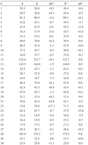
(G 3.38, dd, J 3.4/8.7 Hz), showing typical couplings with vicinal methylene hydrogens, exclude the possibility of hydroxylation at C-19. Therefore, by exclusion, the hydroxyl group was added by the fungus at C-21 or C-22. The comparative analysis of the 13C NMR data for 1 and 2
(Table 1) also shows that the chemical shifts for the carbons at rings A, B and C are very similar. When compared with
1, the 13C chemical shift difference ( G) becomes important
only for some carbons (C-16 to C-22) at rings D and E. A literature survey covering the recent publications on saponin and triterpene molecular structures revealed three compounds interesting for comparison with 2. Thus, the 13C NMR spectrum of 2 was compared with a
16-hydroxyoleanolic acid derived saponin isolated from
Albizia procera,9 a triterpene 21-hydroxyhederagenin,10
and finally with scoparianoside (3), a 22-hydroxyolenolic acid derived saponin, isolated from fruits of Kochia scoparia.11 The best mach found indicated compound
2 to be a 22-hydroxy saponin. The 13C NMR data of 2
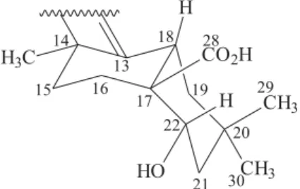
(Table 1) shows good agreement ( G close to zero) with those of 3 considering the signals for carbons at rings D and E. The chemical shift of C-17 (G53.0) appears to be a good guide for the assignment of the hydroxyl group at C-22, since it is at ca.G46.0 – 48.0 in other rings D/E hydroxylated compounds.9-11 In the same way observed for
scoparianoside (3)11, it was not detected H-22/C-28 HBMC
long-range correlation. Probably, the long relaxation time of C-28 and the cis H-22/C-28 spatial relationship (Figure 3) difficult this observation. The shielding effect ( G = -7.3) Figure 2. Sections of the 1H-1H COSY spectra of 1 (A) compared to 2 (B) showing an additional correlation due H-22 (G 4.55) coupled with H-21
(G 1.81).
Table 1.13C NMR dataa of aglicons in saponins 1 and 2and calculated 13C chemical shift difference indicating the positioning of the additional
OH group in 2 and comparison with a model compound (3)
C 1 2 G1b 311 G2c
1 39.3 38.8 -0.5 38.4 -0.4
2 26.5 26.6 +0.1 26.4 -0.2
3 81.3 80.9 -0.4 89.1 +8.2
4 43.8 43.1 -0.7 39.4 -3.7
5 47.9 47.9 0.0 55.5 +7.6
6 18.4 17.9 -0.4 18.3 +0.4
7 33.2 33.2 0.0 32.9 -0.3
8 40.0 39.8 +0.2 39.8 0.0
9 48.5 47.4 -1.1 47.8 +0.4
10 37.2 36.7 -0.5 36.8 +0.1
11 24.0 23.7 -0.3 23.6 -0.1
12 122.6 122.7 +0.1 122.7 0.0
13 145.5 144.0 -1.5 144.0 0.0
14 42.4 42.3 -1.1 42.4 +0.1
15 28.7 27.8 -0.9 27.8 0.0
16 24.0 16.7 -7.3 16.8 +0.1
17 46.9 53.0 +6.1 53.0 0.0
18 42.4 43.3 +0.9 43.4 +0.1
19 47.0 45.7 -1.3 45.8 +0.1
20 31.2 31.4 +0.2 31.4 0.0
21 34.6 43.4 +8.8 43.1 -0.3
22 33.6 70.9 +37.3 71.3 +0.4
23 64.4 63.7 -0.7 27.9 -35.8
24 14.4 14.0 -0.4 16.8 -2.9
25 16.4 15.9 -0.5 15.2 -0.7
26 17.9 17.2 -0.7 17.2 0.0
27 26.4 26.3 -0.1 26.6 +0.3
28 182.0 179.3 -2.7 179.3 0.0
29 33.1 32.5 -0.6 33.3 +0.8
30 23.9 25.0 +1.1 25.0 0.0
a. At 100 MHz in pyridine-d 5.
b. G1 = G2 – G1.c. G2 = G3 – G2. G < 0.5
Hydroxylation of a Hederagenin Derived Saponin by a Xylareaceous Fungus J. Braz. Chem. Soc.
834
of the C-22-hydroxyl over C-16 (G 16.7) and a nOe between H-22 and 3H-29 observed in ROESY spectrum indicates that compound 2 is a 22 -hydroxylated product of 1.
In general, deglycosylations (hydrolysis at the sugar chain) are frequently reported as saponin metabolisms done by microorganisms.12-14 Hydroxylation (monooxygenase
expression) is the reaction that occurred in saponin 1. This biochemistry process may find important technological application since expression of monooxigenase from fungi in saponins is not commonly found in the literature.
Experimental
Equipment
Preparative scale HPLC separations were performed using a Shimadzu equipment (LC-8A pump, CBM-10A communication module) equipped with an UV detector (SPD-6AV) set at 206 nm. HPLC grade acetonitrile (ACN) was obtained from Mallinckrodt, and H2O was purified in a Milli-Q system (Millipore). ESI-MS/MS spectra were acquired in negative ion mode on a triple quadrupole Micromass Quattro LC spectrometer, equipped with a Z-Spray API ion source and a megaflow electrospray probe. NMR spectra were recorded in C5D5N (Aldrich) on a Bruker DRX 400 spectrometer operating at 400 MHz for hydrogen and 100 MHz. Some analysis were also performed using a Bruker AV 500 Ultrashield spectrometer operating at 500 MHz for 1H and 125 MHz for 13C.
Plant material
Fruits of S. saponaria were collected in São Carlos, São Paulo State, Brazil. A voucher specimen (No. 003651) was deposited in the herbarium of the Botanic Department of Universidade Federal de São Carlos, Brazil.
Extraction and isolation of SAP 1
Fruits of S. saponaria were dried under circulating air at a temperature of 50 oC during 48 h. Finely powdered
pericarpe (451.3 g) of dried fruits separated from seeds was extracted with methanol. The methanol extract was concentrated by distillation under reduced pressure and finally lyophilized to eliminate residual water. Part of the lyophilized extract (15.0 of 101.3 g) was stirred with 130 mL of aqueous NaOH 2 mol L-1 for 30 min. The reaction
solutions were acidified with 23 mL of HCl 2 mol L-1. The
resulting solution was passed through an octadecylsilyl (ODS) open column, which was further eluted with pure MeOH. The MeOH solution was evaporated and then part of the extract obtained (ca. 3 g) was repetitively subjected to preparative scale reverse phase HPLC (ca. 150 mg per injection), on an ODS column (Shimadzu Prep K, 2.0×25.0 m), eluted with ACN:H2O (1:1 v/v) at a flow rate of 10 mL min-1, with UV detection at 206 nm.
3-O-( -D-xylopyranosyl)-(1 3)- -L-rhamnopyranosyl-(1 2)- -L-arabinopyranosyl-hederagenin (1)
White amorphous powder; [ ]D25 + 113.4° (MeOH, c
0.001);1H NMR (400 MHz, C
5D5N): Aglicon: G 5.49 (br
s, H-12), 4.29 (m, H-3), 4.28 (d, J 10 Hz, H-23a), 3.93 (d,
J 10 Hz, H-23b), 3.32 (dd, J 3.7/9.0 Hz, H-12), 1.26 (s, 3H-27), 1.14 (s, 3H-24), 1.06 (s, 3H-30), 1.04 (s, 3H-26), 1.01 (s, 3H-25), 0.93 (s, 3H-29), Arabinose: 5.07 (d, J 7 Hz, H-1), 4.62 (m, H-2), 4.00 (dd, J 3.9/8.1 Hz, H-3), 4.14 (m, H-4), 4.30 (m, H-5a), 3.67 (br d, J 10.8 Hz, H-5b), Rhamnose: 6.35 (br s, H-1), 4.92 (m, H-2), 4.83 (dd, J
2.9/9.5 H-3), 4.46 (t, J 9.5 Hz, H-4), 4.85 (dq, J 6.2/9.5 Hz, H-5), 1.55 (d, J 6.2 Hz, 3H-6), Xylose: 5.32 (d, J 7.3 Hz, H-1), 3.94 (t, J 8.8 Hz, H-2), 3.99 (t, J 9.5 Hz, H-3), 4.00 (dt,J 9.5/5.1 Hz, H-4), 4.13 (dd, J 11.0/5.1 Hz, H-5a), 3.56 (dd,J 11.0/9.5 Hz, H-5b); 13C NMR (100 MHz, C
5D5N):
see Table 1; ESIMS (daughter ion scan, 35 eV.): 881 [M-H]
-(100), 749 [M-H-Xyl]- (23), 603 [M-H-Xyl-Rha]- (29), 471
[Hederagenin - H]-(16).
Isolation of fungus
Details of fungus isolation procedures and tentative identification are described elsewhere.7 After their isolation,
the fungus was labeled with the code LaBioMMi217 and deposited at the Laboratory Biochemistry and Micromolecular of Microorganisms – LaBioMMi – of the Chemistry Department of the Federal University of São Carlos, São Carlos, Brazil.
Biotransformation experiments and chemical analysis
Murgu et al. 835 Vol. 19, No. 5, 2008
days. The saponin 1 (100 mg) was dissolved in 20 mL of distilled water and equally distributed in twenty 0.5-liter Erlenmeyer flasks, each containing 150 mL of liquid medium (45 g glucose, 0.48 g NH4NO3, 5.0 g KH2PO4, 1.0 g MgSO4, 0.1 g FeSO4, 0.015 g CuSO4, 0.161 g ZnSO4, 0.01 g MnSO4, and 0.1 g (NH4)2MoO4 dissolved in 1.5 L of distilled water). These Erlenmeyer flasks were autoclaved (121oC, 25 min) and then pieces of the PDA
(potato-dextrose-agar) culture containing mycelium were added to fifteen of the twenty flasks (five were kept as control) and were allowed at 25 0C standing in the dark during 15 days.
The mycelium was separated by gravity filtration and the liquid phase was concentrated under reduced pressure and finally lyophilized. The extract was repetitively subjected to preparative scale separations using the same conditions described above for the isolation of 1. These procedures let to isolation of 18.0 mg of pure saponin 2. The ESI-MS/ MS spectra were acquired in negative ion mode on a triple quadrupole Micromass Quattro LC spectrometer. The desolvation and ion source block temperatures were set, respectively, at 300 and 125oC. Gaseous N
2 was used to
nebulize (80 L/H) and desolvate (350 L/H). The optimal voltages found for the probe and ion source components to produce maximum intensity of the ions [M-H]- were 3.3 kV
for the stainless steel capillary, 38V for the sample cone, and 6V for the extractor cone. The parent/daughter runs (MS/MS) were performed by adding Ar in the collision cell to produce a pressure of 1.5 x 10⁻³ mBar for collisional
induced dissociation (CID). The optimal collisional energy used to decompose the ions [M-H]- was 25-30 eV.
3-O-( -D-xylopyranosyl)-(1 3)- -L-rhamnopyranosyl-(1 2)- -L-arabinopyranosyl-22 -hydroxyhederagenin (2)
White amorphous powder; [ D25 + 98.1° (MeOH, c
0.001);1H NMR (400 MHz, C
5D5N): Aglicon: G 5.50 (br
s, H-12), 4.55 (m, H-22), 4.30 (m, H-3), 4.26 (d, J 10 Hz, H-23a), 3.91 (d, J 10 Hz, H-23b), 3.38 (dd, J 3.4/8.7 Hz, H-12), 1.34 (s, 3H-27), 1.15 (s, 3H-24), 1.09 (s, 3H-30), 1.09 (s, 3H-26), 1.00 (s, 3H-25), 0.98 (s, 3H-29), Arabinose: 5.06 (d, J 7 Hz, H-1), 4.63 (m, H-2), 4.02 (dd, J 3.9/8.1 Hz, H-3), 4.13 (m, H-4), 4.33 (m, H-5a), 3.65 (br d, J 10.8 Hz, H-5b), Rhamnose: 6.31 (br s, H-1), 4.91 (m, H-2), 4.80 (dd,J 2.9/9.5 Hz, H-3), 4.43 (t, J 9.5 Hz, H-4), 4.87 (dq,
J 6.2/9.5 Hz, H-5), 1.58 (d, J 6.2 Hz, 3H-6), Xylose: 5.29 (d,J 7.3 Hz, H-1), 3.91 (t, J 8.8 Hz, H-2), 3.97 (t, J 9.5 Hz, H-3), 4.01 (dt, J 9.5/5.1 Hz, H-4), 4.11 (dd, J 11.0/5.1 Hz, H-5a), 3.55 (dd, J 11.0/9.5 Hz, H-5b); 13C NMR (100 MHz,
C5D5N):see Table 1; ESIMS (daughter ion scan, 35 eV.): 897 [M-H]– (100), 765 [M-H-Xyl]- (29), 747
[M-H-Xyl-OH]– (8), 619 [M-H-Xyl-Rha]- (63), 601
[M-H-Xyl-Rha-OH]– (52), 487 [22-Hydroxy-hederagenin - H]-(78).
Acknowledgments
The authors are gratefull to Fundação de Amparo à Pesquisa do Estado de São Paulo (FAPESP), Conselho Nacional de Desenvolvimento Científico e Tecnológico (CNPq), Coordenação de Aperfeiçoamento de Ensino Superior (CAPES) and Financiadora de Estudos e Projetos (FINEP) for financial support.
References
1. Voutquenne, L.; Ann Pharm Fr.2001,59, 407.
2. Murgu, M.; Rodrigues-Filho, E.; J. Braz. Chem. Soc.2006,17, 1281.
3. Gruiz, K.; Adv. in Exper. Med. Biol.1996,404, 527. 4. Morrissey, J. P.; Osbourn A. E.; App. Environ. Microbiol.1999,
63, 708.
5. Bower, P.; Clarke, B. R.; Lunness, M. J.; Daniels, M. J.; Osbourn, A. E.; Science1995,267, 371.
6. Maor, R.; Shirasu, K.; Curr. Opin. Microbio.2005,8, 399. 7. Amaral, L. S.; Murgu, M.; Rodrigues-Filho, E.; Souza, A. Q.
L.; Sarquis, M. I. M.; World J. Microbiol. Biotech.2008, in press.
8. Grover, R. K.; Roy, A. D.; Roy, R.; Joshi, S. K.; Srivastava, V.; Arora, S. K.; Magn. Reson. Chem.2005, 43, 1072.
9. Melek, F. R.; Miyase, T.; Ghaly, N. S.; Nabil, M.; Phytochemistry
2007,68, 1261.
10. Romussi, G.; Falsone, G.; Arch. Pharm. (Weinheim) 1985,318, 219.
11. Yoshikawa, M.; Shimada, H.; Morikawa, T.; Yoshizumi, S.; Matsumura, N.; Murakami, T.; Matsuda, H.; Hori, K.; Yamahara, J.; Chem. Pharm. Bull.1997,45, 1300.
12. Shin, H. Y.; Park, S. Y.; Sung, J. H.; Kim, D. H.; App. Environ. Microbiol.2003,69, 7116.
13. Woods, K.; W., Hamilton; C. J., Field; R. A.; Carbohyd. Research2004,339, 2325.
14. Carter, J. P.; Spink, J.; Cannon, P. F.; Daniels, M. J.; Osbourn, A. E.; App. Environ. Microbiol.1999,65, 3364.
Received: August 23, 2007 Web Release Date: March 25, 2008
S
u
p
p
le
m
e
nta
ry
Inf
o
rm
a
ti
o
n
J. Braz. Chem. Soc., Vol. 19, No. 5, S1-S7, 2008. Printed in Brazil - ©2008 Sociedade Brasileira de Química 0103 - 5053 $6.00+0.00
*e-mail: [email protected]
Hydroxylation of a Hederagenin Derived Saponin by a Xylareaceous Fungus Found
in Fruits of Sapindus saponaria
Michael Murgu,
aLuiz F. Arruda Santos,
aGezimar D. de Souza,
aCristina Daolio,
aBernd Schneider,
bAntônio Gilberto Ferreira
aand Edson Rodrigues-Filho*
,aaDepartamento de Química, Universidade Federal de São Carlos, CP 676, 13565-905 São Carlos-SP, Brazil
bMax Planck Institute for Chemical Ecology, Beutenberg Campus, Hans-Knöll-Straße 8, D-07745 Jena, Germany
Hydroxylation of a Hederagenin Derived Saponin by a Xylareaceous Fungus Found in Fruits J. Braz. Chem. Soc.
S2
Figure S2. ESI-MS/MS of saponin 1.
Figure S3.1H NMR spectrum (400 MHz, C
Murgu et al. S3 Vol. 19, No. 5, 2008
Figure S4.13C NMR spectrum (100 MHz, C
5D5N;TMS) of saponin 1.
Hydroxylation of a Hederagenin Derived Saponin by a Xylareaceous Fungus Found in Fruits J. Braz. Chem. Soc.
S4
Figure S6.1H NMR spectrum (500 MHz, C
5D5N;TMS) of saponin 2.
Figure S7.13C NMR spectrum (100 MHz, C
Murgu et al. S5 Vol. 19, No. 5, 2008
Figure S8. 2D 1H-1H COSY NMR spectrum (400 MHz, C
5D5N;TMS) of saponin 2.
Figure S9. 2D 1H-1H TOCSY NMR spectrum (500 MHz, C
Hydroxylation of a Hederagenin Derived Saponin by a Xylareaceous Fungus Found in Fruits J. Braz. Chem. Soc.
S6
Figure S10. 2D 1H-1H ROESY NMR spectrum (500 MHz, C
5D5N;TMS) of saponin 2.
Murgu et al. S7 Vol. 19, No. 5, 2008