Article
Printed in Brazil - ©2008 Sociedade Brasileira de Química 0103 - 5053 $6.00+0.00
*e-mail: [email protected]
Solid-Phase Microextraction for Determination of
3-Chloro-4-(dichloromethyl)-5-hydroxy-2[5H]-furanone in Water
Andrea L. Rezemini,
aJorge M. Vaz
band Lilian R. F. Carvalho*
,aaInstituto de Química, Universidade de São Paulo, CP 26077, 05599-970 São Paulo-SP, Brazil
bInstituto de Pesquisas Energéticas e Nucleares, Av. Prof. Lineu Prestes 2242, 05508-900 São Paulo-SP, Brazil
Microextração em fase sólida, usando a derivatização on-line com bis(trimetilsilil)trifluoro-acetamida, cromatografia a gás e espectrometria de massas, foi avaliada para a quantificação de 3-cloro-4-(diclorometil)-5-hidróxi-2(5H)-furanona (MX) em amostras de água. Foram usadas fibras de diferentes polaridades empregando a amostragem por imersão e por headspace. Para o sistema de imersão, foram avaliados vários parâmetros que afetam a extração de MX, como pH, salinidade, temperatura e tempo de extração. O método otimizado (fibra de poliacrilato; 20% Na2SO4; pH 2,0; 60 min; 20 °C) foi aplicado para águas cloradas proveniente de reservatórios de abastecimento de água-amostras naturais e amostras com adição de MX (50 ng L-1 e 100 ng L-1). A recuperação de MX variou de 44 a 72%. A quantificação do MX em amostras de água foi feita por padrão externo empregando o modo de monitoramento de íon selecionado. O coeficiente de correlação (0,98%), o desvio padrão relativo (5%), o limite de detecção (30 ng L-1) e o limite de quantificação (50 ng L-1) foram obtidos a partir da curva analítica.
Solid-phase microextraction, using on-line bis(trimethylsilyl)trifluoroacetamide derivatisation, gas chromatography, and mass spectrometry, was evaluated in the quantification of 3-chloro-4-(dichloromethyl)-5-hydroxy-2(5H)-furanone (MX) in water samples. Fibres encompassing a wide range of polarities were used with headspace and direct immersion sampling. For the immersion procedure, various parameters affecting MX extraction, including pH, salinity, temperature, and extraction time were evaluated. The optimised method (polyacrylate fibre; 20% Na2SO4; pH 2.0; 60 min; 20 °C) was applied for reservoir chlorinated water samples-either natural or spiked with MX (50 ng L-1 and 100 ng L-1). The recovery of MX ranged from 44 to 72%. Quantification of MX in water samples was done using external standard and the selected ion monitoring mode. Correlation coefficient (0.98%), relative standard deviation (5%), limit of detection (30 ng L-1) and limit of quantification (50 ng L-1) were obtained from calibration curve.
Keywords:water analysis, 3-chloro-4-(dichloromethyl)-5-hydroxy-2[5H]-furanone (MX), solid-phase microextraction, GC-MS
Introduction
Although chlorination has been commonly used in the treatment of drinking water, many chlorinated by-products formed during this process pose human health
risks.1 One of the most potent direct-acting mutagens in the
Salmonella typhimurium tester strain TA100 is
3-chloro-4-(dichloromethyl)-5-hydroxy-2(5H)-furanone (MX)2,3
which has been found in chlorinated drinking water from
Finland,4,5 North America,6,7 Netherlands,8 and Japan.9
Although the MX found in chlorinated drinking water was
at low concentrations, ranging from trace amounts to 675 ng L-1,10,11 it has been shown to account for approximately
50% of the total mutagenicity of such water.12 According to
the World Health Organization (WHO), MX concentrations
of 1.8 g L-1 are associated with a 10í5 cancer risk for a 60
kg adult drinking 2 litres of water per day.13,14
The closed tautomeric (pH 3) form of MX presents
higher mutagenicity than does the open form.15-17 Since the
Although quantitative analyses of drinking water have been performed using GC techniques, pre-column derivatisation has been required to obtain accurate analyses
of these polar organic compounds.10,18 It has been reported
that GC with electron-capture9 and MS detects MX in
water.11,15 In such studies, solid-phase extraction11,18 and
liquid-liquid extraction10,15 have been employed prior to
the chromatographic analyses.
We previously described an alternative GC-MS method for quantification of trace levels of MX in chlorinated
water samples.19 Clean-up by solid-phase extraction and
extraction of water samples with dichloromethane were applied prior to the pre-concentration of MX. The analyte was derivatised directly in the GC injector. The resulting MX trimethylsilyl derivative was identified and quantified using MS. Although this method offers a short analysis time, as well as satisfactory detection and quantification limits, the liquid-liquid extraction technique of isolating MX in water requires a large amount of sample and involves the use of fairly large amounts of organic solvents.
In the present study, a method based on GC–MS and solid-phase microextraction (SPME) was developed in order to determine MX in water. As it is a solventless technique, which requires only a few millilitres of sample and allows the pre-concentration step to be omitted,
SPME is an attractive method.20,21 The optimum conditions
defined were applied for the determination of MX in water samples.
Experimental
Chemicals
The MX standard (98%; 5 mg) was obtained in a transparent film form from Sigma (St. Louis, MO, USA).
Stock MX solution (1.5 mg mL-1) was prepared with
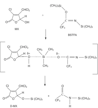
ethyl acetate (HPLC grade) and stored at 20 °C. The derivatisation reagent, bis(trimethylsilyl)trifluoroacetamide (BSTFA), was purchased from Merck (Darmstadt, Germany). An ethyl acetate solution containing 10%
BSTFA was used to obtain the derivatised MX (D-MX),22
shown in Figure 1.
Solid-phase microextraction and derivatisation procedures
The SPME device was tested with various commercially available fibres mounted in the manual SPME fibre holder (Supelco, Bellefonte, PA, USA). The following phase coatings and film thicknesses were used: polydimethylsiloxane (PDMS) at 7 m; PDMS/
divinylbenzene (DVB) at 65 m; carboxen/PDMS at 75 m; carbowax (CW)/DVB at 65 m; and polyacrylate (PA) at 85 m. All of the fibres were pre-conditioned according to the Supelco instruction manual. After each analysis, the fibre was always kept under vacuum in order to eliminate gas bubbles that were occasionally adsorbed onto the fibre surface.
The direct and headspace sampling modes were tested using all fibres described above. For direct extraction, the fibre was exposed for 1 h to a solution of MX in deionised
water (1 g L-1) under magnetic agitation. For the headspace
and direct sampling modes, 2.5 mL and 4.0 mL of MX solution were used, respectively.
The same MX derivatisation procedure was used for all except the CW/DVB fibre. After MX sampling, each SPME fibre was removed from its sample vial and inserted into a vial containing 0.5 mL of the 10% BSTFA solution in ethyl acetate, where it remained for 5 min under magnetic agitation. After adsorption, each fibre was introduced into the GC splitless injection port at 250 °C for 3 min to achieve thermal desorption and in situ derivatisation. For the CW/DVB-coated fibres, the MX was desorbed from the fibre immediately after sampling, and transferred to the injector (the carrier gas flux was stopped during 3 min to dessorb MX from fibre and to transfer to the GC injector and, after this time, the flux was turn on to move MX molecules from GC injector to the column at the same time), and 1 L of the BSTFA was immediately injected to achieve derivatisation. This procedure was used to avoid
the reaction between the derivatisation reagent and the OH groups of polyethylene glycol of the CW stationary phase that would become inactive.
The efficiency of MX extraction using the headspace SPME mode for the PDMS and PA fibres was evaluated by varying the aqueous solution pH (2.0 or 5.7), the extraction
temperature (20 or 60 °C), and the Na2SO4 (20% m/v
or absent). The efficiency of the direct SPME mode for extracting MX from the PA fibres was evaluated by varying
the pH (2.0 or 5.7), the Na2SO4 (20% m/v or absent), and the
extraction time (15, 60, or 240 min). The aqueous solution used was deionised water at pH 5.7.
The calibration curve for D-MX in deionised water was obtained using five solutions at different MX concentrations
(50, 100, 200, 500 and 1000 ng L-1). The following
optimised extraction conditions were used: PA fibre, direct extraction mode, extraction time of 60 min, pH 2.0 and
addition of 20% m/v of Na2SO4.
In all analyses, SPME fibre carryover was achieved using 3 min of splitless time. Blanks were not taken into account as no contaminant was detected during the MX retention time.
Aliquots (30, 50 and 100 ng L-1) of MX were added to
the deionised water samples (n = 2) and chlorinated water samples (n = 2). The solutions obtained were analysed using SPME and GC-MS under optimised conditions.
Gas Chromatographic-Mass Spectrometric analysis
The D-MX was analysed by GC–MS using a model GCMS-QP5000 gas chromatograph (Shimadzu, Kyoto, Japan), the mass selective detector of which was operated in the scan mode (electron impact ionisation at 70 eV). The CLASS-5000 program, version 2.10 (Shimadzu) was used to analyse the data.
The following parameters were set for MS: interface
temperature, 240 °C; mass range, 45-350 m/z; solvent cut
time, 5 min; detector voltage, 1.50 kV; scan interval, 0.5 s in the scan mode.
The GC parameters were as follows: carrier gas, helium
(purity 99.999%); gas flow, constant at 0.8 mL min-1;
total gas flow, 50.3 mL min-1; linear temperature gradient,
40 °C to 310 °C at 20 °C min-1. The split/splitless injector
was operated in splitless mode and was maintained at 250 °C during the introduction of the SPME fibre into the injection port. The splitless valve was closed for 3 min in order to compound desorption from the SPME fibre to the GC injector. A fused-silica open tubular WCOT column coated with a chemically bonded and cross-linking 5% diphenyl 95% dimethylsiloxane stationary phase (HP-5MS: 30 m × 0.25 mm I.D., 0.25- m film thickness) was used.
Results and Discussion
In order to achieve the best MX extraction conditions in water, parameters that affect the equilibration process between phases were evaluated.
Headspace sampling
The headspace sampling is the preferred sampling mode due to its faster equilibration times and lower interference
problems.23 For this reason, it was tested. Using headspace
sampling with all phase coatings evaluated, MX in deionised water was not detected at ambient temperature.
Temperature can affect extraction efficiency. At higher temperatures, the diffusion coefficient increases and the distribution constant decreases, leading to faster equilibration between the analyte and phases. Using PA and PDMS fibres, extractions were performed at 60 °C. Under those conditions, MX was not detected.
Due to the hydrophilic nature of MX, the headspace sampling mode was evaluated by altering the pH and salinity of the aqueous solution. Among all phases studied,
MX was only detected in the 20% m/v Na2SO4aqueous
solution at pH 2.0 when the more polar (PA) fibre coating was used. Nevertheless, the recovery was very poor
(0.12 0.02%).
Although the headspace sampling mode is preferentially used for complex matrices, such as drinking water, our headspace SPME experiments were not very successful. Some extraction parameters were modified to improve the extraction efficiency but MX remained predominantly in the aqueous phase.
Direct immersion sampling
The results of direct SPME extraction using different phase coatings are presented in Table 1. In deionised water, MX was not recovered using the non-polar PDMS and PDMS/DVB fibres under extraction conditions, and the Carboxen/PDMS, CW/DVB, and PA fibres presented very poor recovery. The Carboxen/PDMS and CW/DVB fibres presented similar recovery rates (0.27 and 0.32%, respectively), and the PA fibre presented a slightly greater recovery (1.0%) than did the mixed-phase fibres. However, direct SPME extraction using these fibres might be improved through optimisation of the method. In the present study, the fibre with the highest polarity (the PA fibre) was selected for method optimisation.
BSTFA solution with all fibres, except CW/DVB fibres. In each experiment, a small amount of MX (5%) was transferred from the fibre to the solution. This was evaluated by examining the recovery results obtained.
Optimisation of direct SPME sampling conditions
In order to improve the efficiency of direct SPME extraction using the PA fibre, pH, salinity, and extraction time were adjusted.
Variations in pH and the addition of Na2SO4 and their
effects on MX recovery can be seen in Figure 2. The results
show that lower pH improves MX recovery (41 2%). It
has been suggested that greater quantities of MX migrate towards polar fibres, as the less water-soluble form, furanone,
is predominantly found in the aqueous solution at pH 2.0.24
Furanone is a tautomeric species of the MX chemical equilibrium (Figure 3). In the present study, the addition of
Na2SO4 in order to increase the ionic strength of the solution
in acid medium resulted in the best extraction condition.25
The optimised condition was pH 2.0 and addition of 20%
m/v Na2SO4to the aqueous solution (94 3%).
The various extraction times (15, 60 and 240 min) and their effects on MX recovery are shown in Figure 4. The best results (94 3 and 89 3%, respectively) were observed for longer extraction times (60 and 240 min). The migration of MX from the aqueous phase to the fibre occurred slowly, as PA, a rigid polymer, was used as the
fibre coating.21
Qualitative and quantitative analysis of MX
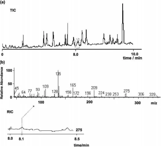
A total ion chromatogram of the D-MX standard
(30 ng L-1; retention time, 8.1 min) in deionised water,
obtained for PA fibre with direct SPME in scan mode, was used to identify MX. The electron impact mass spectrum and some fragment ions for D-MX in the mass spectrum are suggested in Figure 5. It is noteworthy that a chromatographic peak of an unidentified compound in deionised water was observed at the same MX retention time (not shown). Some different D-MX fragment ions indicated the formation of a product. Based on these findings, we can speculate that a BSTFA by-product, a derivatised carboxylic acid, was formed during the derivatised fibre immersion in the deionised water. Since common fragment ions, such as
those at m/z107, 135, and 209, corresponding to the D-MX
chromatographic peak, were observed in the BSTFA
by-product and in the D-MX mass spectra, the ion at m/z 275
was selected for MX quantification.
The five-point calibration curve for D-MX in deionised water obtained from direct SPME of PA fibres under the
Table 1. Behaviour of the fibres in direct SPME
Fibre MX recovery (%), m/z 275
(meana SD)
PDMS
---PDMS/DVB
---Carboxen/PDMS 0.27 0.06
CW/DVB 0.32 0.05
PA 1.0 0.1
aduplicate analysis; fragment ion used for quantification; SD: standard deviation; ---: not recovered; PDMS: polydimethylsiloxane; DVB: divinylbenzene; CW: carbowax; PA: polyacrylate.
Figure 2. Effects that pH and the addition of salt have on extraction
efficiency, [MX] = 1 g L-1, using direct immersion SPME sampling. The recovery (%) is shown at the top of the bar.
Figure 3. Tautomeric equilibrium of MX and its geometric isomers.15
Figure 4. Effect that extraction time has on extraction efficiency,
optimised conditions was obtained through GC-MS used in
the scan detection mode. The r2and RSD were calculated
(0.98 and 5%, respectively) using replicate analysis (n = 2). The detection limit (3:1 signal-to-noise ratio) and the quantification limit (10:1 signal-to-noise ratio), calculated
by using the scan mode, and were found to be 30 ng L-1 and
50 ng L-1, respectively. The GC injector liner was frequently
cleaned to avoid a chromatographic baseline increase, as the PA polymer may bleed in the injector port during thermal desorption. The completeness of MX desorption from fibres was evaluated in all analyses. The life of a given PA fibre was determined to be approximately 50 analyses.
Because of the matrix complexity, data were acquired using the scan detection mode. However, if selected ion
monitoring mode were used, MX concentrations below
30 ng L-1 might be detected.
The proposed method was applied in deionised water sample and chlorinated water samples from two water reservoirs (reservoirs 1 and 2) located in different regions of the city of São Paulo, Brazil. Total ion chromatograms corresponding to non-spiked and spiked chlorinated water samples from reservoir 2 are shown in Figures 6a and 7a, respectively. Mass spectra of non-spiked and spiked chlorinated water samples (Figures 6b and 7b, respectively) indicate that the chromatographic peak at 8 min cannot be attributed to the MX compound, as the spiked deionised water mass spectrum (Figure 5b) differs from that of chlorinated water (Figure 6b). Reconstructed
Figure 5. Electron impact mass spectrum and some fragment ions proposed for D-MX18 for spiked deionised water sample, [MX] = 50 ng L-1.
Figure 6. Chlorinated water sample from reservoir 2 (non-spiked): (a) total ion chromatogram (TIC); (b) mass spectrum of the BSTFA by-product and its
Table 2. Recovery of MX in deionised and chlorinated water samples
Sample Recovery (%)
MX = 30 ng L-1
Recovery (%) MX = 50 ng L-1
Recovery (%) MX = 100 ng L-1
deionised water 28 8 46 7 70 6
chlorinated water1 --- 45 5 72 4
chlorinated water2 --- 44 5 68 5
n = 2; quantification by using SIM mode; 1reservoir 1; 2reservoir 2; --- no recovered
Figure 7. Spiked chlorinated water sample from reservoir 2, [MX] = 50 ng L-1: (a) total ion chromatogram (TIC); (b) D-MX mass spectrum and its
reconstructed ion chromatogram (RIC), m/z 275.
ion chromatograms and their mass spectra (Figures 6b and 7b) also show this different behaviour. The mass spectra corresponding to the chlorinated water samples from reservoirs 1 and 2 were found to be similar (not shown).
By using SPME technique with GC-MS method, MX was found to be below the limit of detection in the chlorinated water samples from both water reservoirs. On the other hand, MX was detected in these samples when they were analysed previously by using liquid-liquid
extraction technique with GC-MS.19
Results of MX recovery from chlorinated and deionised water samples (Table 2) show that the SPME technique with
GC-MS method using SIM mode allows quantifying MX at
concentrations 50 ng L-1. The MX recovery rates ranged
from 44 to 46% and 68 to 72% for spiking of 50 ng L-1 and
100 ng L-1, respectively and, at 30 ng L-1, MX was only
recovered from the deionised water sample (28%). Lower extraction efficiency was observed in lower amount of MX added to the matrix.
Since MX has been found at concentrations ranging
from 13 to 675 ng L-1 in chlorinated water samples
collected in other countries such as Finland,11,26 and North
America,10,27 the method proposed, which offers a detection
chlorinated water samples. A classification of waters into two groups, samples with MX and samples without MX or with MX at concentration below the reference value established by the WHO, may be useful for control of drinking water quality.
Conclusions
A selective method for MX determination in water was developed based on SPME technique with GC-MS. The method presented good sensitivity and satisfactory results of MX recovery from chlorinated water samples. Because SPME is a solventless extraction technique that uses only a few millilitres of sample and does not require the pre-concentration step, the method constitutes an advance in comparison with the conventional analysis methods for MX determination in water. The method proposed may be applied for environmental routine analysis as it detects MX in water samples at concentration well below the reference value established by the WHO. Ultimately, the most interesting improvement of the method, anticipating its implementation as a routine analytical method, is that there is no need for extensive sample manipulation. Furthermore, this SPME method benefits from much shorter analysis time than any extraction method.
Acknowledgments
The authors wish to acknowledge the Brazilian Conselho Nacional de Desenvolvimento Científico e Tecnológico (CNPq, National Council for Scientific and Technological Development) for sponsoring the doctoral fellowship.
References
1. McDonald, T. A.; Komulainen, H.; J. Environ. Sci. Health - Part C2005,23,163.
2. International Agency for Research on Cancer, Chlorinated Drinking-Water; Chlorination By-Products; Some Other Halogenated Compounds; Cobalt and Cobalt Compounds, IARC Monogr. Eval. Carcinog. Risks Humans, Vol. 52, International
Agency for Research on Cancer, Lyon, France, 1991. 3. Langvik, V.; Hormi, O.; Tikkanen, L.; Holmbom, B.;
Chemosphere1991,22, 547.
4. Rantakoko, P.; Yritys, M.; Vartiainen, T.; J. Chromatogr. A 2004, 1028, 179.
5. Langvik, V.; Hormi, O.; Chemosphere1994,28, 1111.
6. Meier, J. R.; Knohl, R. B.; Coleman, W. E.; Ringhand, H. P.; Munch, J. W.; Kaylor, W. H.; Streicher, R. P.; Kopfler, F. C.; Mutat. Res.1987,189, 363.
7. Wright, J. M.; Schwartz, J.; Vartiainen, T.; Maki-Paakkanen, J.; Altshul, L.; Harrington, J. J.; Dockery, D. W.; Environ. Health Perspectives 2002,110, 157.
8. Backlund, P.; Wondergem, E.; Voogd, K.; Jong, A. D.; Sci. Total Environ.1989,84, 273.
9. Ogawa, S.; Kita, H.; Hanasaki, Y.; Fukui, S.; J. Chromatogr.
1993,643, 221.
10. Kronberg, L.; Christman, R. F.; Singh, R.; Ball, L. M.; Environ. Sci. Technol.1991,25, 99.
11. Kronberg, L.; Holmbom, B.; Reunannen, M.; Tikkanen L.; Environ. Sci. Technol.1988,22, 1097.
12. Curiex, F. L.; Munter, T.; Kronberg, L.; Chem. Res. Toxicol.
1997,10, 1180.
13. Charles, M. J.; Chen, G.; Kanniganti, R.; Marbury, G. D.; Environ. Sci. Technol.1999,62, 445.
14. http://www.who.int/water_sanitation_health/dwq/chemicals/ mx.pdf, accessed in February 2008.
15. Charles, J.; Chen, G.; Kanniganti, R.; Marbury, G. D.; Environ. Sci. Technol.1992,26, 1030.
16. Backlund, P.; Kronberg, L.; Tikkanen L.; Chemosphere1988, 17, 1329.
17. Singer, P. C.; Wat. Sci. Technol.1999,40, 25.
18. Smeds, A.; Vartiainen, T.; Maki-Paakkanen, J.; Kronberg, L.; Environ. Sci. Technol.1997,31, 1033.
19. Rezemini, A. L.; Vaz, J. M.; Carvalho, L. R. F.; J. Chromatogr.
A2002,972, 259.
20. Pawliszyn, J.; Solid Phase Microextraction. Theory and
Practice, Wiley-VCH: New York, 1997.
21. Wercinski, S. A. S.; Solid Phase Microextraction. A Practical
Guide, Marcel Dekker: New York, 1999.
22. Knapp, D. R.; Handbook of Analytical Derivatization Reactions, Wiley: New York, 1979.
23. Helaleh, M. I. H.; Fujii, S.; Korenaga T.; Talanta2001,54, 1039.
24. Horth, H.; Fielding, M.; James, H.A.; Thomas, M. J.; Gibson, T.; Wilcox, P.; Production of Organic Chemicals and Mutagens During Chlorination of Amino Acids in Water, Wiley: New York,
1998.
25. Alpendurada, M. F.; J. Chromatogr. A2000,889, 3. 26. Kronberg, L.; Wat. Sci. Tech.1999,40, 31.
27. Richardson, S. D.; Trends Anal. Chem. 2003,22, 666.
Received: March 21, 2007
Web Release Date: April 29, 2008
![Figure 4. Effect that extraction time has on extraction efficiency, [MX] = 1 g L -1](https://thumb-eu.123doks.com/thumbv2/123dok_br/18992603.461170/4.892.95.432.866.1088/figure-effect-extraction-time-extraction-efficiency-mx-l.webp)
![Figure 5. Electron impact mass spectrum and some fragment ions proposed for D-MX 18 for spiked deionised water sample, [MX] = 50 ng L -1 .](https://thumb-eu.123doks.com/thumbv2/123dok_br/18992603.461170/5.892.121.745.156.349/figure-electron-impact-spectrum-fragment-proposed-spiked-deionised.webp)