0103 - 5053 $6.00+0.00
Article
*e-mail: [email protected]
On-Line LC/UV/MS Analysis of Flavonols in the Three Apple Varieties Most Widely
Cultivated in Brazil
Luciana A. Tiberti, a Janete H. Yariwake,*,a Karine Ndjokob and Kurt Hostettmannb
a
Universidade de São Paulo, Instituto de Química de São Carlos, CP 780, 13560-970 São Carlos-SP, Brazil
b
Laboratoire de Pharmacognosie et Phytochimie, École de Pharmacie Genève-Lausanne, Université de Genève, Quai Ernest Ansermet 30, 1211 Genève 4, Genève, Switzerland
Este trabalho apresenta a primeira investigação sobre a estrutura química dos flavonóis presentes em cascas das três variedades de maçãs mais cultivadas no Brasil: Gala, Golden e Fuji. As análises foram feitas por CL/UV/EM (cromatografia líquida de alta eficiência com detector UV com arranjo de diodos acoplada à espectrometria de massas), usando adição pós-coluna de reagentes de deslocamento de UV e também espectrometria de massas de múltiplos estágios (EMn) com ionização “electrospray” no modo negativo. A identificação “on-line” com
base nos dados de CL/UV/EM demonstrou a presença de rutina, hiperosídeo, isoquercitrina, quercitrina e outros três derivados de quercetina-3-O-pentosídeo. As análises por CL/UV/EM também demonstraram que as três variedades de maçã têm perfis cromatográficos semelhantes.
This work describes the first detailed investigation into the chemical structures of flavonols present in the three apple varieties most commonly cultivated in Brazil: Gala, Golden and Fuji. The analyses were carried out by LC/UV/MS (high-performance liquid chromatography coupled to diode array UV detection and mass spectrometry), using post-column addition of UV shift reagents, as well as multiple stage mass spectrometry (MSn) with electrospray ionization in the
negative ion mode. Rutin, hyperoside, isoquercitrin, quercitrin, quercetin and three other quercetin-3-pentoside derivatives were identified through the characterization of on-line-based LC/UV/MS data. LC/UV/MS analysis also revealed that the three apple cultivars have similar chromatographic profiles.
Keywords: apple, flavonols, LC/UV/MS, UV shift reagents
Introduction
Apple (Malus domestica Borkh., Rosaceae) cultivation
stands out as an important and rapidly growing market segment in Brazil. While a few traditional apple varieties have been locally grown, such as Rainha, Soberana and Brazil varieties, other varieties such as Gala, Golden and Fuji have proved superior in terms of growth and productivity,
particularly in Brazil’s southern and central regions.1,2
Previous papers have reported on the chemical composition of apple varieties from the USA and from Europe, identifying the main components as esters of caffeic and coumaric acids, procyanidines and flavonols (quercetin
glycosylated derivatives).3-10 In recent years, an increasing
number of publications have reported the chemistry of flavonols, especially in view of their biological (antibacterial,
antiviral, anti-inflammatory, anti-allergic, anti-trombotic, vasodilatory, anti-mutagenic and neoplastic) properties and their ability to protect against or inhibit the development of
cancer when consumed on a regular basis.11 Notwithstanding
the data available from apple samples cultivated in the Northern hemisphere, it must be pointed out that the production of flavonols may vary in response to different
environmental conditions.11 Therefore, the possibility of
differences in the chemical composition of flavonols from samples cultivated in the Southern hemisphere cannot be excluded. Furthermore, the composition of flavonols in Brazilian apples has still only been partially investigated.
Arabbi et al.12 quantified the flavonol content in Gala, Golden,
and Fuji samples cultivated in Brazil, using aglycone quercetin as a standard, but did not elucidate the structure of these flavonols.
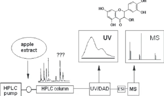
Our research groups have been working on the analysis
the use of on-line LC techniques as a tool for the chemical profiling of plant extracts. The hyphenation (coupling) of high-performance liquid chromatography with UV photodiode array detection (LC/UV) and liquid chromatography with mass spectrometry (LC/MS) provide spectrometric information comparable to those recorded for pure constituents. The molar mass as well as structural information can be obtained by LC/MS/MS or by in-source collision-induced (CID) fragmentation. The application of LC/MS and other LC-hyphenated techniques for the on-line
structural investigation of plant compounds was reviewed.15
In the case of phenolic compounds such as flavonoids and xanthones, on-line UV spectra provide useful information (type of chromophore or pattern of substitution) comple-mentary to those obtained with LC/MS. An important tool is the on-line addition of reagents that induce shifts in the UV
absorption maxima (λmax). LC/UV analysis with post-column
addition of UV shift reagents provides useful information about the oxidation pattern, the position of free phenolic and methoxyl groups, and the position of the linkage of sugar
moieties (O- or C-glycosylation).
In this paper, we report on a study of flavonols from the three apple varieties most commonly cultivated in Brazil (Gala, Golden and Fuji) by LC/UV and tandem mass
spectrometry (LC/MSn), using electrospray ionization (ESI)16
in the negative ion mode. To the best of our knowledge, this is the first report on the characterization and identity of flavonols from samples of these apple varieties cultivated in Brazil using hyphenated techniques.
Experimental
Plant material
Samples of Gala, Golden, and Fuji apple varieties were kindly provided by the Fischer-Fraiburgo Group (Fraiburgo, Santa Catarina, Brazil). The cultivars were grown in the state of Santa Catarina, Brazil. The Gala, Golden, and Fuji samples were harvested within 25 kilometers from each other in February, March, and April, respectively, and were kept in
conventional cold chambers (-20 oC) in a controlled
atmosphere. The apples were peeled manually (1–2 mm thickness) to separate the peels from the pulp. The peels were then dried at 40 °C, grounded into a powder and sieved, and only particles between 1.0 and 2.0 mm were used.
Chemicals
All chemicals and solvents were analytical or HPLC grade. The standards of rutin, hyperoside, isoquercetin, quercetin, and quercetrin were kindly provided by the
Institute of Pharmacognosie and Phytochimie, University of Lausanne, Switzerland. All standards were dissolved in methanol for HPLC analysis.
Extraction of flavonols
Samples of apple peels (1.0 g) were extracted under magnetic stirring with 10 mL of a methanol:water mixture
(1:1) for 30 minutes at 50 oC. The solid residue was filtered
out and the extracts stored at -20 oC before
chromatographic analysis.
LC/UV/MS analysis
On-line LC/UV/MS analysis were performed on a HPLC system with UV photodiode array detector (PAD) coupled to a mass spectrometer. The experimental setup
was previously described and discussed17 (see also
Supplementary Information).A HP-1100 system equipped with binary pumps, an inline degasser, an autosampler, and a PAD detector (Hewllett–Packard, Palo Alto, CA, USA) was used. UV spectra were recorded between 200 and 450 nm and the UV trace was measured at 360 nm. A
Symmetry C18 analytical column (250 × 4.6 mm i.d.;
particle size, 5 µm) fitted with a Novapack RP C18 guard
column (Waters, Millford, MA) were used for the separation. The mobile phase consisted of 0.5% formic acid in deionized water (A) and 0.5% formic acid in acetonitrile:methanol (50:50, v/v) (B). The gradient program was as follows: 25% B to 40% B in 20 min, 40% B to 50% B in 5 min, 50% to 100% B in 5 min followed
by 100% B in 5 min at a flow rate of 1 mL min-1. There
was a 10-min post-run going back to the starting conditions for reconditioning. The column oven temperature was kept
at 40 oC. The injection volume was 10 µL.
The classical shift reagents were prepared according
to the literature.18 The reagents used in post-column
derivatization system were all aqueous solutions, as
follows: strong base, sodium hydroxide (0.02 mol L-1);
aluminum chloride (0.3 mol L-1; with this reagent, the
reaction coil was heated up to 60 oC); sodium acetate (0.5
mol L-1). The solvent delivery system comprised two
M-6000 pumps, a M-720 gradient controller and a U6K injector (Waters). The photodiode array detector HP-1040A (Hewlett-Packard) coupled with an HP-85 personal computer (Hewlett-Packard) was used for recording chromatogram and UV-Vis spectra. For post-column derivatization, an Eldex Model A-30-5-2 (Eldex Labs, Menlo Pak, CA, USA) pump and a reaction coil were employed. Shift reagents were added to the eluent at a
LC/MS detection was performed directly after UV-PAD measurements. Analyses were performed using a Finnigan MAT LCQ (San Jose, CA, USA) ion trap mass spectrometer (IT-MS) equipped with a Finnigan electrospray (ESI) interface, operated under the following
conditions: capillary temperature 225 oC, capillary voltage
–57 V, cone voltage –35 V, spray voltage –2.8 kV and gas
nebuliser flow (N2) 4 L min-1. Product ion mass spectra
were recorded in the range of m/z 150-1000. CID was
performed applying 30% of energy level. Energy levels on the Finnigan IT-MS are given in % and not in eV since
the voltages applied vary according to the m/z value of
precursor ions. The MS/MS method was used in the negative ion mode and was based on scan-dependent type experiments: the most-abundant ion was automatically
selected as precursor ion and fragmented up to the MS4
stage, each successive most-abundant fragment ion being selected again as precursor ion for next step. The instrument parameters were optimized using rutin prior to analysis of apple extracts.
Results and Discussion
General aspects
The elution conditions were established by testing several gradients of three different mobile phases: water-acetonitrile, water–methanol and water–(acetonitrile: methanol 50:50 v/v), to all of which 0.5% formic acid was added to avoid tailing of the chromatographic peaks. The latter proved to be the best mobile phase for separating the apple compounds and also proved suitable for electrospray
ionization. The temperature for this analysis (40 oC) was
optimized considering both separation and resolution of chromatographic peaks.
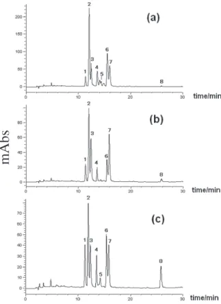
This study focused on the flavonoids present in apple peel extracts, because of the lower concentration of these compounds in the extracts prepared with the whole fruit, which were unsuitable for a LC/MS analysis. The LC/ UV chromatograms (Figure 1) showed similar profiles for the extracts of the three apple varieties, with the exception of the flavonol-3-glycoside corresponding to compound
5, which was not detected in the Golden apple peel extract
(the small peaks with retention time close to the compound
5 are not flavonoids, according to their UV spectra). A
total of eight flavonols (Figure 2) were identified by the on-line combination of LC/UV/MS data, as will be discussed below. Based on the overall on-line data, the apple peel extracts were compared with commercially available standards under the same analytical conditions. The comparison of the retention times, UV and mass
spectra with those obtained from the standards provided
unequivocal confirmation of the identification of rutin (1),
hyperoside (2), isoquercitrin (3), quercitrin (7), and
quercetin (8).
LC/UV/MS analysis
The on-line recording of UV spectra allowed for a rapid attribution of flavonol peaks, since these exhibit
Figure 1. HPLC-UV/PAD chromatograms (λ = 360 nm) of apple peel
ex-tracts. (a) Gala variety; (b) Golden variety and (c) Fuji variety. 1, rutin; 2,
hyperoside; 3, isoquercetin; 4, quercetin-3-O-pentoside; 5, quercetin-3-O -pentoside; 6, quercetin-3-O-pentoside; 7, quercetrin; 8, quercetin (chro-matographic conditions described inExperimental,LC/UV/MS analysis).
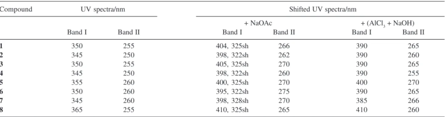
characteristic UV spectra (Table 1), with two maximum
absorption (λmax) bands at 240-280 nm (Band II) and
300-380 nm (Band I). Useful information was also obtained with the post-column addition of UV shift reagents according to a protocol already tested for flavonoids and
xanthones.19,20 The shifted and original UV spectra were
overlaid and compared for each peak, and the shifts were
interpreted according to data reported in the literature.21
These analyses provided important complementary information on the structural features and oxygenation pattern for the on-line identification.
The UV spectra of peaks 1-8 (Table 1) indicated that
all these molecules shared the same chromophore group and were probably flavonols (OH group at position 3).
However, compounds 1-7 showed λmax between 345-355
nm (Band I) and 250-260 nm (Band II), indicating the
presence of a substituted 3-OH group, while compound 8
showed λmax at 365 nm (Band I) and 255 nm (Band II),
suggesting a free 3-OH group.
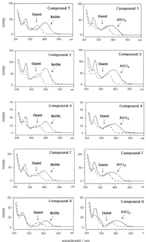
Analyses of UV spectra in HPLC with the post-column
addition of reagents are usually carried out in methanol.20
However, the conditions described herein involve HPLC analyses using an acidic acetonitrile-methanol-water system, which were employed on the shift reagents tested for the different standards of flavonols. The comparison of the on-line UV/PAD spectra with those obtained after the post-column addition of UV shift reagents (Figure 3) allowed for the determination of the substitution pattern on the aglycone, as follows: the addition of NaOAc showed a bathocromic shift in Band I (45-55 nm), revealing a hydroxyl group in position 4’ of the cinnamoyl system, while a bathocromic shift was also observed in Band II (10-15 nm), confirming the presence of free 7-OH groups for all the compounds under investigation. UV spectra in
the presence of AlCl3 exhibited bathocromic shifts in Band
I (40-45 nm) as a result of complexes formed by AlCl3
with 3’,4’-orthodihydroxyl groups in the B-ring, or with 4-keto and 5-hydroxyl groups. These data were observed
for compound 1-8, indicating the substitution of two OH
groups on ring B, i.e., all the compounds studied here
corresponded to quercetin derivatives.
On-line MS and MSn analyses were performed to
ascertain the molar mass of the flavonols under investigation and the latter to obtain detailed structural information. In a previous study, single stage LC/MS using atmospheric pressure chemical ionization (APCI) in the positive ion mode was applied to the investigation of
Basque cider apples of the Goikoetxea variety.10 However,
in the present study, the negative ion mode showed better results in the ESI-MS analysis. ESI process results in limited fragmentation of the deprotonated molecule
[M-H]–, of the flavonol glycosides present in apple
extracts22,23 and therefore, LC/MSn analyses by in-source
CID were also carried out in this study.
The [M-H]– ions were taken as precursors in the
ESI-MS2 analysis and yielded spectra corresponding to
deprotonated aglicone species, [A-H]–, formed due to the
loss of sugar units, as observed for compounds 1-7 (Table
2). As shown for 1, the [M-H]– ion in the ESI-MS spectrum
was observed at m/z 609, while ES-MS2 ion at m/z 301
was attributed to [M-H-146-162]_, with the loss of 308 u
corresponding to a rhamnose (146 u) plus a glucose
(162 u) moiety. For compounds 2 and 3, the [M-H]– ion
Table 2. On line ESI-IT-MSn data obtained in the analysis of apple
ex-tracts (negative ion mode)
Compound tR/ min R [M-H]– Main fragments
MS2MS3MS4
1 11.3 Glu-Rha 609 301 179 151
2 12.0 Gal 463 301 179 151
3 12.4 Glu 463 301 179 151
4 13.6 pentose 433 301 179 151
5 14.3 pentose 433 301 271 271
6 15.5 pentose 433 301 179 151
7 15.9 Rha 447 301 179 151
8 25.8 H 301 nd nd nd
nd: not detected. Gal: galactose; Glu: glucose; Rha: rhamnose.
Table 1. On-line UV data of apple flavonols identified in this study
Compound UV spectra/nm Shifted UV spectra/nm
+ NaOAc + (AlCl3 + NaOH)
Band I Band II Band I Band II Band I Band II
1 350 255 404, 325sh 266 390 265
2 345 250 398, 322sh 262 390 260
3 350 255 405, 325sh 270 390 265
4 345 250 398, 322sh 260 390 255
5 355 260 400, 325sh 270 400 270
6 350 260 395, 322sh 275 390 265
7 345 260 398, 328sh 270 385 266
8 365 255 410, 325sh 265 410 260
was observed at m/z 463 and a similar ion obtained for
compound 1 was observed in the ESI-MS;2 the ion of m/z
301 was attributed to [M-H-162]–, corresponding to a loss
of 162 u (hexose), i.e., galactose and glucose for
compounds 2 and 3, respectively. Compounds 4, 5, and 6
showed identical ESI-MS and ESI-MS2 data with [M-H]–
ion at m/z 433 and a deprotonated aglycone ion
[M-H-132]– at m/z 301, respectively; a loss of 132 u
corres-ponding to a pentose unit (possibly apiose, arabinose or xylose, which are the pentose units most frequently found
in flavonol glycosides). For compound 7, the [M-H]– ion
was observed at m/z 447 and deprotonated aglycone ion
[M-H-146]– at m/z 301, as was observed for compounds
1-6, with a loss of 146 u corresponding to a rhamnose.
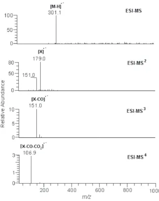
The spectra of compound 8 in the first stage showed the
[M-H]– ion at m/z 301, which was attributed to the
aglycone quercetin, while the ESI-MS2 spectrum showed
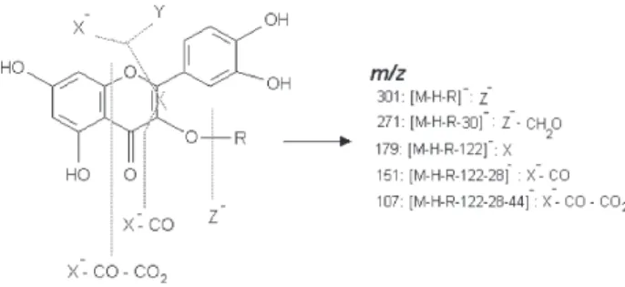
the formation of m/z 179 (X–) ion due to the fragmentation
of rings A and B (Figure 3).
The ESI-MS3 and ESI-MS4 spectra of compounds 1-8
also provided specific information on the fragmentation of rings A and B (Figure 3), which resulted mainly in the
formation of m/z 179 (X–) and m/z 151 (X–-CO) ions. The
elimination of neutral fragment Y (m/z 122, MS3), followed
by loss of CO (m/z 28, MS4), was observed in compounds
1-7. For compound 8, the ESI-MS3 and ESI-MS4 spectra
showed the formation of m/z 151 (X–-CO) and m/z 107
(X–-CO-CO
2) ions. Furthermore, X
– showed losses of 29 u
of CHO–• (neutral radical) and 30 u of CH
2O (neutral loss).
These fragmentation results are in agreement with those
obtained by Justesen,24 who investigated flavonols present
in herbal extracts by APCI-MS/MS in the negative ion mode (fragmentation pathways are shown in Figure 3; for
ESI-MSn spectra, see also Supplementary Information).
Conclusions
The LC-hyphenated technique proved to be a rapid and efficient procedure for the characterization of targeted compounds, using analytical-scale conditions and small
amounts of plant material, and also provided novel information on the chemical composition of the three Brazilian apple varieties (Gala, Golden, and Fuji).
The LC/UV/MS data presented herein showed similarities with the chemical composition of the apple
samples studied here except for compound 5 (a
quercetin-3-O-pentoside), which was not found in the Golden
cultivar. The presence of compounds 1-3 and 7 is in
agreement with data reported for apples grown in North
America and Europe.25-29 On the other hand, aglycone
quercetin (8), which has been reported only once in the
literature,10 was found in the three varieties cultivated in
Brazil. Alonso-Salces et al.10 proposed that this aglycone
has not been reported before, probably due to its low
concentration. However, it would be also possible that 8
results from the hydrolysis of quercetin-O-glycosides
during the sample preparation step, since apple fruit is rich in organic acids.
Furthermore, this study demonstrated, for the first time,
the occurrence of three different quercetin-3-O-pentoside
derivatives in apple peel extracts (Gala and Fuji), while previous reports reported the presence of only two
quercetin-3-O-pentoside derivatives29 (as in the Golden
variety). The differences between the data obtained in this study and that reported in the literature suggest variations in the composition of apple flavonols due to different varieties or growth conditions.
Supplementary Information
Supplementary data are available free of charge at http://jbcs.sbq.org.br, as PDF file.
Acknowledgments
The authors are grateful to Mr. Arioval Pioli (Fischer-Fraiburgo Group, (Fischer-Fraiburgo, SC, Brazil) for furnishing apple samples. This work was supported by FAPESP (L.A.T. fellowship, 98/10271-0 and J.H.Y. research fellowships, 96/3107-3 and 02/00493-2), CNPq and CAPES.
References
1. Centro de Pesquisa Agropecuária de Clima Temperado, Ministério da Agricultura; Coleção Plantar: A Cultura da Maçã, EMPRAPA/ SPI: Brasília, Brasil, 1994, Vol. 19. 2. Dall’orto, F. A. C; Ojima, M.; Barbosa, W.; Castro, J. L.; Rigitano,
O. In Melhoramento da Macieira em São Paulo; Boletim Técnico, Governo do Estado de São Paulo, Secretaria da Agricultura, Instituto de Ciências Agronômicas: Campinas, 1987.
Figure 3. Structure and fragmentation pathways of flavonols by negative
3. Dick, A. J.; Redden, P. R.; Demarco, A. C.; Lidster, P. D.; Grindley, T. B.; J. Agric. Food Chem.1987, 35, 529. 4. Oleszek, W.; Lee, C. Y.; Jaworski, A. W.; Price, K. R.; J. Agric.
Food Chem.1988, 36, 430.
5. Escarpa, A.; González, M. C.; J. Chromatogr., A. 1998, 823, 331.
6. Awad, M. A.; de Jager, A.; van Westing, L. M.; Sci. Hortic.
2000, 83, 249.
7. Awad, M. A.; de Jager, A.; van der Plas, L. H. W.; van der Krol, A. R.; Sci. Hortic. 2001, 90, 69.
8. Schieber, A.; Keller, P.; Carle, R.; J. Chromatogr. A2001, 910, 265.
9. Schieber , A.; Keller, K.; Streker, P.; Klaiber, I.; Carle, R.;
Phytochem. Anal.2002, 13, 87.
10. Alonso-Salces, R. M.; Ndjoko, K.; Queiroz, E. F.; Ioset, J. R.; Hostettmann, K.; Berrueta, L. A.; Gallo, B.; Vicente, F.; J. Chromatogr. A2004, 1046, 89.
11. Robards, K.; Antolovich, M.; Analyst1997, 122, 11R. 12. Arabbi, P. R.; Genovese, M. I.; Lajolo, F.; J. Agric. FoodChem.
2004, 52, 1124.
13. Colombo, R.; Yariwake, J. H.; Queiroz, E. F.; Ndjoko, K.; Hostettmann, K.; J.Chromatogr. A2005, 1082, 51.
14. Colombo, R.; Yariwake, J. H.; Queiroz, E. F.; Ndjoko, K.; Hostettmann, K.; Phytochem.Anal.2006, 17, 337.
15. Wolfender, J.-L.; Hostettmann, K.; J.Chromatogr. A2003,
1000, 437.
16. Crotti, A. E. M; Vessecchi, R.; Lopes, J. L. C.; Lopes, N. P.
Quim. Nova2006, 29, 287.
17. Hostettmann, K.; Wolfender, J.-L.; Terreaux, C.; Pharm. Biol.
2001, 39Supl., 18.
18. Ducrey, B.; Wolfender, J.-L.; Marston, A.; Hosttetmann, K.;
Phytochemistry1995, 38, 129.
19. Hostettmann, K.; Domon, B.; Schaufelberger, D.; Hostettmann, M.; J. Chromatogr.1984, 283, 137.
20. Wolfender, J.-L.; Hostettmann, K.; J. Chromatogr.1993, 647, 191.
21. Markham, K. R.; Techniques of Flavonoid Identification, Academic Press Inc.: London, 1982.
22. Cuyckens, F.; Claeys, M.; J. Mass Spectrom.2004, 39, 1. 23. de la Mora, J. F.; Van Berkel, G. J.; Enke, C. G.; Cole, R. B.
Martinez-Sanchez, M.; Fenn, J. B.; J. Mass Spectrom. 2000, 35, 939.
24. Justesen, U.; J. Chromatogr., A2000, 902, 369.
25. Hollman, P. C. H.; van Trijp, J. M. P.; Buysman, M. N. C. P.; Gaag, M. S. v.d.; Mengelers, M. J. B.; Vries, J. H. M.; Katan, M. B.; FEBS Lett.1997, 418, 152.
26. Knekt, P.; Isotupa, S.; Rissanen, H.; Helliovaara, M.; Jarvinen, R.; Hakkinen, S.; Aromaa, A.; Reunanen, A.; Eur. J. Clin. Nutr.
2000, 54, 415.
27. Tsao, R.; Yang, R.; Young, J. C.; Zhu, H.; J. Agric. FoodChem.
2003, 51, 6347.
28. Tsao, R.; Yang, R.; J. Chromatogr. A2003, 1018, 29. 29. Kahle, K.; Kraus, M.; Richling, E.; Mol. Nutr. Food Res. 2005,
49, 797.
Received: May 31, 2006 Web Release Date: November 24, 2006
0103 - 5053 $6.00+0.00
Supplementary Information
*e-mail: [email protected]
On-line LC/UV/MS Analysis of Flavonols in the Three Apple Varieties Most Widely
Cultivated in Brazil
Luciana A. Tiberti, a Janete H. Yariwake,*,a Karine Ndjokob and Kurt Hostettmannb
a
Universidade de São Paulo, Instituto de Química de São Carlos, CP 780, 13560-970 São Carlos-SP, Brazil
b
Laboratoire de Pharmacognosie et Phytochimie, École de Pharmacie Genève-Lausanne, Université de Genève, Quai Ernest Ansermet 30, 1211 Genève 4, Genève, Switzerland
Figure S2. ESI-MSn spectra of 1 (rutin).
Figure S4. ESI-MSn spectra of 2 (hyperoside) and 3 (isoquercitrin).
Figure S3. UV/PAD and shifted-UV spectra of compounds 1, 2,4, 7, 8
recorded on-line.
Figure S5. ESI-MSn spectra representative of the quercetin-3-O
Figure S7. ESI-MSn spectra of 8 (quercetrin).