Introduction
Functional brain asymmetry refers to how information is processed more or less eficiently by each brain hemisphere in different tasks (Springer, & Deutsch, 1993). Speciically for face recognition, one of the factors that appears to contribute to this asymmetry is the spatial frequency (SF) components of face images. Pre-processing performed by retinal SF channels tuned to different bands of the spectrum (Campbell, & Robson, 1968) supports high-level cognitive operations in the cortex, such as analytical and holistic processing (de Heering, Turati, Rossion, Bulf, Goffaux, & Simion, 2008). Analytical processing refers to the processing of a single feature independently of the context (i.e., eyes, nose, and mouth on a face; Schwarzer, & Zauner, 2003), unlike holistic processing that refers to integrating the features into a gestalt (i.e., in a face, this type of processing interconnects the facial features; Goffaux, & Rossion, 2006; Maurer, Le Grand, & Mondloch, 2002). The availability of the spatial frequency components of a visual image may vary depending on the exposure duration. High spatial frequencies (HSF)
Lina María Perilla-Rodríguez, Rui de Moraes Junior, and Sérgio Sheiji Fukusima, Departmento de Psicologia, Universidade de São Paulo, Ribeirão Preto, Brazil. Correspondence regarding this article should be directed to: Lina María Perilla-Rodríguez, FFCLRP - Universidade de São Paulo, Programa de Pós-Graduação em Psicobiologia, Av. Bandeirantes, 3900, Monte Alegre, Ribeirão Preto, SP, CEP 14040-901, Brasil. Phone: 3602-4448. Fax: +55-16-3633-2660. E-mail: [email protected].
Lateral visual hemiield asymmetry and sex differences in
recognizing low and high spatial frequency iltered faces
Lina María Perilla-Rodríguez, Rui de Moraes Junior, and Sérgio Sheiji Fukusima
Universidade de São Paulo, Ribeirão Preto, SP, Brazil
Abstract
The present study investigated whether low and high spatial frequency filtered images of faces were recognized differently when briefly presented in the right and the left visual fields of men and women. The method of confidence rating was applied to assess pooled Receiver Operating Characteristic curves based on z scores and the d´ parameter of Signal Detection Theory for recognition indices, in addition to response times. The results showed that men better recognized low spatial frequency filtered faces than high spatial frequency filtered faces in both visual fields, suggesting that both the right and left hemispheres in males prioritize low spatial frequencies to recognize faces. The results for women were similar to men only when the faces were shown in the left visual field. When the faces were presented in the right visual field, women better recognized high spatial frequency filtered faces, suggesting that the left hemisphere in females prioritizes high spatial frequencies, whereas the right hemisphere in females prioritizes low spatial frequencies to recognize faces. Keywords: facial recognition, spatial frequencies, brain asymmetry, sex differences, Signal Detection Theory - confidence rating.
Received 22 December 2012; received in revised form 23 October 2013; accepted 29 October 2013. Available online 23 December 2013.
are extracted subsequently to low spatial frequencies (LSF; Goffaux, & Rossion, 2006; Sergent, 1982b). HSF mediate analytical processing, and LSF mediate holistic processing (Boeschoten, Kemner, Kenemans, & Van Engelan, 2005; Hills, & Lewis, 2009).
2004; Peyrin, Chauvin, Chokron, & Marendaz, 2003) and faces (Keenan, Whitman, & Pepe, 1989; Whitman, & Keegan, 1991).
Interest in the human face as an object of study has been increasing since the end of the last century. In addition to its social and evolutionary relevance, behavioral and neuroimaging data indicate that face processing depends more on SF content than on other stimuli (Collin, Liu, Troje, McMullen, & Chaudhuri, 2004; Yue, Tjan, & Biederman, 2006). Thus, it became a model for SF perception in complex visual stimuli. Nevertheless, few studies have investigated the relationship between SF brain asymmetry in face recognition.
Keenan et al. (1989) performed a face recognition task with SF masking in a divided visual ield. The results corroborated Sergent’s hypothesis (1982b), although they presented the faces tachistoscopically only for 10 ms. Researchers have found differential processing between previously learned faces and new faces with face presentation onsets of 200-400 ms (Münte, Brack, Grootheer, Wieringa, Matzke, & Johannes, 1998), 300-600 ms (Paller et al., 2003; Paller, Gonsalves, Grabowecky, Bozic, & Yamada, 2000), 400-600 ms (Yovel, Levy, Grabowecky, & Paller, 2003), 110-600 ms (Barbeau, Taylor, Regis, Marquis, Chauvel, & Liégeois-Chauvel, 2008), and > 200 ms (Münte, Urbach, Düzel, & Kutas, 2000).
Whitman, & Keegan (1991) conducted a study in which pairs of faces were tachistoscopically presented at LSF or HSF in the right visual ield (RVF) or left visual ield (LVF), and the participant provided same-different responses. The results partially supported the SF hypothesis of hemispheric specialization. Presentations in the LH/RVF produced more errors, and this difference was greater for faces in the LSF. In the RH/LVF, faces presented in a LSF had lower response times and lower error rates. Although an exposure time of 200 ms was used, two stimuli were presented simultaneously, possibly impairing face encoding. Additionally, the eccentricity of the face’s inner edge was < 1.3 degrees of visual angle. This distance enabled the stimuli presented to be focused on the binocular convergence area, which may invalidate the technique of divided visual ield. Finally, only men (n = 26) participated in the study.
A sample composed of men shows that researchers do not usually select participants based on sex or are restricted to male participants to avoid gender effects. The literature rarely addresses sex differences in SF hemispheric asymmetry, which may be important, given that sex can modulate patterns of hemispheric dominance (Voyer, 1996). The study by Keenan et al. (1989) discussed above investigated the inluence of sex in their sample of 15 men and 15 women, but no differences were found. Peyrin, Chokron, Guyader, Gout, Moret, & Marendaz (2006a) also used the divided visual ield with the presentation of SF iltered complex stimuli, but they used scenes instead of faces. The results showed an effect of SF asymmetry only in the male sample (12 of 24 participants).
To elucidate this issue, the present study investigated sex differences in hemispheric asymmetry using a face recognition task under the inluence of spatial iltering and assessed accuracy and RTs. We used a modiied divided visual ield method based on the technique used by Tripathy, Levi, Ogmen, & Harden (1995), which sought to quantify any perceived length distortion for vertical bars presented across the blind spot so that we could use a higher exposure time compared with the traditional method and ensure that the face was processed. Moreover, unlike other studies, this experiment was conducted in two phases: memorization and recognition (despite the matching task design, which is widely used in the literature). The short duration of each matching-task trial may impair the transfer of SF band information to short-term visual memory (Gao, & Bentin, 2011). The results presented herein are interpreted in terms of perceptual processes of face processing and coding because the decrease in visual short-term memory retention is not inluenced by the SF spectrum (Gao, & Bentin, 2011). Finally, we mixed Yes-No and conidence rating methods of Signal Detection Theory (SDT; Macmillan, & Creelman, 2005) to calculate the participant’s performance.
Methods
Participants
Forty volunteers (mean age, 27.7 years; standard deviation [SD], 7.57 years; range, 19-52 years), including 20 male students (mean age, 27.4 years; SD, 5.62 years; range, 22-44 years) and 20 female students (mean age, 28.0 years; SD, 9.26 years; range, 19-52) from the University of São Paulo, participated in this study after providing informed consent according to the current rules in Brazil on human experiments (process no. 268/2006 - 2006.1.1368.59.9). All of the participants had normal or corrected visual acuity in both eyes. All of them were right handed, based on a 16-item questionnaire adapted from the Edinburgh Inventory (Oldield, 1971). The mean handedness score on the Edinburgh Inventory was 85 (SD, 10.10) for men and 84 (SD, 9.18) for women.
Stimuli
and itting this image onto a black rectangle (213 × 255 pixels) using Photoshop 7.0 (Adobe). The faces presented for the recognition test were also duplicated on the screen with eccentricity of 3.58 degrees of visual angle (inner edge; Figure 1).
Procedure
The participants were comfortably seated on a chair in a dark room in front of a computer while resting their head on a chinrest. The experiment was completed in two phases: memorization and recognition. The order of stimulus presentation in each phase was randomized by Superlab 2.0 (Cedrus).
In the memorization phase, the participants were required to keep in memory 14 pictures (half female) of uniltered faces. Each face was shown without a time restriction at the center of the computer screen. Four sessions were required to complete this task. In the last session, the subjects were asked to recognize the 14 faces by responding “yes” or “no” to indicate whether the face presented on the monitor had been previously presented. When any misidentiication occurred, the participants were reexposed to the faces until total recognition was achieved. The subjects were not given any explicit instructions about learning strategies. This phase was performed on a computer that was different from the one used for the second phase, although it was in the same room.
The recognition phase was performed immediately after the memorization phase. The latter included 84 trials, with 14 memorized faces (half female) in each iltering condition (14 LSF iltered, 14 HSF iltered, and 14 uniltered), for a total of 42 target faces and 42 distractor stimuli in the same conditions without any stimulus repetition. The participants were irst instructed to centrally place a red cross that was presented until they pressed the spacebar on the keyboard. After showing a face for 300 ms, a blank screen was presented for 500 ms. The participants were then asked to indicate as accurately and quickly as possible whether the face was new (a distractor) or had been presented in the memorization phase (a target). The conidence in their response was rated by pressing one of six keys on a numeric keypad according to a 6-category scale, from category 1 (“I am certain that I do not recognize it”) to category 6 (“I am certain that I recognize it”). The other categories represented intermediate conidence levels for the response. Before the experimental session, the participants completed ive practice trials to ensure they understood the task.
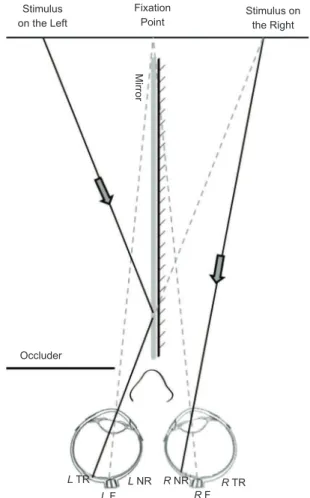
In the second phase, the stimuli were presented to the participant using dichoptic images (Figure 2) proposed by Tripathy et al. (1995). A mirror (30 cm height × 35
cm length × .3 cm width) was placed vertically and perpendicularly to the midline of the monitor using a support attached to the monitor. Through this support, an adjustable black cardboard occluder was placed to prevent direct sight on the left and right half of the monitor by the left and right eye, respectively, but it allowed the observer to perceive the image relected in the mirror through a gap between the occluder and mirror. The back of the mirror was covered with black paper. Even with the occurrence of saccades, this adaptation in the divided visual ield prevented the stimulus presented in one visual hemiield from reaching the ipsilateral nasal retina, given the presence of the occluder. In the hemiield that was not occluded, the information could only reach the nasal retina because of the eccentricity adopted. The stimulus viewed through the gap was mirrored (i.e., inverted) so that the binocular effect could occur, and the participants perceived only one face in the hemiield that was not occluded. The sample was independent for each hemisphere tested, with 20 participants (half female) for the right and 20 participants for the left.
a b c
Figure 2. Dichoptic presentation of the image through the
mirror and occluder. The face presented on the left half was
relected on the mirror and had its image projected on the
temporal retina of the left eye. The right eye viewed the right half of the screen directly, and the image presented in this half was projected on the nasal retina. Thus, all of the information was initially processed in the left hemisphere. This process was inverted to test the right hemisphere. F, fovea; NR, nasal retina; TR, temporal retina; L, left; R, right. Figure based on the model image of Tripathy et al. (1995).
Figure 1. Example of (a) uniltered, (b) low spatial frequency
iltered, and (c) high spatial frequency iltered faces.
Stimulus on the Left
Stimulus on the Right
Occluder
L TR
L F
L NR R NR
R F
R TR
Mirror
Data and statistical analysis
Conidence ratings and mean response times were recorded. Recognition performance was calculated using SDT (Macmillan, & Creelman, 2005). Receiver Operating Characteristic (ROC) curves were calculated using the z scores of the cumulative frequency of responses to the categories of certainty in the face recognition task. The z scores of the responses to the wrongly recognized distracting faces (false alarms; abscissae) were plotted as a function of the z scores in response to the correctly recognized memorized faces (hits; ordinates).
Based on the ROC curves, the sensitivity parameter da (sensitivity parameter of the group) of SDT was calculated for each experimental group. It corresponded to the distance between the distributions of the “noise” and “noise + signal” measured by the root-mean-square standard deviations of these two distributions. In this case, we obtained da according to the equation dα= √2/ (1−β2) × α, where α and β are the magnitude of the
linear coeficient and slope, respectively, of the zROC regression line (Fukusima, & Landeira-Fernandez, 2012). As a more robust indicator of individual sensitivity, the three recognition categories were grouped into just one category. The same was done for the three categories of non-recognition. Thus, calculating the more traditional d’ index (sensitivity parameter of the observer) was possible for each participant. We performed a mixed between-within subjects analysis of variance (ANOVA), with d’ and Response Time with Spatial Frequency (uniltered, LSF iltered, and HSF iltered) as the within-subject factors and Brain Hemisphere (BH; left and right) and Sex (men and women) as the as between-subjects factors. Post hoc comparisons were performed to determine the signiicance of pairwise contrasts by applying Bonferroni correction. An alpha level of .05 was used. These analyses were performed using Systat 13 (Cranes).
Results
Parameters dα and d’
The same pattern of dα of each iltered condition in both hemispheres was achieved, with better recognition of uniltered faces (RH: .94; LH: 1.37), followed by LSF iltered faces (RH: .83; LH: .87) and HSF iltered faces (RH: .47; LH: .80), respectively. The LH/RVF was better in all iltered conditions
When dα was extracted based on sex, men and women had different results. Men recognized uniltered faces better than LSF and HSF iltered faces in the RH/ LVF (uniltered: .95; LSF iltered: .76; HSF iltered: .40) and LH/RVF (uniltered: 1.14; LSF iltered: .94; HSF iltered: .58). The RH/LVF results obtained for women followed the same pattern (uniltered: .93; LSF iltered: .90; HSF iltered: .56). However, in the LH/RVF, the HSF iltered faces were better recognized than LSF iltered faces (uniltered: 1.61; HSF iltered: 1.04; LSF iltered: .83). The dα data for each group were extracted from the ROC curves (Figure 3) and were consistent with the individuals’ d’ average.
Figure 3. ROC curves on z coordinates for each iltering
condition according to hemisphere/visual ield and sex in the
face recognition task. HSF, high spatial frequency; LSF, low
spatial frequency; uniltered, full spatial spectrum.
The ANOVA of d’ revealed a signiicant SF × Sex × BH interaction (F2,35 = 3.993, p = .027). But SF did not interact with Sex or BH independently. Thus, tests of the hypothesis were separated into SF, Sex, and BH analyses. The SF analysis of d’ revealed a signiicant main effect (F2,35 = 10.187, p = .001). Pairwise comparisons showed signiicant differences between recognizing uniltered faces and HSF iltered faces (p < .001).
An ANOVA was performed for each sex. A signiicant effect on SF recognition was found (F2,17
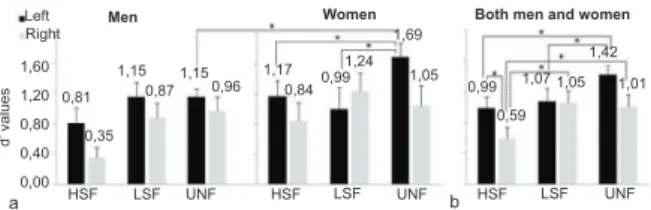
= 5.269, p = .017). Pairwise comparisons showed that uniltered faces were better recognized than HSF iltered faces (p = .013) in males (Figure 4a). Likewise, women showed a signiicant effect on SF recognition (F2,17 = 7.852, p = .004). The uniltered faces were better recognized than HSF iltered faces (p = .015) and LSF iltered faces (p = .035; Figure 4a). Unlike men, a signiicant SF × BH interaction was found for women (F2,17 = 11.754, p = .001). An ANOVA was also conducted to identify differences in face recognition by women according to BH. A signiicant effect of SF on recognition was found only for the LH/RVF (F2,8 = 21.051, p = .001), indicating that uniltered faces were better recognized than HSF iltered faces (p = .022) and LSF iltered faces (p = .002; Figure 5a).
Figure 4. Comparison between sexes and hemispheres in
performance in the face recognition task among iltering
conditions using the d’ parameter (a) and response time (b). HSF, high spatial frequency; LSF, low spatial frequency;
uniltered, full spatial spectrum. *p < .05 (ANOVA). LEFT HEMISPHERE
Two Hemispheres
0,58
HSF LSF UNF HSF LSF UNF
a b
1,00
2474 2301 2549 2343 2165 2122 1,011,12 1,06
1,37
Two Hemispheres Men
Men
Women
3000
2000
1000
0
Women d’ values Response time (msec) 1,60 1,20 0,80 0,40 0,00 LEFT HEMISPHERE RIGHT HEMISPHERE RIGHT HEMISPHERE WOMEN MEN WOMEN MEN z score (False Alarm) z score (False Alarm) z score (False Alarm) Uniltered LSF HSF z score (False Alarm) z score (Hit) z score (Hit) z score (Hit) z score (Hit) 2 1 0 2 1 0 2 1 0 2 1 0 -2 -1 0 1
-2 -1 0 1
-2 -1 0 1
-2 -1 0 1
-1
-1
-1
An ANOVA was then performed for each BH. For the RH/LVF, a signiicant effect of SF on recognition was found (F2,17 = 6.869, p = .007). Pairwise comparisons showed signiicant differences between uniltered faces and HSF iltered faces (p = .019) and between HSF iltered faces and LSF iltered faces (p = .011; Figure 5b). For the LH/RVF, a signiicant effect of SF on recognition was found (F2,17 = 11.406, p = .001). Uniltered faces were better recognized than HSF iltered faces (p = .011) and LSF iltered faces (p = .017; Figure 5b). A signiicant SF × Sex interaction was found (F2,17 = 5.274, p = .017) for the LH/RVF. An ANOVA was performed to identify the interaction between sex and iltering condition for the LH/RVF. We found that women better recognized uniltered faces than men (F1,18
= 6.284, p = .022; Figure 5a).
Men Women
Left Right
1,60
1,20
0,80
0,40
0,00 0,81
0,35
HSF LSF UNF HSF LSF UNF HSF LSF UNF
a b
1,15 1,17
0,840,99 1,24
1,69
1,05 0,99
0,59 1,07 1,05
1,42
1,01 0,87
1,15 0,96
d’
values
Both men and women
Figure 5. Comparison of performance using the d’ parameter
obtained in the face recognition task among iltering conditions.
(a) Men and women for each hemisphere. (b) Performance for left and right hemispheres for both men and women. HSF,
high spatial frequency; LSF, low spatial frequency; uniltered, full spatial spectrum. *p < .05 (ANOVA).
When contrasting individual performance based on the data obtained for HSF iltered faces as the dependent variable, we found signiicant effects of sex (F1,36 =
4.580, p = .039; Figure 4a) and BH (F1,36 = 3.955, p = .054; Figure 5b). A signiicant effect of BH was also found for uniltered faces (F1,36 = 4.601, p = .039).
Response time
The ANOVA performed for RT revealed a signiicant SF × Sex × BH interaction (F2,35 = 3.699, p =
.035), with no interaction with Sex or BH independently. Again, tests of the hypothesis were separated into SF, Sex, and BH analyses. The SF analysis of RT revealed a signiicant main effect (F2,35 = 7.096, p = .003),
indicating that uniltered faces were recognized faster than HSF iltered faces (p = .011) and LSF iltered faces (p = .001).
Separate ANOVAs for each sex showed that women recognized all face stimuli equally, whereas men had signiicant differences (F2,17 = 7.396, p = .005).
Pairwise comparisons showed that uniltered faces were recognized faster than LSF iltered faces (p = .004) and HSF iltered faces (p = .054; Figure 4b).
An ANOVA was performed for each BH. For the RH/LVF, a signiicant effect of SF on recognition was found (F2,17 = 5.412, p = .015). Uniltered faces were recognized faster (p = .011) than LSF iltered faces (Figure 6b). Comparisons of individual performance revealed a Sex × BH interaction for HSF iltered
faces (F1,36 = 4.521, p = .04). This interaction was found speciically for the LH/RVF, with a signiicant difference between sexes (F1,18 = 5.037, p = .038). Women recognized HSF iltered faces faster than men (Figure 6a).
Figure 6. Response time for each iltering condition in
the face recognition task. (a) Men and women for each hemisphere. (b) Performance for left and right hemispheres for both men and women. HSF, high spatial frequency; LSF,
low spatial frequency; uniltered, full spatial spectrum. *p <
.05 (ANOVA).
Discussion
The present study investigated sex differences in hemispheric asymmetry in a face recognition task under the inluence of spatial iltering. The results indicated that women and men differed with regard to both sensitivity and RT as a function of SF and hemispheric asymmetry.
Early studies of hemispheric lateralization with the processing of emotional stimuli, including faces (Proverbio, Brignone, Matarazzo, Del Zotto, & Zani, 2006), facial recognition (Godard, & Fiori, 2010), face discrimination (Hausmann, & Gunturkun, 1999), the recognition of natural scenes with spatial iltering (Peyrin et al., 2006a), an attentional probe task (Davidson, Cave, & Sellner, 2000), and a spatial task (Gur et al., 2000), demonstrated that men are more lateralized than women. In the present study, we did not ind hemispheric specialization in men. They had the same pattern of recognition for both hemispheres when considering SDT (dα and d’) and RTs. The best performance occurred for uniltered faces, followed by LSF iltered faces. The worst performance was found for HSF iltered faces. Among women, no signiicant effect was found for SF hemispheric specialization. Based on the descriptive statistics, we observed an inversion of patterns in the recognition of faces with spatial iltering between BHs, with better recognition of HSF and LSF iltered faces in the LH/RVF and RH/ LVF, respectively, supporting the hypothesis of Sergent (1982b). Furthermore, women had better performance recognizing uniltered faces and were faster recognizing HSF iltered faces with the LH/RVF than men. With regard to differences in RT, the descriptive data, independent of iltering condition, showed that women were also faster recognizing stimuli with the LH/RVF, and men were faster with the RH/LVF. These results are consistent with Roalf, Lowery, & Turetsky (2006), who studied hierarchical stimuli and event-related potentials
Men Women
Left Right
3000
2000
1000
0 2889
2059 2835
22642425 1905 1821
2782
2032 2654
1939
2306 2355242124342459 2182 2105
HSF LSF UNF HSF LSF UNF HSF LSF UNF
a b
Response time (msec)
(ERPs). Their ERP data showed that women were lateralized with LH/RVF dominance, especially for local stimuli. Women also had a RT delay for global stimuli. In that study, men showed similar results between stimulus conditions (i.e., global and local) and hemispheres and had lower RTs to global stimuli. Women have more eficient analytical processing, and they respond more quickly to local stimuli than to global stimuli (Roalf et al., 2006). Moreover, men presented longer RTs when visual processing was performed by the LH/RVF than by the RH/LVF (Godard, & Fiori, 2010).
When the analysis was performed independently of sex, Sergent’s hypothesis (1982b) could be partially corroborated. The main difference between BHs was found in HSF iltered faces. Stimuli with a HSF presented to the LH/RVF were easier to recognize than stimuli presented to the RH/LVF, regardless of sex. Uniltered and LSF iltered faces were better recognized than HSF iltered faces when presented in the RH/LVF. Our results are consistent with many studies that have supported the SF hypothesis of hemispheric specialization (Coubard et al., 2011; Evert, & Kmen, 2003; Keenan et al., 1989; Kitterle et al., 1993; Peyrin et al., 2004; Peyrin et al., 2003; Proverbio et al., 1997; Yamaguchi, Yamagata, & Kobayashi, 2000).
With regard to hemisphere dominance in face recognition, the literature reports an advantage of the RH/LVF (Gazzaniga, 2000; Levy, Trevarthen, & Sperry, 1972; Ramon, & Rossion, 2012; Springer, & Deutch, 1993; Whitman, & Keegan, 1991; Yovel, Tambini, & Brandman, 2008). We did not ind an advantage of the RH/LVF for face recognition with regard to either sensitivity or speed in any of the iltering conditions. One of the reasons why the RH/LVF was not better than the LH/RVF for facial recognition might be that the stimulus exposure time was 300 ms. The advantage in analytical processing of complex stimuli performed by the LH/RVF would be impaired by a short exposure time (Sergent, 1982a). Additionally, holistic processing is more pronounced in the RH/LVF in the early stages of perception (Ramon, & Rossion, 2012). As suggested by previous studies, the stimulus exposure time could be a key factor to establish cerebral asymmetry in visual tasks (Peyrin, Mermillod, Chokron, & Marendaz, 2006b; Sergent, & Hellige, 1986).
The exposure time was based on previous studies of the activity of neural pathways responsible for the recognition of facial identity in both spatial and temporal dimensions. Another reason why a 300 ms exposure time was used in the present study was the absence of external face features. Several studies found that external face features are primordial in processing the identity of unfamiliar faces. Recognizing faces without these elements increases the dificulty, and judgments become slower (Bobes, Martin, Olivares, & Valdes-Sosa, 2000; Caldara, Jermann, Arango, & Van der Linden, 2004).
The present results may have been inluenced by the methodological procedures and task demands.
A longer exposure time increases interhemispheric communication and reduces cerebral asymmetry as well as the presentation of dichoptic images (Lux et al., 2004; Peyrin et al., 2006b). Likewise, a short time restriction favors analytical processing (Hegdé, 2008; Goffaux, Peters, Haubrechts, Schiltz, Jansma, & Goebel, 2011). Despite the differences in RTs, men and women had higher rates of recognition with the LH/RVF.
Generally, when sex and BH were not considered in the analyses, uniltered faces were recognized better and faster than iltered images. This is consistent with the literature, which suggests that faces are more sensitive to SF information (Goffaux et al., 2011). The recognition of uniltered faces only showed statistically signiicant differences compared with the recognition of HSF iltered faces. The fact that uniltered faces and LSF iltered faces showed no statistically signiicant difference could be related to three factors. First, the stimulus presentation was performed on peripheral vision, which is more sensitive to LSFs (Livingstone, & Hubel, 1988). Second, the literature assumes that LSF information supports global processing, which is the automatic coniguration (i.e., default coniguration) of visual attention. In other words, it requires less activation than local processing (Lux et al., 2004). Third, the extraction of LSF information is important because the facial pattern is initially processed holistically (Goffaux, & Rossion, 2006).
One of the main advantages of the present study was the equal distribution of participants of both sexes. A large amount of spatial frequency and hemispheric asymmetry research has not included samples with the same number of men and women (Evert, & Kmen, 2003; Goffaux, Hault, Michel, Vuong, & Rossion, 2005) and usually included samples of only one sex, usually male (Whitman, & Keegan, 1991; Peyrin et al., 2003) or did not report the number of participants of each sex (Hills, & Lewis, 2009; Reinvang, Magnussen, & Greenlee, 2002). For this reason, information about sex differences in hemispheric asymmetry in visual tasks is limited. Another advantage of the present study was the unique technique of image presentation (Tripathy et al., 1995). This adaptation is very affordable, which may thus promote research in laboratories that do not have an eye tracker. Even if saccadic eye movements occur, this technique assures that no visual sweep occurs in the hemisphere’s ipsilateral hemiield. For this reason, using an exposure time of 300 ms was possible, which is not usual in psychophysical experiments that use divided visual ields. A further advantage of this work is the SDT-conidence rating method (Macmillan, & Creelman, 2005) that was used to analyze the participants’ performance. It allows the calculation of a ROC curve and sensitivity that considers the decision criterion for each participant in a single experimental session.
symmetrical than women and recognized LSF iltered faces better than HSF iltered faces. However, descriptive data suggest shorter RTs when the stimuli are presented in the RH/LVF. Women more quickly recognized the stimuli presented in the LH/RVF and HSF iltered faces. Nonetheless, women had better performance in the LH/ RVF in recognizing uniltered faces. When the sex of the participants was not considered, Sergent’s (1982b) hypothesis was partially supported. The HSF iltered faces were better recognized by the LH/RVF, and LSF iltered faces were better recognized than HSF iltered faces in the RH/LVF. In contrast to the literature, the RH/LVF was not better in the recognition task than the LH/RVF. This may be related to the use of a longer exposure time compared with other studies in the literature. We conclude that men and women have different sensitivity and RTs when recognizing faces in cerebral asymmetry research.
Acknowledgements
The authors are grateful to Prof. José Antonio Aznar Casanova for providing the MATLAB codes used to ilter the images and Ana Irene Fonseca Mendes who suggested the use of an adaptation in the divided visual ield method. We also thank the participants for their commitment to the study, Conselho Nacional de Desenvolvimento Científico e Tecnológico (CNPq) and Fundação de Amparo à Pesquisa do Estado de São Paulo (FAPESP) for financial aid.
References
Barbeau, E. J., Taylor, M. J., Regis, J., Marquis, P., Chauvel, P., & Liégeois-Chauvel, C. (2008). Spatio temporal dynamics of face recognition. Cerebral Cortex, 18(5), 997-1009.
Bobes, M. A., Martin, M., Olivares, E., & Valdes-Sosa, M. (2000). Different scalp topography of brain potentials related to expression and identity matching of faces. Cognitive Brain Research, 9(3), 249-260.
Boeschoten, M. A., Kemner, C., Kenemans, J. L., & Engeland, H. V. (2005). The relationship between local and global processing and the processing of high and low spatial frequencies studied by event-related potentials and source modeling. Brain Research, 24(2), 228-236. Caldara, R., Jermann, F., Arango, G. L., & Van der Linden, M. (2004).
Is the N400 category-speciic? A face and language processing
study. Neuroreport, 15(17), 2589-2593.
Campbell, F. W., & Robson, J. G. (1968). Application of Fourier analysis to the visibility of gratings. Journal of Physiology, 197(3), 551-566.
Collin, C. A., Liu, C. H., Troje, N. F., McMullen, P. A., & Chaudhuri, A. (2004). Face recognition is affected by similarity in spatial frequency range to a greater degree than within-category object recognition. Journal of Experimental Psychology: Human Perception and Performance, 30(5), 975-987.
Coubard, O. A., Perez, C., Kazandjian, S., Gaudry, I., Marendaz, C., Guyader, N., Peyrin, C., & Chokron, S. (2011). Visual demand
and visual ield presentation inluence natural scene processing.
Graefes Archive for Clinical and Experimental Ophthalmology, 249(2), 223-232.
Davidson, H., Cave, K. R., & Sellner, D. (2000). Differences in visual
attention and task interference between males and females relect
differences in brain laterality. Neuropsychologia, 38(4), 508-519. de Heering, A., Turati, C., Rossion, B., Bulf, H., Goffaux, V., & Simion,
F. (2008). Newborns’ face recognition is based on spatial frequencies below 0.5 cycles per degree. Cognition, 106(1), 444-454.
Evert, D. L., & Kmen, M. (2003). Hemispheric asymmetries for global and local processing as a function of stimulus exposure duration. Brain and Cognition, 51(1), 115-142.
Fukusima, S. S., & Landeira-Fernandez, J. (2012). Informações complementares sobre a teoria de detecção do sinal aplicada à psicofísica. In J. Landeira-Fernandez, & S.S. Fukusima (Eds.), Métodos em neurociência (pp. 24-33). Barueri: Manole.
Gazzaniga, M. S. (2000). Cerebral specialization and interhemispheric communication: does the corpus callosum enable the human
condition? Brain, 123(7), 1293-1326.
Gao, Z., & Bentin, S. (2011). Coarse-to-ine encoding of spatial
frequency information into visual short-term memory for faces but impartial decay. Journal of Experimental Psychology: Human Perception and Performance, 37(4), 1051-1064.
Godard, O., & Fiori, N. (2010). Sex differences in face processing: are
women less lateralized and faster than men? Brain and Cognition,
73(3), 167-175.
Goffaux, V., Hault, B., Michel, C., Vuong, Q. C., & Rossion, B. (2005). The respective role of low and high spatial frequencies in
supporting conigural and featural processing of faces. Perception,
34(1), 77-86.
Goffaux, V., Peters, J., Haubrechts, J., Schiltz, C., Jansma, B., & Goebel,
R. (2011). From coarse to ine? Spatial and temporal dynamics of
cortical face processing. Cerebral Cortex, 21(2), 467-476.
Goffaux, V., & Rossion, B. (2006). Faces are “spatial”: holistic face perception is supported by low spatial frequencies. Journal of Experimental Psychology: Human Perception and Performance, 32(4), 1023-1039.
Gur, R. C., Alsop, D., Glahn, D., Petty, R., Swanson, C. L., Maldjian, J. A., Turetsky, B. I., Detre, J. A., Gee, J., & Gur, R. E. (2000). An fMRI study of sex differences in regional activation to a verbal and a spatial task. Brain and Language, 74(2), 157-170.
Han, S., Weaver, J. A., Murray, S. O., Kang, X., Yund, E. W., & Woods, D. L. (2002). Hemispheric asymmetry in global/local processing: effects of stimulus position and spatial frequency. NeuroImage, 17(3), 1290-1299.
Hausmann, M., & Gunturkun, O. (1999). Sex differences in functional cerebral asymmetries in a repeated measures design. Brain and Cognition, 41(3), 263-275.
Hegdé, J. (2008). Time course of visual perception: coarse-to-ine
processing and beyond. Progress in Neurobiology, 84(4), 405-439. Hills, P. J., & Lewis, M. B. (2009). A spatial frequency account
of the detriment that local processing of Navon letters has on face recognition. Journal of Experimental Psychology: Human Perception and Performance, 35(5), 1427-1442.
Keenan, P. A., Whitman, R. D., & Pepe, J. (1989). Hemispheric asymmetry in the processing of high and low spatial frequencies: a facial recognition task. Brain and Cognition, 11(2), 229-237. Kitterle, F. L., Christman, S., & Conesa, J. (1993). Hemispheric
differences in the interference among components of compound gratings. Perception and Psychophysics, 54(6), 785-793.
Levy, J., Trevarthen, C., & Sperry, R. W. (1972). Reception of bilateral
chimeric igures following hemispheric deconnexion. Brain, 95(1),
61-78.
Livingstone, M., & Hubel, D. (1988). Segregation of form, color, movement, and depth: anatomy, physiology, and perception. Science, 240(4853), 740-749.
Lux, S., Marshall, J. C., Ritzl, A., Weiss, P. H., Pietrzyk, U., Shah, N. J., Zilles, K., & Fink, G. R. (2004). A functional magnetic resonance imaging study of local/global processing with stimulus
presentation in the peripheral visual hemiields. Neuroscience,
124(1), 113-120.
Macmillan, N., & Creelman, C. (2005). Detection theory: a user’s guide, 2nd edition. Mahwah, N.J.: Lawrence Erlbaum Associates. Maurer, D., Le Grand, R., & Mondloch, C. J. (2002). The many faces of
conigural processing. Trends in Cognitive Sciences, 6(6), 255-260.
Mendes, A. I. F., Arrais, K. C., & Fukusima, S. S. (2009). Faces prototípicas provenientes de amostras populacionais de uma região brasileira. Psicologia: Reflexão e Crítica, 22(2), 261-268. Münte, T. F., Brack, M., Grootheer, O., Wieringa, B. M., Matzke, M., &
Johannes, S. (1998). Brain potentials reveal the timing of face identity and expression judgments. Neuroscience Research, 30(1), 25-34. Münte, T. F., Urbach, T. P., Düzel, E., & Kutas, M. (2000).
Oldield, R. C. (1971). The assessment and analysis of handedness:
the Edinburgh Inventory. Neuropsychologia, 9, 97-113.
Paller, K. A., Gonsalves, B., Grabowecky, M., Bozic, V. S., & Yamada, S. (2000). Electrophysiological correlates of recollecting faces of known and unknown individuals. NeuroImage, 11(2), 98-110.
Paller, K. A., Ranganath, C., Gonsalves, B., LaBar, K. S., Parrish, T.B., Gitelman, D. R., Mesulam, M. M., & Reber, P. J. (2003). Neural correlates of person recognition. Learning and Memory, 10(4), 253-260.
Peyrin, C., Baciu, M., Segebarth, C., & Marendaz, C. (2004). Cerebral regions and hemispheric specialization for processing spatial frequencies during natural scene recognition: an event-related fMRI study. Neuroimage, 23(2), 698-707.
Peyrin, C., Chauvin, A., Chokron, S., & Marendaz, C. (2003). Hemispheric specialization for spatial frequency processing in the analysis of natural scenes. Brain and Cognition, 53(2), 278-282. Peyrin, C., Chokron, S., Guyader, N., Gout, O., Moret, J., & Marendaz,
C. (2006a). Neural correlates of spatial frequency processing: a neuropsychological approach. Brain Research, 16(1073-1074), 1-10.
Peyrin, C., Mermillod, M., Chokron, S., & Marendaz, C. (2006b). Effect of temporal constraints on hemispheric asymmetries during spatial frequency processing. Brain and Cognition, 62(3), 214-220.
Proverbio, A. M., Brignone, V., Matarazzo, S., Del Zotto, M., & Zani, A. (2006). Gender differences in hemispheric asymmetry for face processing. BMC Neuroscience, 7, 44.
Proverbio, A. M., Zani, A., & Avella, C. (1997). Hemispheric asymmetries for spatial frequency discrimination in a selective attention task. Brain and Cognition, 34(2), 311-320.
Ramon, M., & Rossion, B. (2012). Hemisphere-dependent holistic processing of familiar faces. Brain and Cognition, 78(1), 7-13. Reinvang, I., Magnussen, S., & Greenlee, M. W. (2002). Hemispheric
asymmetry in visual discrimination and memory: ERP evidence for the spatial frequency hypothesis. Experimental Brain Research, 144(4), 483-495.
Roalf, D., Lowery, N., & Turetsky, B. I. (2006). Behavioral and
physiological indings of gender differences in global-local visual
processing. Brain and Cognition, 60(1), 32-42.
Sergent, J. (1982a). About face: left-hemisphere involvement in processing physiognomies. Journal of Experimental Psychology: Human Perception and Performance, 8(1), 1-14.
Sergent, J. (1982b). The cerebral balance of power: confrontation
or cooperation? Journal of Experimental Psychology: Human
Perception and Performance, 8(2), 253-272.
Sergent, J., & Hellige, J. B. (1986). Role of input factors in visual-ield
asymmetries. Brain and Cognition, 5(2), 174-199.
Springer, S. P., & Deutsch, G. (1993). Left brain, right brain, 4th edition. New York: W.H. Freeman.
Schwarzer, G., & Zauner, N. (2003). Face processing in 8-month-old
infants: evidence for conigural and analytical processing. Vision
Research, 43, 2783-2793.
Tripathy, S., Levi, D., Ogmen, H., & Harden, C. (1995). Perceived length across the physiological blind spot. Visual Neuroscience, 12(2), 385-402.
Voyer, D. (1996). On the magnitude of laterality effects and sex differences in functional lateralities. Laterality, 1(1), 51-83. Whitman, R. D., & Keegan, J.F. (1991). Lateralization of facial
processing: a spatial frequency model. International Journal of Neuroscience, 60(3-4), 177-185.
Yamaguchi, S., Yamagata, S., & Kobayashi, S. (2000). Cerebral asymmetry of the “top-down” allocation of attention to global and local features. Journal of Neuroscience, 20(9), RC72.
Yovel, G., Levy, J., Grabowecky, M., & Paller, K. A. (2003). Neural
correlates of the left-visual-ield superiority in face perception
appear at multiple stages of face processing. Journal of Cognitive Neuroscience, 15(3), 462-474.
Yovel, G., Tambini, A., & Brandman, T. (2008). The asymmetry of the fusiform face area is a stable individual characteristic that underlies
the left-visual-ield superiority for faces. Neuropsychologia,
46(13), 3061-3068.
Yue, X., Tjan, B. S., & Biederman, I. (2006). What makes faces