Article
0103 - 5053 $6.00+0.00*e-mail: [email protected]; [email protected]
Synthesis and Enzymatic Evaluation of the Guanosine Analogue 2-Amino-6-mercapto-
7-methylpurine Ribonucleoside (MESG). Insights into the Phosphorolysis Reaction
Mechanism based on the Blueprint Transition State: S
N1 or S
N2?
Brenno A. D. Neto,*,a Alexandre A. M. Lapis,b Paulo A. Netz,c John Spencer,d Silvio L. P. Dias,c Silvia M. Tamborim,c Luiz A. Basso,e Diógenes S. Santose and Jairton Dupont*,c
aLaboratory of Medicinal and Technological Chemistry, University of Brasilia (IQ-UnB),
72919-970 Brasilia-DF, Brazil
bUniversidade Federal do Pampa, Unipampa, 96412-420 Bagé-RS, Brazil
cLaboratory of Molecular Catalysis-Institute of Chemistry -UFRGS, Av. Bento Gonçalves, 9500,
91501-970 Porto Alegre-RS, Brazil
dSchool of Science, University of Greenwich at Medway, Chatham Maritime, ME4 4TB, UK
eCentro de Pesquisas em Biologia Molecular e Funcional, Tecnopuc, PUC, 90610-001 Porto Alegre-RS, Brazil
A modiicação experimental para a síntese do MESG (2-amino-6-mercapto-7-metilpurina ribonucleosídeo) 1 foi realizada com sucesso e sua caracterização total apresentada. ESI(+)-MSMS em alta resolução foram realizados indicando que a clivagem nucleosídica como principal e um possível mecanismo SN1. Cálculos ab initio baseados em estados de transição blueprint corroboram com a proposta de um mecanismo SN1 e descartam a possibilidade de um mecanismo SN2. Ensaios com a enzima purina nucleosídica fosforilase (PNP, tanto humana como de M. tuberculosis) indicam a eiciência do substrato na reação de fosforilação do MESG e permitem a determinação de fosfato inorgânico em tempo real em ensaios biológicos.
A modiied experimental procedure for the synthesis of MESG (2-amino-6-mercapto-7-methylpurine ribonucleoside) 1 has been successfully performed and its full characterization is presented. High resolution ESI(+)-MSMS indicates both the nucleoside bond cleavage as the main fragmentation in the gas phase and a possible SN1 mechanism. Ab initio transition state calculations based on the blue print transition state support this mechanistic rationale and discard an alternative SN2 mechanism. Assays using purine nucleoside phosphorylase (PNP) enzyme (human and M. tuberculosis sources) indicate its eficiency in the phosphorolysis of MESG and allow the quantitative determination of inorganic phosphate in real time assay.
Keywords: MESG, PNP enzyme, ESI, tuberculosis
Introduction
2-Amino-6-mercapto-7-methylpurine ribonucleoside 1 (MESG) is a very important substrate for the continuous spectrophotometric assay of inorganic phosphate and for measuring phosphate release kinetics in biological systems.1 In addition, 1 has been employed in the discovery
of purine nucleoside phosphorylase (PNP) enzyme inhibitors.2 It has been established that this molecule
is an important substrate for PNP and the kinetics of its phosphorolysis (or hydrolysis) can be conveniently
followed spectrophotometrically in the range of 355-360 nm (Scheme 1).3 The PNP-catalyzed phosphorolysis of the
guanosine analogue MESG (λmax = 330 nm at pH 7.6) releases the free base 2-amino-6-mercapto-7-methylpurine 2 (λmax = 360 nm at pH 7.6). 1 has also been used to monitor the activities of several ATPases.4
PNP is an enzyme of great importance and plays a role in clinical medicine,5 especially because this enzyme
is associated with profound immunodeiciency in T-cell function.6,7 Further interest in this enzyme has signiicantly
is therefore a target for the development of drugs to treat immunological disorders, such as rheumatoid arthritis, psoriasis, inflammatory bowel disorders and multiple sclerosis, and T-cell proliferative disorders, such as organ transplant rejection, T-cell lymphoma and T-cell leukemia.
Hence, an eficient and appropriate methodology for synthesis and characterization of 1 is essential to our research interests in biological systems.9-13 A previously
described synthesis of this compound is found in the literature14 although the old methodology employed has
inherent technical problems. Furthermore, it is surprising that for this important substrate, its physical-chemical properties have hitherto not been described. Based on our interest in the study ofPNP enzymes15 and in the chemistry
of biologically active compounds,16-19 we describe herein an
adapted synthesis and full spectroscopic characterization of 1. An insight into the phosphorolysis mechanism is also discussed based onelectrospray ionization (tandem) mass spectrometry analysis.
Experimental
General
All chemicals were purchased from commercial sources. All calculations were carried out using Gaussian 98, with optimization using ab initio RHF calculation with basis sets 3-21G and 6-31G**. The transition state calculations, assuming anSN2 (irst) and an SN1 (latter) mechanisms,
were carried out with the same basis and using a QST2 optimization. Both MESG isomers (basic pH tautomer and acidic pH tautomer, see Scheme 1) were considered, as well as the corresponding free-base isomers. ESI mass and tandem mass spectra in positive ion modes were acquired using a Micromass (Manchester-UK) QTof instrument of ESI-QqTof coniguration with 7.000 mass resolving power in the TOF mass analyzer. The following typical operating conditions were used: 3kV capillary voltage, 20 V cone voltage, and dessolvation gas temperature of 110 °C.
General procedure to the synthesis of MESG 1
In a Fischer-Porter reactor, under argon atmosphere, 2-amino-6-chloro-purine ribonucleoside (4.00 g, 13.25 mmol) was dissolved in dry dimethylformamide (10 mL). Methyl iodide (4 mL, (9.12 g), 64.25 mmol) was added and the mixture was stirred overnight (T = 30 °C). Excess methyl iodide was removed under high vacuum. Part of the DMF is also removed during this procedure, but it guarantees the total removal of methyl iodide. Thiourea (2.00 g, 26.27 mmol) was added under an argon atmosphere and the mixture was stirred for an additional hour. Afterwards, pure dimethylamine was slowly added dropwise until the solution became neutral (naked eye observation by color change). This can also be tested with pH indicator paper, but was not necessary. The mixture was directly poured into stirred acetone (500 mL) to give a yellow precipitate. The solid was collected by iltration
and dried in vacuum yielding compound 1 (4.73 g, 81%). Depending on the source of 2-amino-6-chloro-purine ribonucleoside, compound 1 was further chromatographed in silica eluted with ethyl acetate/1-propanol/water (5:2:1; v/v). The compound was dried to a yellow solid and stored desiccated at −80 °C. MESG can be stored at −80 °C over a long period. Decomposition occurs at room temperature. Solutions (water or commonly used buffers) are more stable than pure 1. The elemental analysis must be collected immediately after puriication. 1H NMR analysis was as
expected. 13C NMR (APT) (DMSO-d
6): d ppm 183.8, 162.4,
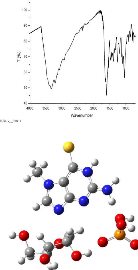
159.7, 146.3, 118.3, 88.3, 85.6, 74.0, 69.5, 60.6, 36.8. FTIR (KBr, νmax/cm-1): 3338, 1603, 1540, 1260, 1041. Elemental
Anal. Calc. for C11H16IN5O4S: C, 29.94; H, 3.65; N, 15.87. Found: C, 29.90; H, 3.55; N, 15.99.
Results and Discussion
The modiied synthesis of 1 (Scheme 2) was performed by addition of methyl iodide directly in a Fischer-Porter reactor, under an argon atmosphere, to 2-amino-6-chloro-purine ribonucleoside, and stirred sealed overnight, affording the imidazolium intermediate. Untreated methyl iodide was removed under high vacuum. Thiourea was directly added in the reactor and the reaction mixture was stirred for an additional hour. The inconvenience of using “methanolic ammonia” from the original procedure14 can
be avoided by direct dimethylamine addition dropwise until the solution became neutral. The acidity change is evident by color change (from yellow to light yellow) and was corroborated with pH indicator paper. Freshly formed compound 1 was then puriied and stored at −80 °C.
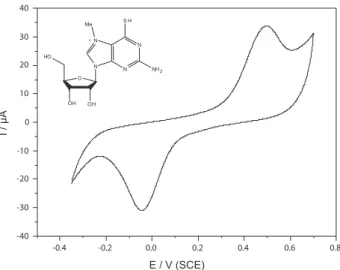
The electrochemical behaviour of 1 was investigated by cyclic voltammetry. The cyclic voltammogram (CV) of 1 is presented in Figure 1.
Non-symmetric redox couples for well deined cathodic and anodic peaks are observed at –0.044 and 496 mV (vs. SCE), respectively. As the structure suggests, the reduction of 1 is facile mainly because of the presence of an iminium-type nitrogen. The peak separation value observed during the charge transfer process (∆Ep) is 540 mV. The large difference observed in both charge transfer processes suggests a slow process at the surface of the platinum electrode. The current ratio of the anodic and cathodic peaks (Ipa/Ipc) is slightly smaller than unity (0.7), signifying that the electrochemical process is quasi-reversible.
Scheme 2. Adapted MESG synthesis.
Figure 1. Cyclic voltammogram of compound 1 (1 mmol L−1 solution)
in a 100 mmol L−1 solution of KCl (20 mL at pH 7.2) recorded at a scan
The enzymatic promoted phosphorolysis reaction of 1 catalyzed by PNP may proceed through a transition state blueprint (Scheme 3), as previously proposed for inosine, the natural enzyme substrate, and recently reviewed.20
It is possible to initiate a discussion whether the mechanism may proceed through an SN1 or SN2 mechanism for PNP substrates.21-23 A total inversion of the chiral
centre in the ribose-1-phosphate product indicates an SN2 mechanism; however, many studies conducted by Schramm’s group indicate an SN1 mechanism. Based on our experience studying reaction mechanisms using mass spectrometry24 (including enzymatic biotransformation12
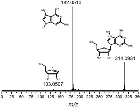
study), we decided to perform a test to gain insight into the mechanism of the phosphorolysis reaction of 1 promoted by PNP enzyme by monitoring the chemically mimicked reaction by electrospray ionization mass spectrometry (ESI). When mixing inorganic phosphate and MESG (water solution), on-line electron ionization (tandem) mass spectrometry monitoring the high resolution ESI(+)-MS was recorded after 10 min of vortex stirring (Figure 2).
We can observe in Figure 2 (A) the presence of 1 (m/z 314.0931), the free base (m/z 182), the protonated ribose-1-phosphate and also a signal at m/z 133.0507. This signal was attributed to an oxonium cation derived from ribose moiety of 1, which is formed when mixing the reagents. Owingto the very low intensity of the signal, an ESI(+)-MSMS could not be recorded, but the high resolution mass points irmly to the online interception of the species. The ESI(+)-MSMS characterization of 1 (Figure 2, B) guarantees that the species is derived from the synthesized substrate. The mild transfer to the gas phase using ESI techniques indicates that
Scheme 3. Proposed transition state blueprint for the phosphorolysis reaction of MESG 1.
in fact, the mechanism of the phosphorolysis reaction may proceed through a SN1 pathway, as suggested by others.21
According to the calculation results (supplementary information, Table S1), the basic pH tautomers are energetically more stable than the acidicpH tautomers. Taking the isolated molecules of reactants and products as reference, the calculated activation energies, regardless of the isomers or level of calculation, would be negative. Taking into account the molecules of reactants and products as being near to one another, which is more representative of the condensed phase, the effective activation energies would be approximately 100 kJ mol−1 in the RHF 3-21G level, and
larger than 65 kJ mol−1 in the highest calculation level (RHF
6-31G**), which indicate that the SN2 mechanism, under these conditions, would be highly improbable. Indeed, it can be energetically discarded. The results clearly indicate that the SN1 pathway would be preferred. In fact, in the enzymatic environment, the idea of an SN1 mechanism is fairlyplausible (Scheme 4, see supplementary information for the colour version).
One furthertest was performed using freshly synthesized compound 1. An enzymatic assay promoted by human PNP accomplished the phosphorolysis reaction (Figure 3). When we used a Mycobacterium tuberculosis PNP source, similar results were obtained.
It is clear from the data displayed in Figure 3 that freshly synthesized compound 1 has a very good eficiency to be used as a biosensor for inorganic phosphate measurements by human PNP-catalyzed phosphorylase reaction. Reproducible data can be obtained after storing compound 1 at very low temperatures (−80 °C) even after months of storage.
Conclusions
In summary, we have described an improved and simple synthesis of MESG and its complete characterization. We have also demonstrated the value of mass spectrometry in furnishing mechanistic insights into important catalytic phosphorolysis reactions such as that of MESG. Our indings, supported by calculations, point to an SN1 mechanism instead of an SN2. Moreover, an improved synthesis and full characterization of MESG has been described.
Acknowledgments
Thanks are due to FAPERGS, PETROBRAS, CAPES and CNPq for inancial support.
Supplementary Information
Supplementary material including Tables and NMR spectra is available free of charge at http://jbcs.sbq.org.br, as pdf ile.
References
1. Webb, M. R.; Proc. Natl. Acad. Sci. U. S. A. 1992, 89, 4884. 2. Cheng, J. M.; Farutin, V.; Wu, Z. J.; Jacob-Mosier, G.; Riley,
B.; Hakimi, R.; Cordes, E. H.; Bioorg. Chem.1999, 27, 307. 3. Kulikowska, E.; Bzowska, A.; Wierzchowski, J.; Shugar, D.;
Biochim. Biophys. Acta1986, 874, 355.
4. Rieger, C. E.; Lee, J.; Turnbull, J. L.; Anal. Biochem.1997, 246, 86.
Scheme 4. Calculated blueprint transition state for the phosphorolysis reaction catalyzed by PNP enzyme. Note that it is based on an SN1 mechanism.
Figure 3. Changes in absorbance at 360 nm due to the phosphorylase-catalyzed reaction of 1 (200 µmol L−1) with inorganic phosphate (from
a stock solution of KH2PO4 50 mmol L−1 ) promoted by human PNP in
5. Montgomery, J. A.; Expert Opin. Invest. Drugs1994, 3, 1303. 6. Markert, M. L.; Immunodeic. Rev.1991, 3, 45.
7. Giblett, E. R.; Ammann, A. J.; Sandman, R.; Wara, D. W.; Diamond, L. K.; Lancet1975, 1, 1010.
8. Stoop, J. W.; Zegers, B. J. M.; Hendrickx, G. F. M.; Siegenbeekvanheukelom, L. H.; Staal, G. E. J.; Debree, P. K.; Wadman, S. K.; Ballieux, R. E.; N. Engl. J. Med.1977, 296, 651.
9. Neto, B. A. D.; Lapis, A. A. M.; Mancilha, F. S.; Vasconcelos, I. B.; Thum, C.; Basso, L. A.; Santos, D. S.; Dupont, J.; Org. Lett.2007, 9, 4001.
10. Neto, B. A. D.; Lopes, A. S.; Wust, M.; Costa, V. E. U.; Ebeling, G.; Dupont, J.; Tetrahedron Lett.2005, 46, 6843.
11. Pinto, A. C.; Lapis, A. A. M.; da Silva, B. V.; Bastos, R. S.; Dupont, J.; Neto, B. A. D.; Tetrahedron Lett.2008, 49, 5639. 12. Czekster, C. M.; Lapis, A. A. M.; Souza, G.; Eberlin, M.
N.; Basso, L. A.; Santos, D. S.; Dupont, J.; Neto, B. A. D.; Tetrahedron Lett.2008, 49, 5914.
13. Pilli, R. A.; Robello, L. G.; Camilo, N. S.; Dupont, J.; Lapis, A. A. M.; Neto, B. A. D.; Tetrahedron Lett.2006, 47, 1669. 14. Broom, A. D.; Milne, G. H.; J. Heterocycl. Chem.1975, 12,
171.
15. Silva, R. G.; Pereira, J. H.; Canduri, F.; de Azevedo, W. F.; Basso, L. A.; Santos, D. S.; Arch. Biochem. Biophys.2005, 442, 49.
16. Spencer, J.; Gaffen, J.; Grifin, E.; Harper, E. A.; Linney, I. D.; McDonald, L. M.; Roberts, S. P.; Shaxted, M. E.; Adatia, T.; Bashall, A.; Bioorg. Med. Chem.2008, 16, 2974.
17. Spencer, J.; Rathnam, R. P.; Patel, H.; Anjum, N.; Tetrahedron
2008, 64, 10195.
18. Russowsky, D.; Neto, B. A. D.; Tetrahedron Lett.2004, 45, 1437.
19. Russowsky, D.; Neto, B. A. D.; Tetrahedron Lett.2003, 44, 2923.
20. Basso, L. A.; da Silva, L. H. P.; Fett-Neto, A. G.; Junior, W. F. D.; Moreira, I. D.; Palma, M. S.; Calixto, J. B.; Astoli, S.; dos Santos, R. R.; Soares, M. B. P.; Santos, D. S.; Mem. Inst. Oswaldo Cruz 2005, 100, 575.
21. Kline, P. C.; Schramm, V. L.; Biochemistry1995, 34, 1153. 22. Kline, P. C.; Schramm, V. L.; Biochemistry1993, 32, 13212. 23. Kline, P. C.; Schramm, V. L.; Biochemistry1992, 31, 5964. 24. Santos, L. S.; Neto, B. A. D.; Consorti, C. S.; Pavam, C. H.;
Almeida, W. P.; Coelho, F.; Dupont, J.; Eberlin, M. N.; J. Phys. Org. Chem.2006, 19, 731.
Received: March 4, 2009
Supplementary Information
0103 - 5053 $6.00+0.00
*e-mail: [email protected] and [email protected]
Synthesis and Enzymatic Evaluation of the Guanosine Analogue
2-Amino-6-mercapto-7-methylpurine Ribonucleoside (MESG). Insights into the Phosphorolysis Reaction
Mechanism based on the Blueprint Transition State: S
N1 or S
N2?
Brenno A. D. Neto,*,a Alexandre A. M. Lapis,b Paulo A. Netz,c John Spencer,d Silvio L. P. Dias,c Silvia M. Tamborim,c Luiz A. Basso,e Diógenes S. Santose and Jaïrton Dupont*,c
aLaboratory of Medicinal and Technological Chemistry, University of Brasilia (IQ-UnB),
72919-970 Brasilia-DF, Brazil
bUniversidade Federal do Pampa, Unipampa, 96412-420 Bagé-RS, Brazil
cLaboratory of Molecular Catalysis-Institute of Chemistry -UFRGS, Av. Bento Gonçalves, 9500,
91501-970 Porto Alegre-RS, Brazil
dSchool of Science, University of Greenwich at Medway, Chatham Maritime, ME4 4TB, UK
eCentro de Pesquisas em Biologia Molecular e Funcional, Tecnopuc, PUC-RS, Brazil
Table S1. MESG calculations energies
Structure 321G 631G**
1 MESG1 – 1387.3591 – 1394.9106
2 MESG2 – 1387.3892 – 1394.9176
3 PHOSPHATE (H2PO4–) – 637.9914 – 641.4856
4 (1+3) MESG1 + PHOSPHATE – 2025.3505 – 2036.3962
5 (2+3) MESG2 + PHOSPHATE – 2025.3806 – 2036.4032
6 REACTANTS 1 –2025.5167 –2036.5672 7 REACTANTS 2 – 2025.5711 – 2036.5594
8 TRANSITION STATE 1 – 2025.4802
9 TRANSITION STATE 2 – 2025.5258 – 2036.5340
Structure 321G 631G**
10 PRODUCTS 1 – 2025.5386 – 2036.5670
11 PRODUCTS 2 – 2025.5751 – 2036.5734 12 (14+16) FREE BASE 1 + RIB–1–P – 2025.5147 – 2036.5494
13 (15+16) FREE BASE 2 + RIB–1–P – 2025.5514 – 2036.5620
14 FREE BASE 1 (ch = 0) – 896.2612 – 901.0708
15 FREE BASE 2 (ch = 0) – 896.2979 – 901.0834 16 RIBOSE–1–PHOSPHATE – 1129.2535 – 1135.4786
17 (10–6) ∆H
R1 (P–R)
– 0.0219 – 57.50kJmol–1
+ 0.0002 0.52 kJmol–1
18 (11–7) ∆HR2 (P–R) – 0.004 – 10.50 kJmol–1
– 0.014 – 36.76 kJmol–1
19 (12–4) ∆HR1 (P–R) (isolated) – 0.1642 – 431.1 kJmol–1
– 0.1532 – 402.2 kJmol–1
20 (13–5) ∆HR2 (P–R) (isolated)
– 0.1708 – 448.4 kJmol–1
– 0.1588 – 416.9 kJmol–1
21 (8–4) EA1 – 0.1297
– 340.0kJmol–1 –
22 (9–5) EA2 – 0.1452
– 381.2kJmol–1
– 0.1308 – 343.4kJmol–1
23 (8–6) EA1’
+ 0.0365 + 95.83kJmol–1 –
24 (9–7) EA2’
+ 0.0453 +118.9kJmol–1
+ 0.0254 + 66.69kJmol–1
25 (1–2) ∆Hinterconversion, isol (R1–R2) + 0.0301 + 79.0 kJmol–1
+ 0.007 + 18.4 kJmol–1
26 (14–15) ∆Hinterconversion, isol (P1–P2) + 0.0367 + 96.4 kJmol–1
+ 0.0126 + 33.1 kJmol–1
27 FREE BASE 1 (ch = +1) – 896.6645 – 901.4640
Figure S1. High resolution ESI(+)MSMS
Figure S3. FTIR (KBr, ν
max/cm-1)