HOX
gene analysis in the osteogenic differentiation of human mesenchymal
stem cells
Song Wha Chae
1*, Bo Keun Jee
1*, Joo Yong Lee
1, Chang Whan Han
2, Yang-Whan Jeon
3, Young Lim
4,
Kweon-Haeng Lee
1,5, Hyoung Kyun Rha
1and Gue-Tae Chae
61
Neuroscience Genome Research Center, The Catholic University of Korea, Seoul, Republic of Korea.
2Department of Orthopedic Surgery, Daejeon St. Mary’s Hospital, College of Medicine,
The Catholic University of Korea, Seoul , Republic of Korea.
3
Department of Psychiatry, Our Lady of Mercy Hospital, The Catholic University of Korea, Incheon,
Republic of Korea.
4Department of Occupational and Environmental Medicine, St. Mary’s Hospital,
The Catholic University of Korea, Seoul, Republic of Korea.
5
Department of Pharmacology, The Catholic University of Korea, Seoul, Republic of Korea.
6Institute of Hansen’s Disease, The Catholic University of Korea, Seoul, Republic of Korea.
Abstract
Human bone marrow-derived mesenchymal stem cells (hMSCs) have the capacity to differentiate into osteoblasts during osteogenesis. Several studies attempted to identify osteogenesis-related genes in hMSCs. AlthoughHOX genes are known to play a pivotal role in skeletogenesis, their function in the osteogenesis of hMSCs has not yet been investigated in detail. Our aim was to characterize the expression of 37HOX genes by multiplex RT-PCR to identify the ones most probably involved in osteogenic differentiation. The results showed that the expression pat-terns of fourHOX genes were altered during this process. In particular, the expression levels of HOXC13 and HOXD13 were dramatically changed. Real-time PCR and Western blot analysis were performed in order to further analyze the expression ofHOXC13 and HOXD13. The qRT-PCR results showed that transcription of HOXC13 was up-regulated by up to forty times, whereas that ofHOXD13 was down-regulated by approximately five times after osteogenic differentiation. The Western blot results for the HOXC13 and HOXD13 proteins also corresponded well with the real-time PCR result. These findings suggest that HOXC13 and HOXD13 might be involved in the osteogenic differentiation of hMSCs.
Key words: human mesenchymal stem cells,HOXgenes, osteogenic differentiation, stem cell differentiation, gene profiling.
Received: October 26, 2007; Accepted: January 22, 2008.
Introduction
Bone marrow-derived stem cells can be divided into two major types: hematopoietic stem cells and nonhemato-poietic, or mesenchymal, stem cells. Human bone mar-row-derived mesenchymal stem cells (hMSCs) have the capacity for self-renewal and multilineage differentiation. Under the appropriate conditions, they can also give rise to mesenchymal tissues such as muscle, bone, fat, and carti-lage (Pittengeret al., 1999). Due to their ability to differen-tiate into osteoblasts, chondrocytes, adipocytes, tenocytes
and myoblasts, hMSCs hold promise for clinical applica-tions in regenerative medicine (Songet al., 2006).
Because osteoblastic cells play a major role in the processes of normal bone growth, remodeling and fracture repair, many researchers have used the process of osteo-genesis to study the differentiation and characteristics of stem cells (Kraus and Kirker-Head, 2006). To obtain osteo-blastic cells, MSCs are incubated with a mixture medium containing dexamethasone,β-glycerophosphate and ascor-bic acid for a period of 2~3 weeks (Bobiset al., 2006).
HOXgenes were initially identified by their homo-logy with the Drosophila HOM genes (Levineet al., 1984; Acamporaet al., 1989; Duboule and Dolle, 1989). These genes encode homeodomain transcription factors related to anterior-posterior axis patterning that takes place during embryonic development (van den Akkeret al., 2001). The
www.sbg.org.br
Send correspondence to Gue-Tae Chae and Kweon-Haeng Lee. Institute of Hansen’s Disease, The Catholic University of Korea, 505, Banpo-dong, Seocho-ku, Seoul, Republic of Korea. E-mail: [email protected].
*These authores contributed equally to this work.
homeodomain contains a 180-bapair homeobox se-quence that encodes a conserved 60 amino acid region and acts as a DNA-binding domain via a helix-turn-helix motif (Gehringet al., 1994). In vertebrates, 39HOXgenes have been identified. These are distributed over four homolo-gous HOX clusters termed HOXA, B, C, and D. These loci are located on four different chromosomal locations and are comprised of nine to eleven genes (Akin and Nazarali, 2005). It is well known that HOX proteins participate in many common developmental processes during normal embryogenesis. Several reports have indicated that HOX
genes play a regulatory role in skeletogenesis (Goff and Tabin, 1997; Kanzler et al., 1998; van den Akker et al., 2001; Remacleet al., 2004).
AlthoughHOXgenes are known to play an essential role in skeletal development and bone formation, there is no report regarding the screening of HOX groups that are in-volved in the osteogenesis of hMSCs. Thus, in the present study, the expression profile ofHOXgenes during osteo-genic differentiation of hMSCs was investigated by multi-plex PCR and the results showed significant changes in the expression of four of them during this process. Of these four genes, the expression of HOXC13 and HOXD13
showed the most dramatic changes. Therefore, the expres-sion levels of HOXC13 and HOXD13 were evaluated by qRT-PCR and Western blot analysis, and the results were similar to the multiplex PCR result. This suggests that the other two genes (HOXA1andHOXC11) are also involved in osteogenesis.
Materials and Methods
Research protocol
The research protocol was reviewed and approved by the human ethical care committee at St. Mary’s Hospital, Catholic University in Daejeon, Republic of Korea. The hMSCs were isolated from the bone marrow of six individ-uals, as described below (Choiet al., 2006). All experi-ments were performed with hMSCs obtained after the third cell passage.
Flow cytometric analysis (FACS) of hMSCs
hMSCs were analyzed by FACS-Calibur (Becton Dickinson, San Jose, CA) as previously described (Choiet al., 2006). FACS analysis was performed using fluorescein isothiocyanate (FITC)-conjugated anti-CD11b, CD29, CD34, CD45, CD73 and CD105 antibodies (BD Bioscience, San Diego, CA) to confirm that the phenotype of the hMSCs was maintained after expansion in the culture. The samples were incubated with antibodies against each surface marker for 30 min, and this treatment was followed by FACS.
Osteogenic differentiation
To induce osteogenic differentiation, hMSCs at the third passage were plated with Dulbecco’s modified
Ea-gle’s medium (DMEM) containing 10% fetal bovine serum (FBS) in a 250-ml tissue culture flask (Nunc, Roskilde, Denmark). The cells were then incubated at 37 °C in 5% CO2for 24 h. The medium was replaced with high-glucose DMEM containing 10% FBS, 0.1 µM dexamethasone, 10 mM β-glycerophosphate, and 0.3 mM ascorbic acid (Sigma, St. Louis, MO) for osteogenic differentiation. This osteogenic medium was replaced every 2 days for 21 days.
Alkaline phosphatase (ALP) staining
About 3 x 10⁵cells were seeded onto each well of a 6-well plate. After incubation for 12 h at 37 °C in 5% CO2, the medium was replaced with osteogenic differentiation medium, replaced again every 2 days for periods of 10 and 21 days. The 10-day and 21-day differentiated and undif-ferentiated hMSCs were washed twice with ice-cold PBS (phosphate buffered saline), fixed with 2% paraformal-dehyde/ 0.1 M sodium cacodylate for 10 min, and washed with 0.1 M cacodylic acid. The cells were incubated with ALP substrate solution (5 mg naphthol AS-TR phosphate in 25 mL water plus 10 mg Fast red TR in 24 mL of 0.1 M Tris buffer, pH 9.5) for 1 h at room temperature. Cells were photographed using a Nikon TE-300 (Tokyo, Japan) in-verted light microscope.
von Kossa staining
Approximately 3 x 10⁵cells were seeded onto each well of a 6-well plate. After incubation for 12 h at 37 °C in 5% CO2, the medium was replaced with osteogenic differ-entiation medium and thereafter replaced every 2 days for periods of 10 and 21 days. Day-10 and day-21 differenti-ated and undifferentidifferenti-ated hMSCs were washed with dis-tilled water, fixed with 4% formalin, and then treated with 5% silver nitrate. Then the cells were exposed to UV light for 1 h, 5% thiosulfate was added, and the cells were placed at room temperature after a washing step with distilled wa-ter. The samples were photographed with a Nikon TE-300 (Tokyo, Japan) inverted light microscope.
RNA isolation and reverse transcriptase-polymerase chain reaction (RT-PCR)
RT-PCR analysis was performed as described by Jee
products were run on a 1% agarose gel and then analyzed under UV light after staining with ethidium bromide. The gel was photographed and then quantitatively measured by scanning densitometry. The experiments were performed with three different RNA samples.
Immunoblotting analysis
Immunoblotting analysis was performed as previ-ously described (Jeeet al., 2007). After incubation with osteogenic differentiation medium for 21 days, the cells were lysed on ice for 30 min in RIPA buffer (50 mM Tris-HCl pH 7.5, 0.15 M NaCl, 1% NP-40, 0.1% SDS, 1 mM DTT and 1 mM PMSF) containing a mixture of pro-tease inhibitors (Roche, Mannheim, Germany). The insolu-ble materials were separated using 10% polyacrylamide gels containing 10% sodium dodecyl sulfate (SDS), 1.5 M Tris-HCl, 0.035% N, N, N’, N’-tetra-methylenediamine and 7 mg ammonium persulfate. The separated proteins were transferred to a nitrocellulose membrane (Whatman, Dassel, Germany) at 36 mA in a transfer buffer that con-tained 39 mM glycine, 48 mM Tris base, 0.037% SDS, and 20% methanol. The membranes were sequentially incu-bated with anti-OPN (osteopontin), OCN (osteocalcin), HOXC13 monoclonal antibodies (mAb; Abnova, Taipei, Taiwan), and HOXD13 mAb (Santa Cruz Biotechnology, Santa Cruz, CA) at 1:1000 dilutions. A horseradish peroxi-dase-conjugated anti-rabbit IgG was used as a secondary antibody at a dilution of 1:1500 (Pierce Biotechnology, Rockford, IL). Detection was performed with an electro-chemiluminescence detection reagent (Amersham Biosci-ences, Uppsala, Sweden). In some cases, the Western blots were stripped and re-blotted with antibody, according to the manufacturer’s instructions.
Multiplex PCR ofHOXgenes
Multiplex PCR was performed using the GeneXP Hu-man HOX Assay Kit (Seegene, Seoul, Republic of Korea). After the induction of osteogenic differentiation for 21 days, total RNA was isolated from the cells using an
RNeasy Mini Kit (QIAGEN, Valencia, CA). Two micro-grams of total RNA were reverse-transcribed in order to synthesize cDNA using an AccuPower RTPReMix kit (Bioneer, Inc., Rockville, MD). The synthesized cDNAs were used as templates for multiplex PCR,according to the manufacturer’s instructions (http://www.seegene.com). PCR was carried out under the following conditions: 1 cy-cle at 94 °C for 15 min, followed by 40 cycy-cles of 94 °C for 0.5 min, 63 °C for 1.5 min, and 72 °C for 1.5 min, and a fi-nal cycle at 72 °C for 10 min.GAPDHwas used as an inter-nal control. Electrophoresis was carried out on a 2% aga-rose gel. The multiplex PCR products were analyzed with the Alpha EaseFC software (Alpha Innotech, San Leandro, CA). The experiment was performed six times on each indi-vidual (Table 2).
Real-time quantitative PCR analysis (qPCR)
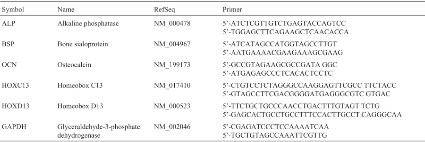
After the induction osteogenic differentiation for 21 days, total RNA was isolated using an RNeasy Mini Kit (QIAGEN, Valencia, CA). Two micrograms of total RNA were reverse-transcribed in order to synthesize cDNA, us-ing an AccuPower RTPReMix kit (Bioneer, Inc., Rock-ville, MD). For relative quantification, the reactions were performed in a total volume of 20µL, containing 15µL of LightCycler®FastStart DNA Master SYBR Green 1 (Ro-che Diagnostics, Mannheim, Germany), 10 ng of cDNA, and 10 pmol of each primer. Real-time quantitative PCR was carried out with specific primers, in a LightCycler In-strument (Roche Diagnostics, Mannheim, Germany). The samples were analyzed in triplicate. The primer sequences used are listed in Table 1.GAPDHwas used as an internal control. For quantification, the data were analyzed using the LightCycler analysis software (Roche Diagnostics, Mannheim, Germany). Relative quantification of target gene expression was evaluated using the comparative CT method (Wanget al., 2004). TheΔCTvalue was determined by subtracting the target CTof each sample from its respec-tiveGAPDHCTvalue. Calculation ofΔCTinvolves using the meanΔCTvalue of the control gene as an arbitrary
con-Table 1- Primer sequences.
Symbol Name RefSeq Primer
ALP Alkaline phosphatase NM_000478 5’-ATCTCGTTGTCTGAGTACCAGTCC 5’-TGGAGCTTCAGAAGCTCAACACCA
BSP Bone sialoprotein NM_004967 5’-ATCATAGCCATGGTAGCCTTGT 5’-AATGAAAACGAAGAAAGCGAAG
OCN Osteocalcin NM_199173 5’-GCCGTAGAAGCGCCGATA GGC 5’-ATGAGAGCCCTCACACTCCTC
HOXC13 Homeobox C13 NM_017410 5’-CTGTCCTCTAGGGCCAAGGAGTTCGCC TTCTACC 5’-GTAGCCTTCGACGGGGATGAGGGCGTC GTGAC
HOXD13 Homeobox D13 NM_000523 5’-TTCTGCTGCCCAACCTGACTTTGTAGT TCTG 5’-GAGCACTGCCTGCCTTTCCACTTGCCT CAGGGCAA
GAPDH Glyceraldehyde-3-phosphate dehydrogenase
stant to subtract from all otherΔCT mean values. Fold-changes in gene expression of the target gene were equiva-lent to 2-ΔΔCT. The values obtained were then entered into a Student’sttest.Pvalues less than 0.05 were considered significant.
Statistical analysis
To investigate differentially expressed HOX genes during osteogenic differentiation from hMSCs, the data ob-tained from multiplex PCR were examined by variance analysis (ANOVA) with SPSS 12.0 software for Windows (SPSS, Chicago, IL). Tukey’s HSD test was used for post hoc comparisons. For all statistical tests, an error probabil-ity of p < 0.05 was regarded as significant.
Results
Characterization of hMSCs
In an effort to explore the characterization of hMSCs, flow cytometry was used to examine the expression of the surface antigens CD11b, CD29, CD34, CD45, CD73, and CD105 in the isolated hMSCs. The isolated hMSCs were submitted to FACS analysis and found to be positive for CD29 (68 ± 2.5%), CD73 (96.9 ± 2.7%) and CD105 (91.5±2.5%), and negative for CD11b, CD34 and CD45. These results show that the hMSCs were successfully
iso-lated and that the culture-expanded hMSCs maintained their phenotype (Figure 1).
Osteogenic differentiation
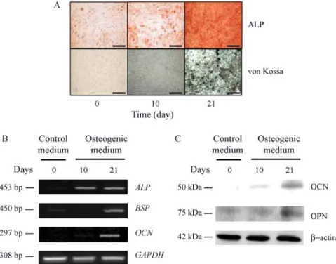
ALP and von Kossa staining were used to examine the differentiation of hMSCs into osteoblasts in the osteo-genic medium. Although ALP staining at day 10 showed a weak color signal, the intensity of ALP activity increased remarkably by day 21. The intensity of von Kossa staining also peaked at day 21 (Figure 2A). RT-PCR was performed using osteogenic markers to confirm hMSC osteogenesis (Table 1). The mRNA expression levels of the osteogenic markers, which included bone sialoprotein (BSP), OCN and ALP, were significantly higher at day 21 than at day 0 (Figure 2B). Immunoblot analysis was performed using OCN and OPN in order to obtain further confirmation of osteogenesis. The results of von Kossa staining and RT-PCR were identical to the result observed with ALP and showed that the expression of the OCN and OPN proteins increased as differentiation progressed (Figure 2C). All of the corresponding results confirmed that the hMSCs were successfully differentiated into osteoblasts.
Analysis ofHOXgene expression using multiplex PCR
Multiplex PCR was used to assess the expression levels ofHOXgenes during osteogenic differentiation. The
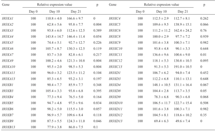
expres-Table 2-HOXmultiplex PCR. A total of thirty-sevenHOXgenes were examined using multiplex RT-PCR. The relative expression values are expressed as mean ± SEM.Pvalues were calculated using ANOVA.
Gene Relative expresion value p Gene Relative expresion value p
Day 0 Day 10 Day 21 Day 0 Day 10 Day 21
HOXA1 100 110.8 ± 4.0 166.6 ± 9.7 0 HOXC4 100 112.5 ± 2.9 112.7 ± 8.1 0.262
HOXA2 100 62.8 ± 5.6 95.8 ± 7.7 0.004 HOXC5 100 109.6 ± 9.5 138.9 ± 15.1 0.066
HOXA3 100 93.8 ± 6.0 112.6 ± 12.5 0.389 HOXC6 100 111.2 ± 11.2 162.4 ± 24.2 0.76
HOXA4 100 143.8 ± 14.7 146.4 ± 11.4 0.054 HOXC8 100 100.0 ± 2.9 97.7 ± 7.2 0.939
HOXA5 100 74.4 ± 5.1 92.7 ± 12.7 0.226 HOXC9 100 101.6 ± 3.8 100.3 ± 7.1 0.982
HOXA6 100 105.7 ± 8.7 130.3 ± 12.5 0.119 HOXC10 100 93.8 ± 4.8 90.1 ± 3.3 0.644
HOXA7 100 83.7 ± 3.0 82.8 ± 6.1 0.217 HOXC11 100 136.6 ± 9.6 100.6 ± 9.0 0.01
HOXA9 100 100.2 ± 4.6 121.3 ± 10.8 0.804 HOXC12 100 118.1 ± 5.3 130.8 ± 10.5 0.095
HOXA10 100 95.5 ± 2.0 98.5 ± 5.3 0.804 HOXC13 100 91.3 ± 5.3 191.0 ± 10.5 0
HOXA11 100 96.0 ± 3.2 123.5 ± 11.2 0.104 HOXD1 100 106.7 ± 6.2 94.0 ± 7.4 0.452
HOXA13 100 85.3 ± 6.5 93.2 ± 3.1 0.197 HOXD3 100 112.2 ± 6.8 110.1 ± 13.1 0.648
HOXB1 100 90.4 ± 7.7 85.9 ± 7.7 0.348 HOXD4 100 148.1 ± 10.5 131.1 ± 16.4 0.067
HOXB3 100 105.4 ± 3.3 93.8 ± 6.8 0.395 HOXD8 100 104.4 ± 2.8 113.7 ± 3.5 0.05
HOXB4 100 77.3 ± 9.4 76.5 ± 5.0 0.164 HOXD9 100 78.3 ± 6.8 90.3 ± 6.8 0.068
HOXB5 100 94.7 ± 4.8 97.5 ± 9.6 0.834 HOXD10 100 106.5 ± 11.7 122.7 ± 15.4 0.508
HOXB6 100 98.2 ± 5.0 115.5 ± 3.0 0.057 HOXD11 100 101.6 ± 3.8 100.3 ± 7.1 0.982
HOXB7 100 96.9 ± 5.7 109.6 ± 8.4 0.118 HOXD12 100 104.5 ± 8.1 118.6 ± 10.2 0.35
HOXB9 100 87.5 ± 5.5 124.3 ± 11.0 0.046 HOXD13 100 69.4 ± 6.3 49.6 ± 7.4 0
sion patterns of the 37HOXgenes were screened at day 0, day 10, and day 21 in both undifferentiated and differentiated hMSCs. The expression of the 37HOXgenes at the level of transcription is listed in Table 2. The HOXA1, HOXA4, HOXA6, HOXA9, HOXA11, HOXB9, HOXC5, HOXC6,
HOXC12, HOXC13, HOXD4, and HOXD10 genes were up-regulated under the osteogenesis-induced condition. On the other hand,HOXB4andHOXD13were down-regulated during osteogenesis. The otherHOXgenes showed no signifi-cant changes in their mRNA expression levels.
Figure 1- Phenotypic characterization of hMSCs using flow cytometric analysis. FACS analysis showed that the cells were negative for CD11b, CD34 and CD45 expression and positive for CD29, CD73 and CD105, which are phenotypes currently known to be characteristic of hMSCs. The gray line indi-cates the control of the CD marker isotypes.
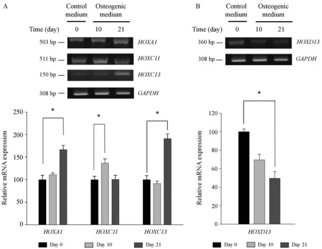
Statistical analysis revealed that four HOX genes showed significant differences in expression at the tran-scription level. TheHOXA1,HOXC11andHOXC13genes were found to be up-regulated. The expression ofHOXC13
was unaltered between day 0 and day 10 and only increased after day 10. The expression ofHOXA1gradually increased for 21 days, but the increase in the expression ofHOXC13
was more dramatic. The mRNA level ofHOXC11 fluctu-ated during osteogenesis. The expression of HOXC11
increased during the first 10 days of osteogenic differentia-tion, but then decreased over the next 11 days (Figure 3A). The expression ofHOXD13was down-regulated during the osteogenesis of hMSCs. The mRNA level ofHOXD13 de-creased gradually over the 21-day period (Figure 3B).
Expression ofHOXC13andHOXD13
The expression ofHOXC13andHOXD13showed the most dramatic change after 21 days of differentiation. The expression ofHOXC13increased by approximately 91%, whereas that ofHOXD13decreased by 50% after osteo-genesis. Real-time quantitative PCR and immunoblotting
analysis were carried out in order to further confirm the in-creased expression ofHOXC13andHOXD13. The results of qPCR showed that the expression ofHOXC13was five times higher at day 10 and forty-two times higher at day 21 than in the undifferentiated state, respectively, whereas the mRNA expression of HOXD13 showed a five-fold de-crease at day 10 (Figure 4). These qPCR results of theHOX
genes were in agreement with those of multiplex PCR. The expression levels of these twoHOXgenes were then submitted to immunoblot analysis to further evaluate their protein level in the osteogenic differentiation of hMSCs. The results showed increased expression of the HOXC13 protein and decreased expression of the HOXD13 protein after 21 days of differentiation (Figure 5). This result was in agreement with those of multiplex and real-time PCR.
Discussion
Many factors are known to regulate osteogenesis (Bo-biset al., 2006). The important factors involved in osteoge-nic regulation include bone morphogenetic protein (BMP),
Figure 3-HOXmultiplex PCR. (A) The mRNA expression ofHOXA1,HOXC11andHOXC13increased during osteogenesis. TheHOXC13gene showed the most significant up-regulation. The expression ofHOXA1gradually increased during osteogenic differentiation. The expression ofHOXC11
transforming growth factor (TGF), insulin-like growth fac-tor (IGF), brain-derived growth facfac-tor (BDGF), fibroblast growth factor (FGF), leptin and parathyroid hormone-rela-ted peptide (PTHrP). These proteins regulate the expres-sion of signals needed for bone remodeling. In addition, many reports have suggested that various transcription fac-tors participate in osteogenesis. Among them, Cbfa1/
Runx2, Osterix,ΔFosB, Fra-1, Aj18, Osf1, Msx2, Dlx5 and TWIST have been shown to play pivotal roles.
Several studies have also reported thatHOXgenes are involved in osteogenesis. These reports showed that HOXA2 plays several important roles in the process of skeletogenesis (Gendron-Maguireet al., 1993; Rijliet al., 1993; Kanzleret al., 1998). Another study found, using quantitative RT-PCR (Dobrevaet al., 2006), that the ex-pression of HOXA2 was up-regulated during osteogenesis. HOXA10 has been shown to contribute to osteogenic lin-eage determination (Hassanet al., 2007). HOXC8 was re-ported to be involved in the regulation of osteogenesis through bone morphogenic protein (BMP) pathways (Juan
et al., 2006). However, no significant changes in the ex-pression ofHOXA2,HOXA10andHOXC8were observed in the present study. The differences in these results may be due to the fact that HOXA2 may have been induced during mouse embryogenesis, and HOXA10 and HOXC8 expres-sion were likely induced by BMP. However, in the present study, mesenchymal stem cells were used, and osteogenic differentiation was inducedin vitrousing dexamethasone, β-glycerophosphate and ascorbic acid.
Knowledge regarding the expression patterns of the
HOXgenes during osteogenic differentiation may reveal the signal pathway of osteogenesis and may also help in the potential therapeutic application of hMSCs. However, a re-port regarding the expression profile ofHOXgenes during osteogenesis has not yet been published. In the present re-port, 37HOXgenes were investigated in order to determine their expression patterns during the osteogenesis of hMSCs. For this purpose, we performed multiplex PCR, real-time PCR and Western blot analysis. Based on the re-sults, we suggest that fourHOXgenes,HOXA1,HOXC13,
HOXC11andHOXD13, might be involved in the osteo-genic differentiation of hMSCs.HOXA1 is a key gene in skull development, and it is a retinoic acid (RA) direct tar-get gene (Ijichi and Ijichi, 2002). Mice with mutations in the HOXA1 hexapeptide motif show skeletal defects (Re-macleet al., 2004). Similar results were reported by Marti-nez-Ceballoset al.(2005), who showed that the disruption of theHOXA1gene results in abnormal ossification of the skull. Andrewset al.(1994) reported that osteogenic pro-tein-1 (OP-1), a member of the TGF-β superfamily, induces HOXA1. In addition, recent microarray analyses revealed that BSP and Col1a1, both key markers of osteo-genesis, are the target molecules of HOXA1 (Martinez-Ceballos et al., 2005). The results of multiplex PCR showed that HOXA1 was significantly increased during osteogenesis. The results of the present study and those of previous reports suggest that HOXA1 is an important factor involved in the osteogenesis of hMSCs.
In the present study, the expression of HOXC13
showed the largest increase. However, there are no previ-ous reports suggesting a relationship betweenHOXC13and osteogenesis. Kulessaet al.(2000) reported that the
over-Figure 4- Real-time PCR analysis ofHOXC13andHOXD1. The data were presented as fold changes relative to day 0. The mRNA expression of
HOXC13was five times higher on day 10 and 42 times higher on day 21 compared to the expression in a control. The expression ofHOXD13 de-creased rapidly at day 10 and slowly inde-creased at day 21. The real-time PCR data were normalized withGAPDHexpression. Asterisk (*) indi-cates a significant increase between two samples (p < 0.05).
expression of the BMP inhibitor resulted in the down-regulation ofHOXC13expression in mutant mice. Based on the findings of the present study, it seems likely that
HOXC13contributes to the osteogenesis of hMSCs via the BMP pathway.
The HOXC11 gene encodes a transcription factor known to be involved in the definition of segment identities along the anterio-posterior axis. The expression of
HOXC11is detected in the mesenchyme posterior to the re-gion forming the femur and fibula (Hostikka and Capecchi, 1998). There is a report suggesting that HOXC11 is in-volved in chondrogenesis, which is regulated by BMP2 and BMP7 (Papenbrocket al., 2000). However, there is no clear evidence thatHOXC11 contributes to osteogenesis, thus
HOXC11may be related to the osteogenesis of hMSCs. In particular, HOXC11 may only be involved in the early stages of the osteogenic process from the hMSCs stage to the osteoblast progenitor cell stage, and not from the osteo-blast progenitor cell stage to the osteoosteo-blast stage, once the expression level drops after day 10 (Figure 3A).
Williamset al.(2005) recently demonstrated that the interaction between the mouse HOXD13 protein and Smad1 might reciprocally antagonize the expression of Runx2, which is a key molecule in mammalian osteo-genesis (Williamset al., 2005). This implies that the ex-pression of HOXD13 may decrease as osteogenesis progresses, which is in agreement with the results of the present study. In light of previous reports onHOXD13and of the present results, it is likely that the decrease in HOXD13 expression during osteogenesis is required for the promotion of osteogenic differentiation (Shi et al., 1999; Yanget al., 2000; Liuet al., 2004; Williamset al., 2005; Liet al., 2006).
There are few studies regarding theHOXgenes in-volved in the differentiation of hMSCs. The results of the present study show that the mRNA expression levels of fourHOXgenes noticeably changed during the osteogenic differentiation of hMSCs. Although the roles of the four genes in the osteogenic differentiation of hMSCs have yet to be clarified, the present study represents a first step eluci-dating the relationship betweenHOXgene expression and the differentiation of hMSCs, making part of the signalling pathway in osteogenic differentiation from hMSCs. Func-tional studies, such as a gene siRNA-mediatd gene silenc-ing or gene transfection, are needed in order to further investigate the role of theHOXgenes in osteogenic differ-entiation.
Acknowledgments
This research was supported by a grant from the Se-oul Research and Business Development Program (10548) funded by the Seoul Metropolitan Government, Republic of Korea.
References
Acampora D, D’Esposito M, Faiella A, Pannese M, Migliaccio E, Morelli F, Stornaiuolo A, Nigro V, Simeone A and Bon-cinelli E (1989) The human HOX gene family. Nucleic Acids Res 17:10385-10402.
Akin ZN and Nazarali AJ (2005) Hox genes and their candidate downstream targets in the developing central nervous sys-tem. Cell Mol Neurobiol 25:697-741.
Andrews PW, Damjanov I, Berends J, Kumpf S, Zappavigna V, Mavilio F and Sampath K (1994) Inhibition of proliferation and induction of differentiation of pluripotent human em-bryonal carcinoma cells by osteogenic protein-1 (or bone morphogenetic protein-7). Lab Invest 71:243-251.
Bobis S, Jarocha D and Majka M (2006) Mesenchymal stem cells: Characteristics and clinical applications. Folia Histochem Cytobiol 44:215-230.
Choi CB, Cho YK, Prakash KV, Jee BK, Han CW, Paik YK, Kim HY, Lee KH, Chung N and Rha HK (2006) Analysis of neu-ron-like differentiation of human bone marrow mesenchy-mal stem cells. Biochem Biophys Res Commun 350:138-146.
Dobreva G, Chahrour M, Dautzenberg M, Chirivella L, Kanzler B, Farinas I, Karsenty G and Grosschedl R (2006) SATB2 is a multifunctional determinant of craniofacial patterning and osteoblast differentiation. Cell 125:971-986.
Duboule D and Dolle P (1989) The structural and functional orga-nization of the murine HOX gene family resembles that of Drosophila homeotic genes. EMBO J 8:1497-1505. Gehring WJ, Qian YQ, Billeter M, Furukubo-Tokunaga K, Schier
AF, Resendez-Perez D, Affolter M, Otting G and Wuthrich K (1994) Homeodomain-DNA recognition. Cell 78:211-223.
Gendron-Maguire M, Mallo M, Zhang M and Gridley T (1993) Hoxa-2 mutant mice exhibit homeotic transformation of skeletal elements derived from cranial neural crest. Cell 75:1317-1331.
Goff DJ and Tabin CJ (1997) Analysis of Hoxd-13 and Hoxd-11 misexpression in chick limb buds reveals that Hox genes af-fect both bone condensation and growth. Development 124:627-636.
Hassan MQ, Tare R, Lee SH, Mandeville M, Weiner B, Monte-cino M, van Wijnen AJ, Stein JL, Stein GS and Lian JB (2007) HOXA10 controls osteoblastogenesis by directly ac-tivating bone regulatory and phenotypic genes. Mol Cell Biol 27:3337-3352.
Hostikka SL and Capecchi MR (1998) The mouse Hoxc11 gene: Genomic structure and expression pattern. Mech Dev 70:133-145.
Ijichi S and Ijichi N (2002) Minor form of trigonocephaly is an au-tistic skull shape? A suggestion based on homeobox gene variants and MECP2 mutations. Med Hypotheses 58:337-339.
Jee BK, Lee JY, Lim Y, Lee KH and Jo YH (2007) Effect of KAI1/CD82 on the beta1 integrin maturation in highly mi-gratory carcinoma cells. Biochem Biophys Res Commun 359:703-708.
Juan AH, Lei H, Bhargava P, Lebrun M and Ruddle FH (2006) Multiple roles of hoxc8 in skeletal development. Ann N Y Acad Sci 1068:87-94.
Kanzler B, Kuschert SJ, Liu YH and Mallo M (1998) Hoxa-2 re-stricts the chondrogenic domain and inhibits bone formation during development of the branchial area. Development 125:2587-2597.
Kraus KH and Kirker-Head C (2006) Mesenchymal stem cells and bone regeneration. Vet Surg 35:232-242.
Kulessa H, Turk G and Hogan BL (2000) Inhibition of Bmp sig-naling affects growth and differentiation in the anagen hair follicle. EMBO J 19:6664-6674.
Levine M, Rubin GM and Tjian R (1984) Human DNA sequences homologous to a protein coding region conserved between homeotic genes of Drosophila. Cell 38:667-673.
Li X, Nie S, Chang C, Qiu T and Cao X (2006) Smads oppose Hox transcriptional activities. Exp Cell Res 312:854-864. Liu Z, Shi W, Ji X, Sun C, Jee WS, Wu Y, Mao Z, Nagy TR, Li Q
and Cao X (2004) Molecules mimicking Smad1 interacting with Hox stimulate bone formation. J Biol Chem 279:11313-11319.
Martinez-Ceballos E, Chambon P and Gudas LJ (2005) Differ-ences in gene expression between wild type and Hoxa1 knockout embryonic stem cells after retinoic acid treatment or leukemia inhibitory factor (LIF) removal. J Biol Chem 280:16484-16498.
Papenbrock T, Visconti RP and Awgulewitsch A (2000) Loss of fibula in mice overexpressing Hoxc11. Mech Dev 92:113-123.
Pittenger MF, Mackay AM, Beck SC, Jaiswal RK, Douglas R, Mosca JD, Moorman MA, Simonetti DW, Craig S and Marshak DR (1999) Multilineage potential of adult human mesenchymal stem cells. Science 284:143-147.
Remacle S, Abbas L, De Backer O, Pacico N, Gavalas A, Gofflot F, Picard JJ and Rezsohazy R (2004) Loss of function but no
gain of function caused by amino acid substitutions in the hexapeptide of Hoxa1in vivo. Mol Cell Biol 24:8567-8575. Rijli FM, Mark M, Lakkaraju S, Dierich A, Dolle P and Chambon
P (1993) A homeotic transformation is generated in the rostral branchial region of the head by disruption of Hoxa-2, which acts as a selector gene. Cell 75:1333-1349.
Shi X, Yang X, Chen D, Chang Z and Cao X (1999) Smad1 inter-acts with homeobox DNA-binding proteins in bone morpho-genetic protein signaling. J Biol Chem 274:13711-13717. Song L, Webb NE, Song Y and Tuan RS (2006) Identification and
functional analysis of candidate genes regulating mesen-chymal stem cell self-renewal and multipotency. Stem Cells 24:1707-1718.
van den Akker E, Fromental-Ramain C, de Graaff W, Le Mouellic H, Brulet P, Chambon P and Deschamps J (2001) Axial skel-etal patterning in mice lacking all paralogous group 8 Hox genes. Development 128:1911-1921.
Wang G, Brennan C, Rook M, Wolfe JL, Leo C, Chin L, Pan H, Liu WH, Price B and Makrigiorgos GM (2004) Balanced-PCR amplification allows unbiased identification of geno-mic copy changes in minute cell and tissue samples. Nucleic Acids Res 32:e76.
Williams TM, Williams ME, Heaton JH, Gelehrter TD and Innis JW (2005) Group 13 HOX proteins interact with the MH2 domain of R-Smads and modulate Smad transcriptional acti-vation functions independent of HOX DNA-binding capa-bility. Nucleic Acids Res 33:4475-4484.
Yang X, Ji X, Shi X and Cao X (2000) Smad1 domains interacting with Hoxc-8 induce osteoblast differentiation. J Biol Chem 275:1065-1072.
Assistant Editor: Klaus Hartfelder