SUBTYPING OF CHILEAN METHICILLIN-RESISTANT STAPHYLOCOCCUS AUREUS STRAINS CARRYING
THE STAPHYLOCOCCAL CASSETTE CHROMOSOME MEC TYPE I
Gustavo Medina¹, Carola Otth¹, Laura Otth¹, Heriberto Fernández¹, Celeste Muñoz¹, María Cruz², Ángela Zaror², Ruby Henriquez², Maria Arce², Myra Wilson¹*
¹
Instituto de Microbiología Clínica, Facultad de Medicina, Universidad Austral de Chile, Valdivia, Chile; ²Sección Microbiología, Laboratorio Central. Hospital Clínico Regional Valdivia.
Submitted: January 17, 2011; Approved: June 07, 2012.
ABSTRACT
The cassette chromosome mec (SCCmec) present in methicillin-resistant Staphylococcus aureus (MRSA) has two essential components, the ccr gene complex and the mec gene complex. Additionally, SCCmec has non-essential components called J regions which are used for MRSA subtyping. This study was performed to determine subtypes MRSA strains carrying SCCmec type I based on polymorphism of regions located downstream of the mecA gene. A total of 98 MRSA strains carrying SCCmec type I isolated from patients hospitalized at the County Hospital of Valdivia (Chile) between May 2007 and May 2008, were analyzed by multiplex PCR designed to amplify the mecA gene and 7 DNA hypervariable regions located around the mecA gene. MRSA strains were classified into seventeen genotypes accordingly to amplification patterns of DNA hypervariable regions. Five genotypes showed amplification patterns previously described. The remaining twelve genotypes showed new amplification patterns. Genotypes 18 and Genotype 19 were the most frequently detected. Regions HVR, Ins117 and pI258 stand out as being present in more than 60% of tested isolates. The acquisition of hypervariable regions by MRSA is a continuous horizontal transfer process through which the SCCmec have been preserved intact, or even may give rise to new types and subtypes of SCCmec. Therefore it is possible to infer that most MRSA strains isolated at the County Hospital of Valdivia (Chile) were originated from two local clones which correspond to Genotype 18 and Genotype 19.
Key words: Subtypified MRSA, polymorphism MRSA, SCCmec MRSA.
INTRODUCTION
Methicillin-resistant Staphylococcus aureus (MRSA) was first isolated in England in 1961 shortly after the development
of methicillin (8). Since then, MRSA has become the most prevalent pathogen causing hospital infection throughout the world, with increased incidence in many countries (2).
MRSA genome has integrated a mobile genetic
element called staphylococcal cassette chromosome mec
(SCCmec), which harbors the mecA gene responsible for
methicillin resistance. This gene encodes PBP2a, an additional
ß-lactam-resistant penicillin-binding protein (4). SCCmec is a
unique genomic island found only in staphylococcal species
that have two essential components, the ccr gene complex (ccr)
and the mec gene complex (mec) (2, 5). The ccr gene
complex is composed of ccr genes and surrounding open
reading frames (ORFs). The mec gene complex is composed of
the mecA gene, regulatory genes, and insertion sequences
upstream or downstream of mecA gene (6, 7).
Remaining parts of SCCmec are called J regions (J1, J2 and
J3), which constitute nonessential components of SCCmec. In
some cases, these regions carry additional antibiotic resistance
determinants (5). J1 is the region between the chromosomal left
junction and the ccr complex; J2 is the region between the ccr
complex and the mec complex and J3 is the region between the
mec complex and the chromosomal right junction. Variations in
the J regions are used for subtyping MRSA strains (9).
Currently, different genetic methods have been developed to
be applied in molecular epidemiologic characterization of MRSA
strains, being pulsed-field electrophoresis (PFGE) the technique
of choice (14). On the other hand, through multiplex PCR
technique it is possible to analyze the polymorphic downstream
of mecA gene. This genetic polymorphism has been used as
an epidemiological marker and has also been the basis of
studies related to the evolutionary origin and subtyping of
methicillin resistance in S. aureus (3).
The aim of this study was to determine subtypes of MRSA
strains carrying SCCmec type I, through the implementation of
a multiplex PCR that allows the detection of mecA gene and
7 DNA hypervariable regions located around the mecA gene.
MATERIALS AND METHODS
Clinical isolates
Ninety eight clinical isolates of MRSA previously typified as SCCmec type I and unrelated to nosocomial outbreaks were studied. All of them were isolated from patients hospitalized at the County Hospital of Valdivia (Chile)
between May 2007 and May 2008. Strains phenotyping was performed using the semi-automated microbiological diagnosis system Dried Gram Positive ID Type 2 panels
(Microscan®) and SCCmec genotyping was performed as described previously (17). The mecA-positive S. aureus ATCC 49476, which contains HVR, pT181, pI258, mecR1 and IS256 regions was used as control.
DNA hypervariable regions Subtyping: A single colony was taken from a Muller Hinton agar plate and suspended in 100 µL of sterile nuclease free water. The suspension was incubated at 100°C for 10 min for DNA extraction. After centrifugation at 20,000g for 2 min, 3 µL of the supernatant was taken and directly added to 25 µL of amplification mixture.
Oligonucleotides sequences used for the amplification of mecA gene and 7 DNA hypervariable regions are listed in Table 1 (3, 17).
The amplification protocols originally described by Huygens et al., and Wilson et al., were modified due to the similar size of PCR amplicon (3, 15). The analysis of each strain was performed in four individual reactions. i) The first reaction included primers to amplify mecA gene, pI258 (I) and mecR1 regions. ii) The second reaction included primers to amplify pI258 (II) and IS256 regions. iii) The third reaction included primers to amplify pUB110 and pT181 regions. iv) The fourth reaction included primers to amplify HVR and Ins117 regions.
The PCR mixture consisted of 3 µL of cell lysate, 0.2 mM concentrations of each deoxynucleoside triphosphate (dNTPs), 0.5 µM concentrations of each primer, 1 Uof DFS
Taq DNA polymerase (Invitrogen®), 10X PCR buffer and 1.5 mM MgCl2 contained in a total volume reaction of 25
Table 1. Primers used for subtyping MRSA. The analysis of each strain was performed in four individual reactions.
PCR amplicon GenBank
Primers Oligonucleotides sequences Target
size (pb) accession nº.
HVRPF TGCAACATCTAACTCCAACC
300 HVR AF181950
HVRP2 TGGAGCTTGGGACATAAATG
DF4 TAACATGCTGTTTTAACC
331 pUB110 M19465
MR1 TGAACGTGGCTCTGACCG MDVF1 GCTTGGGTAACTTATCATGG
215 Ins117 AF181950
IS117R1 CTAAATATAGTAAATTACGG DF1 CACGAGATGAAATGATTTGG
255 pT181 JO1964
DR1 GCATCTGCATTATCTTTACG DF2 ATAGAAAGGAAAAAACATGG
295 pI258 ( I ) L29436
DR2 TTTATACGTAAACCAGTCGG
EF1 CAAAGTGTAAGTAACCCG
270 pI258 ( II L29436
ER1 TATACGTAAACCAGTCGG AF1 TGATATGGGTATTTGG
406 mecR1 AF142100
AR1 TTTTTCACAGTCATTGTCC DF3 ACTAATGGAAAATCAACG
371 IS256 M18086
DR3 TTTTTTTCTGATAATAAACG MecA147F GTGAAGATATACCAAGTGATT
147 mecA X52593
MecA147R ATGCGCTATAGATTGAAAGGAT
First reaction included primers MecA147F - MecA147R, DF2-DR2, and AF1-AR1 Second reaction included primers EF1-ER1, DF3-DR3
Third reaction included primers DF4-MR1, DF1-DR1
Fourth reaction included primers HVRPF-HVRP2, MDVF1-IS117R1
RESULTS
The present study showed that all MRSA strains, previously typified as SCCmec type I, were classified into seventeen genotypes according to amplification patterns of DNA hypervariable regions (Table 2).
Genotypes 2, 6, 14, 15 and 16 showed amplification patterns previously describedby Huygens et al. and Wilson et al. A serial number, starting with the genotype 18, was assigned to the remaining twelve new amplification patterns (Table 2).
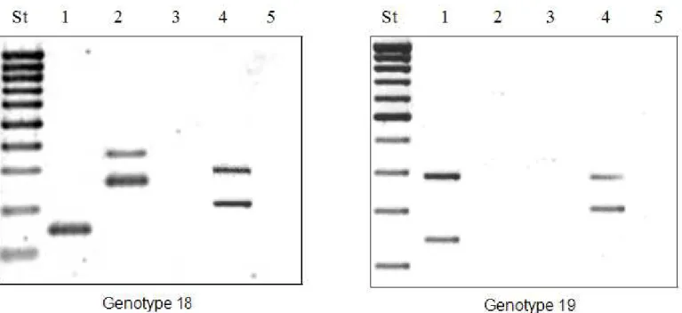
The most frequent amplification patterns found were genotypes 18 and genotype 19 with 24,5% and 20,4% respectively (Figure 1 and Table 2). On the other hand, five strains were classified into genotype 29 which did not detect any of the DNAhypervariable regions (Table 2).
Table 2. Classification of genotypes according to amplification patterns of DNA hypervariable regions and their frequency. DNA hypervariable regions
Genotype HVR pUB110 Ins117 pT181 pI258 mecR1 IS256 Frequency %
2 + - + - - - - 10.2
6 + - - - 4.0
14 + - + + - - 2.0
15 + - - - + - - 8.2
16 + - - - + - + 4.0
18 + - + - + - + 24.5
19 + - + - + - - 20.4
20 + - + + + - + 6.1
21 + - - - + 5.1
22 + - + + - - + 3.0
23 + - + - - - + 2.0
24 - - - - + - + 1.0
25 - + + - - - - 1.0
26 + + + - + - - 1.0
27 + - - + + - + 1.0
28 + - + + + - - 1.0
29 - - - 5.0
98 MRSA strains isolated from patients hospitalized at the County Hospital of Valdivia (Chile), previously typified as SCCmec type I, were subtypified into seventeen genotypes according to amplification patterns of 7 DNA hypervariable regions. The genotypes 2, 6, 14, 15 and 16 showed amplification patterns previously described. A serial number, starting with the genotype 18, was assigned to the remaining twelve new amplification patterns. Amplification patterns corresponding to genotypes 18 and genotype 19 were the most frequent.
DISCUSSION
SCCmec typing is one of the most important molecular tools available forunderstanding the epidemiology and clonal strain relatedness of MRSA (14). However, due to the very complex and diverse structure of the SCCmec element, SCCmec subtyping is a powerful tool applicable to clinical and epidemiological surveillance purposes (10). Based on the horizontal transfer of SCCmec and the polymorphism of regions located "downstream" of the mecA gene, we suggest thatgenotypes identified through the presence of hypervariable regions can be classified as subtypes of MRSA strains previously typified as SCCmec type I.
In the present study ninety eight MRSA strains isolated from patients hospitalized at the County Hospital of Valdivia (Chile), were subtypified into seventeen genotypes according to amplification patterns of 7 DNA hypervariable regions located around the mecA gene.
Seventeen genotypes detected in our environment contrasts with the five genotypes previously identified by Wilson et al., who detected only five genotypes of MRSA strains isolated from patients hospitalized at the County Hospital of Valdivia (Chile) between March 2004 and December 2005 (15). This situation is because in our study we included a greater number of strains and we identified hypervariable regions not detected previously.
The new amplification patterns detected in this study were ranked between genotype 18 and genotype 29. There was a predominance of genotype 18 and genotype 19 with 24.5% and 20,4% respectively (Figure 1 and Table 2). From these data we could infer that most of MRSA strains were originated from two local clones. In fact, we suggest that strains belonging to genotype 18 are different fromthose belonging to genotype 19, does not possess the IS256 region. Therefore, we infer that the strains belonging to genotype 18 come from a strain belonging to genotype 19, in which the IS256 region was integrated.
IS256 region, located downstream of a fragment 2 Kb called dcs (downstream constant segment), is an insertion
sequence that can be independent or as part ofthe transposon Tn4001. This transposon carries the aacA-aphD gene, which encodes resistance to aminoglycoside (1, 11). IS256 region was detected in 46.9% of MRSA strains. These results are different from those obtained by Wilson et al.,who detected this region in 9.4% of MRSA strains (15).
The increase in the prevalence of IS256 region is probably due to a clonalexpansion of some MRSA strains that possess this region in their SCCmec.
Moreover, this situation reflects the constant genomics evolution of MRSA strains in our environment. In two years (2005 - 2007), almost half of strains incorporated the IS256 region in their SCCmec. This is worrying because IS256 region allows the insertion of Tn4001 encoding resistance to aminoglycoside (11).
Ins117 region is a short sequence of 117 bp, flanked by two 15 bp direct repeats, contained within orfX region (11). This region was detected in 69.4% of MRSA strains. These results are different from those obtained by Wilson et al., who did not detect this region in MRSA strains (15). The increase in the prevalence of Ins117 region is probably due to a clonal expansion of some MRSA strain that possess this region in their SCCmec as happened with IS256 region. This is also worrying because Ins117 region, along with IS431, allows the insertion of plasmidpUB110 which encodes resistance to tetracycline and aminoglycoside (11, 12).
pUB110 region is flanked by IS431 and was integrated during the period when mecDNA was being formed and prior to the emergence of the first outbreaks of MRSAinfections in European hospitals in the early 1960s (11, 12). This region was detected in 2% of MRSA strains. In the previous study of Wilson et al. MRSAstrains carrying pUB110 region were not detected (15). Spread of strainspossessing pUB110 region would be a problem due to the resistance that this region encodes. Moreover, pUB110 region is present in subtypes SCCmec IA, II-A, II-b, II-A, II-B and II-C. MRSA strains showed this region can be classified as SCCmec subtype IA (16).
repeat unit elements (DRUs) located between IS431mec and
mecA (13). This region was detected in 92.9% of MRSA strains.
A similar situation is reported by Wilson et al (15). This fact
reflects the high degree of conservation of the HVR region at
the strains isolated in ourhospital environment.
pI258 and pT181 regions are plasmid flanked by IS431 that
encodes resistance tomercury and tetracycline respectively (11).
pI258 region was detected in 69.4% of our strains. Previously
Wilson et al. detected the pI258 region in 81% of their
MRSA strains (15). pT181 region was detected in 13.3% of
our strains. These results are different from those obtained by
Wilson et al. who detected this region in 41.5% of their MRSA
strains (15).
Located upstream of mecA gene lies the mecR1 gene, that
encodes the protein MecR1, which activates the mecA gene
transduction generating the synthesis of PBP2a (2, 16). This
region was not detected in any of our MRSA strains, as it was
previously reported by Wilson et al (15). This situation is
because mecR1 gene ischaracterized by suffering deletions. This
characteristic is highly conserved among strains isolated in our
environment (2).
Finally, based on the results obtained in this study and
the results obtainedpreviously by Wilson et al (15) we suggest
that acquisition of hypervariable regions by MRSA is a
continuous horizontal transfer process through which the
SCCmec has been preserved intact, or even may give rise to
new types and subtypes of SCCmec. This means that MRSA
strains could maintain or increase theirresistance, but in no case it
would decrease.
Continue surveillance studies are needed to make annual
checkups to determinethe prevalence of MRSA subtypes in our
environment, as well as controlling the emergence of new
subtypes. On the other hand, it would allow retrospectivestudies
to detect evolutionary changes and would establish an accurate
antimicrobial therapy, which would shorten the hospitalization
stay, resulting in asignificant decrease in health costs caused by
MRSA infections.
ACKNOWLEDGEMENTS
This work was supported by Direction of Research and
Development of the Universidad Austral de Chile
(DID-UACh-S-2007-63 and S-2010-02).
REFERENCES
1. Deplano, A.; Vaneechoutte, M.; Verschraegen, G.; Struelens, M. (1997) Typing of Staphylococcus aureus and Staphylococcus
epidermidis Strains by PCR Analysis of Inter-IS256 Spacer Length
Polymorphisms. J Clin Microbiol. 35 (10), 2580 – 2587.
2. Hiramatsu, K.; Longzhu, C.; Kuroda, M.; Ito, T. (2001) The emergence and evolution of methicillin-resistant Staphylococcus
aureus. Trends in Microbiology. 9 (10), 486 - 493.
3. Huygens, F.; Nimmo, G.; Schooneveldt, J.; Munckhof, W.; Giffard,
P. (2002) Genotyping of Methicillin-Resistant Staphylococcus aureus
by Assaying for the Presence of Variable Elements Associated with
mecA. J Clin Microbiol. 40 (8), 3093 – 3097.
4. Ito, T.; Katayama, Y.; Asada, K.; Mori, N.; Tsutsumimoto, K.; Tiensasitorn, C.; Hiramatsu, K. (2001). Structural comparison of three types of staphylococcal cassette chromosome mec integrated in the chromosome in methicillin-resistant Staphylococcus aureus.
Antimicrob Agents Chemother. 45 (5), 1323 - 1336.
5. Ito, T.; Katayama, Y.; Hiramatsu, K. (1999). Cloning and nucleotide
sequence determination of the entire mecA of pre-methicillin-resistant Staphylococcus aureus N315. Antimicrob Agents Chemother. 43 (6), 1449 - 1458.
6. Ito, T.; Ma, X.; Takeuchi, F.; Okuma, K.; Yuzawa, H.; Hiramatsu, K. (2004). Novel type staphylococcal cassette chromosome mec driven by a novel cassette chromosome recombinase, ccrC. Antimicrob Agents
Chemother. 48 (7), 2637 - 2651.
7. Ito, T.; Okuma, K.; Ma, X.; Yuzawa, H.; Hiramatsu, K. (2003).
Insights on antibiotic resistance of Staphylococcus aureus from its whole genome: genomic island SCC. Drug Resist Updat. 6 (1), 41 - 52.
8. Jevons, M. (1961). “Celbenin”-resistant staphylococci. Br Med J. 1: 124-125.
9. Milheiriço, C.; Oliveira, D.; Lancastre, H. (2007). Mutiplex PCR strategy for subtyping the staphylococcal cassette chromosome mec
type IV in methicillin- resistant S. aureus: “SCCmec IV multiplex”. J
Antimicrob Chemother. 60 (1), 42 - 48.
10. Oliveira, D.; Lencastre.; H. (2002). Multiplex PCR strategy for rapid identification of structural types and variants of the mec element in methicillin-resistant Staphylococcus aureus. Antimicrob Agents
Chemother. 46 (7), 2155 – 2161.
11. Oliveira, D.; Wu, S.; Lencastre, H. (2000). Genetic organization of the downstream region of the mecA element in methicillin-resistant S. aureus isolates carrying different polymorphisms of this region. Antimicrob Agents Chemother. 44 (7), 1906 - 1910.
Non-b-Lactam Antibiotics, Including Tobramycin. J Clin Microbiol. 39 (2), 779 – 781.
13. Ryffel, C.; Bucher, R.; Kayser, F.; Berger-Bachi, B, (1991). The
Staphylococcus aureus mec Determinant Comprises an Unusual
Cluster of Direct Repeats and Codes for a Gene Product Similar to the
Escherichia coli sn-Glycerophosphoryl Diester Phosphodiesterase. J
Bacteriol. 173 (23), 7416 - 7422.
14. Schmitz, F.; Steiert, M.; Tichy, H.; Hofmann, B.; Verhoef, J.; Heinz, H.; Köhrer, K.; Jones, M. (1998). Typing of methicillin-resistant
Staphylococcus aureus isolates from Düsseldorf by six genotypic
methods. J Med Microbiol. 47, 341 – 351.
15. Wilson, M.; Otth, C.; Medina, G.; Otth, L.; Fernández, J.; Arce, M.;
Zaror, A.; Lizama, V.; Gil, M.; von Chrismar, A. (2007) Genotipos
de Staphylococcus aureus con fenotipo meticilino resistente, aislados
de pacientes del Hospital Base de Valdivia. Rev Méd Chile. 135: 596-601.
16. Yinduo, Ji. (2007). Methicillin-Resistant Staphylococcus aureus
(MRSA) Protocols. Department of veterinary and biomedical sciences, University of Minnesota, St. Paul, MN.
17. Zhang, K.; McClure, J.; Elsayed, S.; Louie, T.; Conly, J. (2005) Novel Multiplex PCR Assay for Characterization and Concomitant Subtyping of Staphylococcal Cassette Chromosome mec Types I to V in Methicillin-Resistant Staphylococcus aureus. J Clin Microb. 43 (10), 5026-5033.