REVIEW ARTICLE
published: 28 October 2014 doi: 10.3389/fphys.2014.00406
Revisiting cAMP signaling in the carotid body
Ana R. Nunes
1*, Andrew P. Holmes
2, Sílvia V. Conde
1, Estelle B. Gauda
3and Emília C. Monteiro
11
CEDOC, Chronic Diseases Research Center, NOVA Medical School/Faculdade de Ciências Médicas, Universidade Nova de Lisboa, Lisboa, Portugal
2School of Clinical and Experimental Medicine, University of Birmingham, Birmingham, UK
3Neonatology Research Laboratories, Department of Pediatrics, Johns Hopkins Medical Institutions, Johns Hopkins University, Baltimore, MD, USA
Edited by:
Rodrigo Iturriaga, Pontificia Universidad Católica de Chile, Chile
Reviewed by:
Ana Obeso, University of Valldolid, Spain
Julio Alcayaga, Universidad de Chile, Chile
*Correspondence:
Ana R. Nunes, Chronic Diseases Research Center, NOVA Medical School/Faculdade de Ciências Médicas, Universidade Nova de Lisboa, Campo Mártires da Pátria, 130, 1169-056 Lisboa, Portugal e-mail: [email protected]
Chronic carotid body (CB) activation is now recognized as being essential in the
development of hypertension and promoting insulin resistance; thus, it is imperative to
characterize the chemotransduction mechanisms of this organ in order to modulate its
activity and improve patient outcomes. For several years, and although controversial,
cyclic adenosine monophosphate (cAMP) was considered an important player in initiating
the activation of the CB. However, its relevance was partially displaced in the 90s by
the emerging role of the mitochondria and molecules such as AMP-activated protein
kinase and O
2-sensitive K
+channels. Neurotransmitters/neuromodulators binding to
metabotropic receptors are essential to chemotransmission in the CB, and cAMP is central
to this process. cAMP also contributes to raise intracellular Ca
2+levels, and is intimately
related to the cellular energetic status (AMP/ATP ratio). Furthermore, cAMP signaling is
a target of multiple current pharmacological agents used in clinical practice. This review
(1) provides an outline on the classical view of the cAMP-signaling pathway in the CB
that originally supported its role in the O2/CO2
sensing mechanism, (2) presents recent
evidence on CB cAMP neuromodulation and (3) discusses how CB activity is affected by
current clinical therapies that modify cAMP-signaling, namely dopaminergic drugs, caffeine
(modulation of A
2A/A
2Breceptors) and roflumilast (PDE4 inhibitors). cAMP is key to any
process that involves metabotropic receptors and the intracellular pathways involved in CB
disease states are likely to involve this classical second messenger. Research examining
the potential modification of cAMP levels and/or interactions with molecules associated
with CB hyperactivity is currently in its beginning and this review will open doors for future
explorations.
Keywords: cAMP signaling, carotid body, pharmacology, phosphodiesterase inhibitors, adenylyl cyclase, adenosine, dopamine, antipsychotics
INTRODUCTION
Adequate homeostatic regulation of arterial oxygen (P
aO
2),
car-bon dioxide (P
aCO
2), pH and blood glucose are important
pro-cesses in physiology. Highly specialized chemosensory type I cells
of the mammalian carotid bodies (CBs) sense acute changes in
P
aO
2, P
aCO
2and pH, and, upon stimulation, release
neurotrans-mitters (NTs) that either inhibit or activate chemosensory fibers
projecting into the central nervous system (CNS). The functional
consequence of CB stimulation is the initiation of important
cardiovascular, respiratory and metabolic reflexes. These reflexes
include an increase in minute ventilation, a sympathetically
medi-ated elevation in heart rate and peripheral vasoconstriction and
an augmentation in adrenaline release from the adrenal medulla,
with the latter leading to an increase in arterial blood glucose
concentration.
Recently, interest in CB physiology has attracted
consider-able attention because of its emerging associations with chronic
cardiovascular disease (
McBryde et al., 2013
). CB
dysfunc-tion and increases in chemoafferent discharge promote
neu-rogenic hypertension in sleep disordered breathing (
Prabhakar
and Peng, 2004
), chronic heart failure (
Schultz et al., 2013
) and
essential hypertension (
Abdala et al., 2012; McBryde et al., 2013
).
Moreover, the CB is a principal regulator in initiating insulin
resistance in animal models of prediabetes and metabolic
syn-drome (
Ribeiro et al., 2013
). Therefore, the modulation of CB
function may be necessary to prevent and treat some of these
con-ditions. A good understanding on the modulation of the cellular
processes occurring downstream of the CB transduction
machin-ery, may not only promote drug development that modify CB
chemodischarge to prevent or treat disease, but will also increase
the awareness that CB chemodischarge can be an inadvertent side
effect of drugs used to treat other diseases.
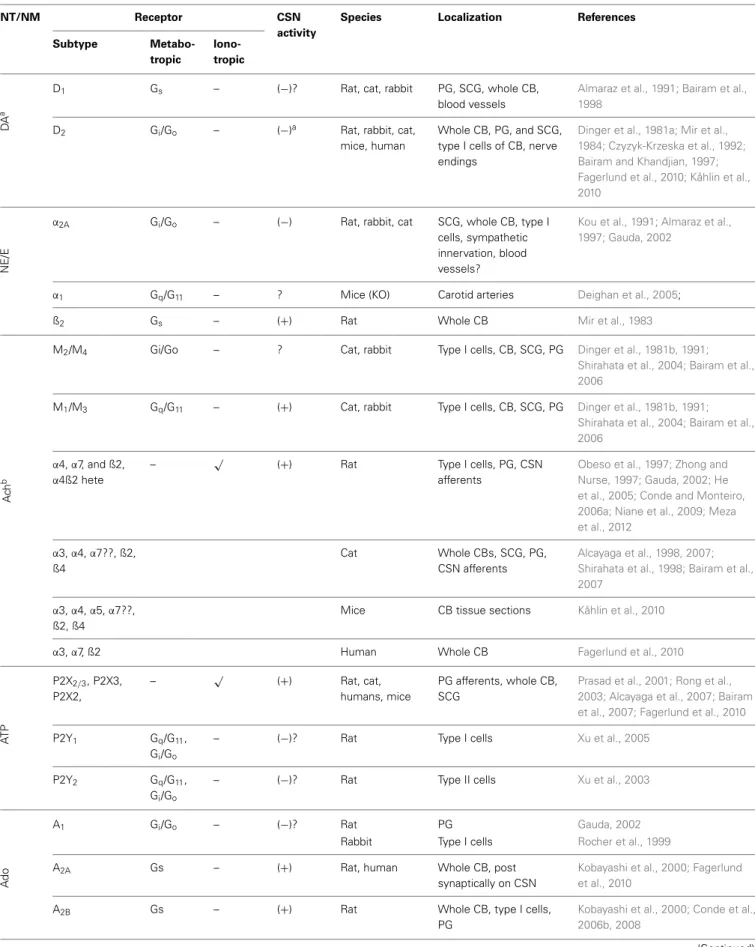
CB type I cells contain molecular sensors that, when activated,
trigger transduction cascades that produce cellular
depolariza-tion, Ca
2+influx and NT and/or neuropeptide secretion. The
list of characterized NTs/neuromodulators (NMs) and
respec-tive receptors in the CB has increased considerably over the
last 20 years (
Table 1
). These NTs/NMs have the potential
to activate metabotropic and ionotropic receptors located on
type I cells (autoreceptors), on afferents of the carotid sinus
nerve (CSN, post-synaptic receptors), or both, exerting either
excitatory or inhibitory actions (
Table 1
). The activation of
Table 1 | Receptors in the carotid body.
NT/NM Receptor CSN
activity
Species Localization References
Subtype
Metabo-tropic
Iono-tropic
DA
a
D1 Gs – (−)? Rat, cat, rabbit PG, SCG, whole CB,
blood vessels
Almaraz et al., 1991; Bairam et al., 1998
D2 Gi/Go – (−)a Rat, rabbit, cat,
mice, human
Whole CB, PG, and SCG, type I cells of CB, nerve endings
Dinger et al., 1981a; Mir et al., 1984; Czyzyk-Krzeska et al., 1992; Bairam and Khandjian, 1997; Fagerlund et al., 2010; Kåhlin et al., 2010
NE/E
α2A Gi/Go – (−) Rat, rabbit, cat SCG, whole CB, type I
cells, sympathetic innervation, blood vessels?
Kou et al., 1991; Almaraz et al., 1997; Gauda, 2002
α1 Gq/G11 – ? Mice (KO) Carotid arteries Deighan et al., 2005;
ß2 Gs – (+) Rat Whole CB Mir et al., 1983
Ac
h
b
M2/M4 Gi/Go – ? Cat, rabbit Type I cells, CB, SCG, PG Dinger et al., 1981b, 1991;
Shirahata et al., 2004; Bairam et al., 2006
M1/M3 Gq/G11 – (+) Cat, rabbit Type I cells, CB, SCG, PG Dinger et al., 1981b, 1991;
Shirahata et al., 2004; Bairam et al., 2006
α4,α7, and ß2,
α4ß2 hete
– √ (+) Rat Type I cells, PG, CSN
afferents
Obeso et al., 1997; Zhong and Nurse, 1997; Gauda, 2002; He et al., 2005; Conde and Monteiro, 2006a; Niane et al., 2009; Meza et al., 2012
α3,α4,α7??, ß2, ß4
Cat Whole CBs, SCG, PG,
CSN afferents
Alcayaga et al., 1998, 2007; Shirahata et al., 1998; Bairam et al., 2007
α3,α4,α5,α7??, ß2, ß4
Mice CB tissue sections Kåhlin et al., 2010
α3,α7, ß2 Human Whole CB Fagerlund et al., 2010
AT
P
P2X2/3, P2X3,
P2X2,
– √ (+) Rat, cat,
humans, mice
PG afferents, whole CB, SCG
Prasad et al., 2001; Rong et al., 2003; Alcayaga et al., 2007; Bairam et al., 2007; Fagerlund et al., 2010
P2Y1 Gq/G11,
Gi/Go
– (−)? Rat Type I cells Xu et al., 2005
P2Y2 Gq/G11,
Gi/Go
– (−)? Rat Type II cells Xu et al., 2003
Ad
o
A1 Gi/Go – (−)? Rat PG Gauda, 2002
Rabbit Type I cells Rocher et al., 1999
A2A Gs – (+) Rat, human Whole CB, post
synaptically on CSN
Kobayashi et al., 2000; Fagerlund et al., 2010
A2B Gs – (+) Rat Whole CB, type I cells,
PG
Kobayashi et al., 2000; Conde et al., 2006b, 2008
Nunes et al. cAMP signaling in the carotid body
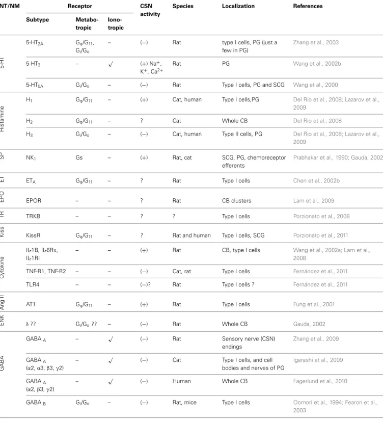
Table 1 | Continued
NT/NM Receptor CSN
activity
Species Localization References
Subtype
Metabo-tropic
Iono-tropic
5-H
T
5-HT2A Gq/G11,
Gi/Go
– (−) Rat type I cells, PG (just a
few in PG)
Zhang et al., 2003
5-HT3 – √ (+) Na+,
K+, Ca2+
Rat PG Wang et al., 2002b
5-HT5A Gi/Go – (−) Rat Type I cells, PG and SCG Wang et al., 2000
Hist
amine
H1 Gq/G11 – (+) Cat, human Type I cells,PG Del Rio et al., 2008; Lazarov et al.,
2009
H2 Gq/G11 – ? Cat Whole CB Del Rio et al., 2008
H3 Gi/Go – (−) Cat, human Type II cells, PG Del Rio et al., 2008; Lazarov et al.,
2009
SP NK1 Gs – (+) Rat, cat SCG, PG, chemoreceptor
efferents
Prabhakar et al., 1990; Gauda, 2002
ET ETA Gq/G11 – ? Rat Type I cells Chen et al., 2002b
EPO EPOR – – ? Rat CB clusters Lam et al., 2009
TR TRKB – – ? ? Type I cells Porzionato et al., 2008
Kiss KissR Gq/G11 – ? Rat and human Type I cells, SCG Porzionato et al., 2011
Cy
to
k
in
e
IL-1B, IL-6Rx, IL-1RI
– – (+) Rat CB, type I cells Wang et al., 2002a; Lam et al.,
2008
TNF-R1, TNF-R2 – – (−) Cat, rat Type I cells Fernández et al., 2011
TLR4 – – (−)? Rat Type I cells ? Fernández et al., 2011
Ang
II
AT1 Gq/G11 – (+) Rat Type I cells Fung et al., 2001
ENK δ?? Gi/Go?? – (−) Rat Whole CB Gauda, 2002
GA
BA
GABAA – √ (−) Rat Sensory nerve (CSN)
endings
Zhang et al., 2009
GABAA
(α2,α3,β3,γ2)
– √ (−) Cat Type I cells, and cell
bodies and nerves of PG
Igarashi et al., 2009
GABAA
(α2,β3,γ2)
– √ (−) Human Whole CB Fagerlund et al., 2010
GABAB Gi/Go – (−) Rat, mice Type I cells Oomori et al., 1994; Fearon et al.,
2003
aMainly inhibitory, but excitatory in rabbit (Iturriaga et al., 2009).bAlthough less characterized in rat, the nAchRα3,4,5,7, and ß2,4 are present in type I cell,α7
in the CNS afferents andα3,4,7, and ß2,4 in PG; the mAchR M1 and M2 are in type I cells, M1 in CSN afferents and M1 and M2 in PG neurons of cat and rabbit (for a revision,Shirahata et al., 2007). ?, suggested, but no direct evidences/not known; NT/NM, neurotransmitters/neuromodulators; DA, dopamine; NE/E, norepiniphrine/epiniphrine; NE, norepiniphrine; Ach, acetylcholine; ATP, adenosine triphosphate; Ado, adenosine; 5-HT, serotonine; GABA, gamma-aminobutyric acid; ENK, enkephalins; SP, substancia P; ET, endothelins; TR, trophin; AngII, Angiotensin; AC, adenylyl cyclase;+, excitatory;−, inhibitory; CB, carotid body; SCG, superior cervical ganglion; PG, petrosal ganglion.
excitatory postsynaptic receptors is translated into an increase
of CSN action potential frequency, and it is this signal that is
conveyed to the CNS. Stimulation of excitatory autoreceptors
induces an increase in [Ca
2+]
iand subsequent further release
of NTs/NMs.
Retrograde communication between petrosal ganglion (PG)
neurons and CB type I and type II cells is another source of
NTs/NMs release in the CB. PG neurons present
catecholamin-ergic traits (
Katz et al., 1983; Katz and Black, 1986
) and
cate-cholamines (CAs) are released from cultured PG neurons upon
stimulation (
Iturriaga et al., 2003
). Moreover, nitridergic
auto-nomic neurons located in the glossopharyngeal and carotid nerve
may also modulate the CB function (
Campanucci et al., 2012
).
The pannexin-1 channel opening have been recently shown to be
important in reciprocal cross-talk pathways between type I and
type II cells, particularly in purinergic transmission (ATP and
Ado) (
Nurse, 2014
).
The specific NT profile, receptor expression and cellular
effects changes with early postnatal development, and in some
cases exhibits interspecies variability [e.g., dopamine (DA) exerts
inhibitory effects on the CB in most species, except rabbit,
(
Iturriaga et al., 2009
)].
Despite the numerous different NTs/NMs released from the
type I cell, even under basal conditions, a convergence upon a
common signaling pathway could confer the overall CB
excitabil-ity and establish its sensitivexcitabil-ity to physiological stimuli. Cyclic
adenosine monophosphate (cAMP) is a common downstream
signaling molecule of numerous receptors expressed in the type
I cells, and is coupled to cellular energetic status (AMP/ATP
ratio). This article therefore aims to summarize how changes
in CB cAMP levels in physiology, pathology and following
pharmacological intervention may be central to alterations in
type I cell excitability leading to chemoafferent discharge and
cardiorespiratory and metabolic reflex responses.
CLASSICAL UNDERSTANDING OF cAMP-SIGNALING
PATHWAY IN THE CAROTID BODY
An involvement of cAMP in the CB chemotransduction (late
70s-early 80s) was originally prompted by the identification of
secreted NTs and their receptor-mediated effects on CB
chemore-ceptor responses (
Table 1
). These secreted NTs included DA
(
Gonzalez and Fidone, 1977
), acetylcholine (ACh) (
Eyzaguirre
et al., 1965
), noradrenaline (NA), substance P, serotonin and
prostaglandin E
2(
Pérez-García et al., 1993
for early references),
and adenosine (Ado) (
McQueen and Ribeiro, 1981; Monteiro and
Ribeiro, 1987; Conde and Monteiro, 2004
), acting through
spe-cific G-protein coupled receptors (
Table 1
). For those NTs that
confer an excitatory response, cAMP levels were increased, while
for those that confer an inhibitory response cAMP levels were
decreased.
Fitzgerald and co-workers, were the first to identify cAMP
in the CB cat homogenates (
Fitzgerald et al., 1977
). Subsequent
studies showed that injection of isoprenaline increased cAMP
accumulation in the rat CB (
Mir et al., 1983
) and elevated
chemoafferent discharge frequency in cat and rabbit models
via stimulation of beta-adrenoreceptors (
Folgering et al., 1982
).
Moreover, administration of dibutyryl cyclic AMP (db-cAMP,
cAMP analog) was found to mimic the excitatory effect of
adenosine on chemosensory discharge (
McQueen, 1983
).
Following these findings, a new wealth of evidence emerged
supporting a role for the cAMP in CB chemotransduction and/or
chemotransmission (
Wang et al., 1989, 1991a; Fidone et al.,
1990; Pérez-García et al., 1990; Cachero et al., 1996; Summers
et al., 2002
). Multiple investigations reported rises in CB cAMP
accumulation following hypoxia exposure (
Fidone et al., 1990;
Pérez-García et al., 1990
), an effect that appeared to be specific
for chemoreceptor tissue (
Wang et al., 1989
) and was
depen-dent on NT release (
Pérez-García et al., 1990
). Activation of
adenylyl cyclases (AC), by forskolin (FSK), potentiated CB CA
secretion and CSN discharge frequency over a range of O
2ten-sions from 30 to 0%, in the intact rabbit CB preparation (
Almaraz
et al., 1991; Wang et al., 1991a
). In addition, FSK and db-cAMP
both inhibited the type I cell O
2-sensitive K
+current,
emphasiz-ing similarities between excitatory cAMP and hypoxic signalemphasiz-ing
cascades (
López-López et al., 1993
). Hypercapnia exposure also
elevated cAMP content (
Pérez-García et al., 1990
) and FSK
aug-mented the hypercapnic CA release (
Pérez-García et al., 1991
). In
isolated rabbit CB type I cells, cAMP analogs potentiated inward
Ca
2+current in a manner that was comparable with hypercapnia
(
Summers et al., 2002
). Despite these data, it was not universally
accepted that endogenous cAMP was physiologically relevant in
the CB.
Delpiano et al. reported, using an
in vitro
preparation of the
cat CB, that anoxia exposure induced only small increases in
cAMP levels (
Delpiano and Acker, 1991
). Furthermore, severe
whole body hypoxia exposure caused both increases and decreases
in CB cAMP accumulation (
Delpiano and Acker, 1991
), and
short periods of hypoxia (2.5–5 min) failed to alter the cAMP
levels in rat CB (
Mir et al., 1983
). K
+and Ca
2+currents,
both important in hypoxic chemotransduction, were shown to
be insensitive to an array of cAMP analogs in the rat CB
type I cells (
Hatton and Peers, 1996
); inwardly rectifying Cl
−current is directly activated by cAMP (
Carpenter and Peers,
1997
). The inter-experiment variability, differences in species and
age, in CB dissection methods, O
2and CO
2stimulus
inten-sity, duration of incubation periods, CB preparations (
in vitro
,
in vivo
, whole CB vs. isolated cells or carotid sinus nerve (CSN)
preparations) and cAMP detection methods (radioimmunoassay,
enzyme-immunoassay, protein binding saturation assays) have
all been credited for the discrepancies reported in the
litera-ture regarding the relevance of cAMP signaling in CB function
(
Table 2
).
Thus, there was still a requirement to further characterize and
better understand the physiological significance of cAMP in the
CB. To consider cAMP signaling as a physiological modulator
of the chemoreceptor activity, disruption of cAMP generation,
metabolism, or its intracellular effectors would need to be
syn-onymous with functional modification of basal CB activity and/or
its responses to hypoxia/hypercapnia. [cAMP]
iis tightly regulated
Nunes
et
al.
cAMP
signaling
in
the
carotid
body
Table 2 | Effects of different work conditions on cAMP levels in the carotid body.
[cAMP] Species Anesthesia Preparation Basal conditions Stimulus Technique Units References
O2 CO2 Time Inc.media O2 CO2 Time Inc. media
(%) (%) (min) (%) (%) (min)
=a Cat 12
month
Na-pentob. Whole CB, In vivo, In vitro
R.A. R.A. n/a n/a 5 n/a 3/10/20 n/a RIA pmol/CB Delpiano et al.,
1984
30 3 60/120 Locke’s 0 3 2 Locke’s+[IBMX]
0.8 mM
↑ Cat
12month
Na-pentob. Whole CB, In vitro
35 4 180 Locke’s modified 3 4 2 Locke’s modified
+[IBMX] 0.8 mM
RIA pmol/CB Delpiano and
Acker, 1991
↑ Rabbit adult Na-pentob. Whole CB,
In vitro
100 0 30 Tyrodes’s 5 0 10 Tyrodes’s RIA pmol/mg
tissue
Wang et al., 1989
↑ Rat adult Na-pentob. CB slices,In
vitro
100 0 30 Locke’s+
[theophyline] 10 mM
4 0 10 Locke’s+
[theophylline] 10 mM
Immune-reactivity
% positive cells
Wang et al., 1991b
↑ Rabbit adult Na-pentob. Whole CB,
In vitro
100/95 0/5 30 Tyrodes’s 0/5/7/10 5/20 10 Tyrodes’s+[FSK]
0.01 mM, [IBMX] 0.5 mM and [Ca2+] 2 mM
RIA pmol/mg
tissue
Pérez-García et al., 1990
↑ Rabbit adult CO2 Whole CB,
In vitro
21 5 20 HCO−
3
enriched-medium
21 10 5 HCO−
3
enriched-medium
EIA pmol/µg
protein
Summers et al., 2002
↑ Rabbit adult Na-pentob. Whole CB,
In vitro
20 5 30 Tyrodes’s
modified+ [HCO−
3] 24 mM
7 5 10 Tyrodes’s modified
+[HCO−
324 mM
+[IBMX] 0.5 mM
RIA pmol/mg
tissue
Cachero et al., 1996
↑ Rabbit adult Na-pentob. Whole CB,
In vitro
100 0 30 Tyrode’s
modified
5 0 10 Tyrode’s modified RIA pmol/mg
tissue
Chen et al., 1997
= Rat adult Urethane Whole CB,
In vivo
R.A R.A. n/a n/a 5 0 2/5 n/a Protein
binding
pmol/CB Mir et al., 1983
= Rat (3, 12,
24 months)
Na-pentob. Whole CB, In vitro
95 5 15 Tyrodes’s
modified
95/20/10/5 5 30 Tyrodes’s modified +[IBMX] 0.5 mM
EIA pmol/mg
tissue
Monteiro et al., 2011
= Rat adult Na-pentob. Whole CB,
In vitro
20/95 5 15 Tyrodes’s
modified
5 5 30 Tyrodes’s modified
+[IBMX] 0.5 mM
EIA pmol/mg
tissue
Nunes et al., 2010
aSmall increases in cAMP levels were observed in hypoxia only in the absence of IBMX. Inc., incubation; pentob., pentobarbital; R.A., room air; n/a, not applicable; RIA, radioimmunoassay; Locke’s (in mM):
NaCl 128, KCL 5.6, CaCl2122.1, D-glucose 5.5, NaHCO310 and Hepes 7; Tyrode’s (in mM):NaCl 112, KCl 4.7, CaCl22.2, MgCl21.1, Na-glutamate 42, Hepes 5, glucose 5.6, pH 7.4; Tyrode’s modified solution (in
mM):NaCl 140, KCl 5, CaCl22, MgCl21.1, Hepes 10, glucose 5.5, pH 7.42; HCO−3 medium (in mM): NaCl 117, KCl 4.5, CaCl22.5, MgCl21, sucrose 10, glucose 11, HCO−323, pH 7.42; CB, carotid body; EIA, Enzyme Immuno Assay; IBMX, Isobutyl-1-methylxanthine.
www
.fr
ontiersin.or
g
October
20
1
4
|
V
olume
5
|A
rticle
406
|
CO
2and HCO
−3(depending on specific AC isoforms- see below).
[cAMP]
ican also be modified by direct diffusion from one cell
to another through gap junctions (
Bevans and Harris, 1999
)
or through transport to the extracellular milieu where it
pro-duces regulatory functions in multiple tissues (for a review
Bankir
et al., 2002; Hofer and Lefkimmiatis, 2007
). Downstream effectors
classically include protein kinase A (PKA) (
Taylor et al., 1990
),
cyclic nucleic gated ion channels (
Craven and Zagotta, 2006
) and
exchange proteins activated by cAMP (EPACs) (
De Rooij et al.,
1998
).
Recently, better research tools have become available to more
accurately detect intracellular cAMP and its regulation, thereby
allowing us re-examine the enzymatic regulation of cAMP within
the CB and its intracellular targets, during normoxia, hypoxia and
hypercapnia conditions.
NOVEL FINDINGS CHARACTERIZING THE ENZYMATIC
REGULATION OF cAMP ACCUMULATION IN THE CAROTID
BODY
Over the last decade, the enzymes involved in the
cAMP-pathway signaling in the CB have been identified, and their
activity modulated by natural stimuli. Novel findings have been
recently reported as to how O
2/CO
2exposure affects the CB
cAMP-signaling.
THE ROLE OF ADENYLYL CYCLASES IN THE CAROTID BODY ACTIVITY
The AC are enzymes that catalyze the synthesis of cAMP
through the cyclization of ATP. There are two main classes:
the classic NT-sensitive transmembrane (tmAC) and the more
recently described soluble adenylyl cyclase (sAC) (for a review see
Kamenetsky et al., 2006
). The activity of the former is primarily
influenced by extracellular signals (e.g., NTs, hormones,
pharma-cological agents) and is further subclassified in terms of G-protein
associations, Ca
2+related signaling pathways (
Halls and Cooper,
2011
) and more recently by CO
2interactions (
Townsend et al.,
2009; Cook et al., 2012
). sAC is regulated directly by HCO
−3and
Ca
2+, in a pH independent manner, as being shown primarily
in testis and further extended to other organs as described below
(
Chen et al., 2000; Jaiswal and Conti, 2003
).
The presence of the different AC mRNA transcripts was
only reported recently; intact rat CBs (16-17 postnatal days)
express tmAC1, tmAC2, tmAC3, tmAC4, tmAC6 and tmAC9,
with tmAC1, tmAC4 and tmAC6 exhibiting the highest fold
expression level (
Nunes et al., 2013
). sAC mRNA has also been
identified, and is expressed at greater levels in the CB than in
non-chemosensitive neuronal tissues (
Nunes et al., 2009
). The
studies on AC mRNA were performed in whole CB
(contain-ing type I and type II cells, vessels, nerve end(contain-ings, etc.). Whether
there is a specific and clearly distinguished physiological function
for each AC isoform, in the CB is unknown. Many agents that
target cAMP signaling pathways are also likely to non-selectively
act on the respective G-protein coupled receptors or PDE, thus
making individual AC targeting challenging. However, in other
tissues, individual AC isoforms do demonstrate unique
func-tionality and this has led to increased interest in identifying
specific AC isoforms as potential drug targets (
Pierre et al., 2009
).
Genetic association studies have been valuable in unraveling the
importance of specific AC in physiology and disease. For instance,
a polymorphism of AC6 have been associated with alterations
in blood pressure and heart rate regulation in humans (
Hodges
et al., 2010
). Point mutations of the AC3 gene are also associated
with decreased insulin release in animal models of type 2
dia-betes (
Abdel-Halim et al., 1998
). Correlating specific mutations
in tmAC genes with CB dysfunction and hypertension across
patient populations may help refine CB disease related research.
This data is currently unavailable but could be invaluable given
the emerging relevance of the CB in cardiovascular system
pathology.
Although ATP binding to ionotropic receptors likely mediates
excitatory chemodischarge to hypoxia, DA and Ado are two key
participants in modifying type I cell and/or post-synaptic cAMP
via their modification of tmAC activity. Hypoxia-induced raises
in type I cell [Ca
2+] and
3H-DA neurosecretion are depressed in
the presence of specific D
2receptor agonists (
Benot and
López-Barneo, 1990; Carroll et al., 2005; Conde et al., 2008
), an effect
that is associated with a reduction in CB cAMP content in both
conditions. Deficiency of D
2receptors in adult mice blunts type
I cell neurosecretion, but not CSN responses to hypoxia,
pos-sibly consistent with opposing pre-synaptic and post-synaptic
neuromodulation (
Prieto-Lloret et al., 2007
). Systemic inhibition
of Ado receptors decreases, but does not abolish, the CB
medi-ated acute phase of the hypoxic ventilatory response (
Lee et al.,
2005
). Using
in vitro
CB preparations, Conde et al. reported that
blocking Ado receptors depresses hypoxic induced CA release and
chemoafferent activity, an effect that is greater in milder rather
than severe hypoxic conditions (
Conde et al., 2006b, 2012a
). D
2receptors are negatively coupled to AC while Ado A
2Bare
posi-tively coupled to AC. Blockage of Ado A
2Breceptors counteract
the decrease in cAMP elicited by D
2receptor activation
sug-gesting an A
2Band D
2autoreceptor interaction accounting for
overall [cAMP]
iin the type I cell (
Conde et al., 2008
). In acutely
dissociated type I cells, Ado A
2Areceptor inhibition abolishes
the [Ca
2+]
ielevations evoked by Ado (
Xu et al., 2006
). Since
both A
2Aand A
2Breceptors exert their actions through
excita-tion of tmACs (reviewed in
Ribeiro and Sebastião, 2010
), it is
the increase in [cAMP]
i, that is most likely to account for its
overall chemostimulatory function. Accordingly, directly
inhibit-ing tmACs with SQ22536, does indeed depress hypoxic induced
CA-secretion (
Rocher et al., 2009
).
These findings do not, however, confine CB cAMP content to
the regulation of DA and Ado. Essentially any NT/receptor
sys-tem that is coupled to tmAC will alter cAMP levels in the CB,
including histamine/H
1and H
3receptors (
Del Rio et al., 2008,
2009; Thompson et al., 2010
), adrenaline/
β
-adrenergic receptors
(
Mir et al., 1983; Hauton et al., 2013
), pituitary adenylate
cyclase-activating protein (PACAP)/PAC
1receptor (
Xu et al., 2007; Roy
et al., 2013
), among others (also see
Table 1
).
sAC activity has been described in numerous tissues where
changes in HCO
−3/CO
2are essential to their function. For
Nunes et al. cAMP signaling in the carotid body
endothelium it plays a role in the activation of the cystic
fibro-sis transmembrane conductance regulator (
Sun et al., 2004
). sAC
mRNA has now been identified in the whole CB, and although
the sAC mRNA cellular localization has not been demonstrated,
it is expressed at greater level in the intact organ than in other
non-chemosensitive neuronal tissues (
Nunes et al., 2009, 2013
).
The physiological role of sAC in CO
2sensing was only recently
studied in the CB chemoreceptors and its function appears
somewhat equivocal. Contrary to those that reported rises in
cAMP content, and PKA dependent Ca
2+current during
iso-hydric hypercapnia (
Pérez-García et al., 1990; Summers et al.,
2002
), observations from our laboratory indicate that
increas-ing the HCO
−3/CO
2ratio from 24mM /5% (normocapnia) to
44mM/10% (isohydric hypercapnia) does not alter cAMP
con-tent, PKA activity or CSN discharge frequency and, under these
conditions, these assays were insensitive to the sAC inhibitor KH7
(
Nunes et al., 2013
). We did however show that KH7 decreased
cAMP content under basal conditions and we speculated that
sAC contributes more to normocapnic rather than hypercapnic
[cAMP]
i, at least in the rat CB. Examining the extent of sAC
HCO
−3saturation in normocapnia/normoxia and whether this
alters type I cell [Ca
2+]
i, chemoafferent frequency or responses
to hypoxia/acidosis may thus be an important area for future
investigation.
THE ROLE OF PHOSPHODIESTERASES IN THE CAROTID BODY ACTIVITY
PDE catalyze the hydrolysis of the 3-cAMP phosphate bonds
of adenosine 3,5—cyclic monophosphate to AMP. According to
the pharmacological principle that the regulation of the second
messenger degradation can often make a more rapid and larger
percentage change in concentration than comparable regulation
of the rates of synthesis, PDEs are important modulators of cAMP
levels, preventing uncontrolled diffusion of cAMP through the
cell and consequently contributing to the formation of
local-ized pools or gradients of cAMP within the cell (
Lugnier, 2006
).
There are 11 known distinct PDE isoforms, each displaying
unique substrate affinity and variable adjustment to endogenous
co factors and pharmacological inhibitors (
Bender and Beavo,
2006
).
Uncharacterized PDE was first identified in the CB in 1977
by Hanbauer and Lovenberg, and these studies provided the first
evidence for an O
2dependent cAMP affinity (
Hanbauer and
Lovenberg, 1977
). From then on, studies aiming to indirectly
assay CB AC activity and manipulate cAMP levels in responses
to different oxygen concentrations have been performed in the
presence of the xanthine 3-isobutyl-1-methylxantine (IBMX), a
non-selective PDE inhibitor (
k
i=
1–10 mM,
Dousa, 1999
and
IC
50=
2–50 mM,
Bender and Beavo, 2006
) with potential to
block Ado receptors (
k
i=
7.28 mM,
Daly et al., 1991
). A
particu-lar limitation of IBMX is its inability to inhibit PDE7 and PDE8
(
Lugnier, 2006
).
The PDE4 isoform was recently proposed as a major
regu-lator of cAMP-hydrolyzing activity in the rat CB (
Nunes et al.,
2010
). PDE4 comprises four subtypes (PDE4A, PDE4B, PDE4C,
and PDE4D) with at least 35 known splice variants (
Bender
and Beavo, 2006
). Selective pharmacological inhibition of PDE4
increases CB cAMP content in normoxia and causes even greater
rises during hypoxia. These increases are however considerably
lower than those observed in the presence of IBMX, suggesting
a physiological role of additional isoforms (
Nunes et al., 2010
).
However, at the time of this review, no functional data
relat-ing PDE4 activity with CB responses to hypoxia or hypercapnia
has been published; functional studies are necessary to further
strengthen the position of PDE4 in the CB. Intriguingly, given
that PDE4 activity increases with chronic hypoxia in O
2-sensitive
pulmonary arteries and blood (
Maclean et al., 1997; Spoto et al.,
1998; Millen et al., 2006
), a compensatory role for PDE4 to
counter CB hyperactivity, although speculative, is plausible. That
said, the consequences of chronic hypoxia or chronic
intermit-tent hypoxia exposure on PDE activity in the CB remains to be
explored.
THE ROLE OF cAMP EFFECTORS IN THE CAROTID BODY ACTIVITY: PROTEIN KINASE A, EXCHANGE PROTEIN ACTIVATED BY cAMP AND CYCLIC NUCLEOTIDE GATED CHANNELS
PKA is the classical downstream effector of cAMP. It is a
holo-tetrameric serine/theonine kinase composed of two regulatory
and two catalytic subunits. Four cAMP molecules bind to the
reg-ulatory subunits, each with two cAMP binding sites. The cAMP
binding promotes the dissociation of the catalytic subunits that
bind ATP to become catalytically active and phosphorylate serine
and threonine residues in intracellular targets such as A-kinase
anchoring proteins (AKAPS) and ion channels. AKAPs tether
PKA to particular cellular organelles and to the plasma
mem-brane confining the PKA signaling to a small pool within the cells
(
Beene and Scott, 2007
). In the nucleus, PKA can
phosphory-late transcription factors, such as cAMP response element binding
protein (CREB), and thus regulate gene expression.
A physiological role for PKA in CB chemotransmission is at
present controversial. In dissociated rabbit type I cells, PKA
inhi-bition by PKA inhibitor (PKAi) diminishes the rise in L-type Ca
2+current in response to isohydric hypercapnia (
Summers et al.,
2002
). However, we reported that PKA activation status, as
mea-sured by Fluorescent Resonance Energy Transfer (FRET) based
reporters, is unaltered during isohydric hypercapnia in isolated
rat type I cells (
Nunes et al., 2013
). Multiple blockers of PKA have
no effect on hypoxic CA-secretion in the intact rat CB
prepa-ration (
Rocher et al., 2009
). In contrast, acute rises in type I
cell [Ca
2+]
ievoked by Ado and PACAP are essentially abolished
by PKA inhibition with H89 (10
µ
M) (
Xu et al., 2006, 2007
).
Sustained plateau CSN activity mediated by PACAP is inhibited
by only 41% in the presence of H89, in a preparation including
the carotid bifurcation- CB-CSN-superior cervical ganglion (
Roy
et al., 2013
). Furthermore, type I cell activation by methylcholine
is sensitive to tmAC inhibition but not H89 inhibition, which
suggests that cAMP signaling cascades in the CB are
indepen-dent of PKA activation (
Thompson and Wyatt, 2011
). Whether
these multiple discrepancies reflect fundamental species
differ-ences, different preparations (whole CB vs. cultures or type I
cells) or other unidentified experimental factors are unclear, but
precaution must be taken when using H89 due its reported
non-specific inhibitory effect (
Lochner and Moolman, 2006
). In
cel-lular preparations, it is likely that transmitters released from type
I cells are lost to the superfusate and so their potential excitatory
or inhibitory autoregulation of the type I cell chemosensitivity to
hypoxia or hypercapnia is not apparent. Additionally, the
contri-bution of retrograde communication (PG neurons to CB cells)
should be also taken in account (
Katz et al., 1983; Katz and Black,
1986; Iturriaga et al., 2003
) as well as the new concept of tripartite
sensory synapse between type I, type II and PG neurons (
Nurse,
2014
). Also, we now know that the contribution of the NTs to the
hypoxic chemosensitivity in the CB depends on hypoxic intensity
meaning that different hypoxic intensities will evoke the release
of different NTs (
Conde et al., 2012a
) and therefore the
differ-ences observed in PKA activation can reflect distinct hypoxic
intensities/mediators involved.
Different effects of PDE4 inhibitors on cAMP accumulation
induced by hypoxia (
Nunes et al., 2010
), could suggest a different
degree of PDE phosphorylation induced by differences in PKA
activity mediated by hypoxia (
Bender and Beavo, 2006
).
Complex cAMP driven mechanisms through PKA and
extra-cellular signal regulated kinase (ERK) mediated phosphorylation
can modify PDE4 specific isoforms activity and subsequently alter
the sensitivity to selective inhibitors (
Bender and Beavo, 2006
).
Thus, determining whether acute hypoxia causes PDE4
activa-tion by PKA or ERK mediated phosphorylaactiva-tion would be of
interest.
EPAC is a guanine nucleotide exchange factor (GEF) for the
RasGTPase homologs, Rap1 and Rap2. EPAC is composed of two
regions: an N-terminal regulatory region containing a
cAMP-binding site and a C-terminal catalytic region, with GEF activity
(
De Rooij et al., 1998
). In the inactive conformation, EPAC is
folded and the regulatory domain functions as an auto-inhibitory
domain. cAMP binding unfolds the protein, allowing Rap to bind
(for a review
Gloerich and Bos, 2010
). Rap GTPases cycle between
an inactive GDP-bound and an active GTP-bound state, with
GEFs mediating the exchange of GDP for GTP. GTPase-activating
proteins then convert Rap to the inactive form. The activated
Rap-GTP activates a variety of different mechanisms in the
cell: promotes integrin-mediated cell adhesion, gap junction
for-mation and ERK
1/2MAPK-mediated protein phosphorylation,
stimulates phospholipase C-
ε
which hydrolyzes PIP2 to
gener-ate diacylglycerol, and the Ca
2+mobilizing second messenger IP
3(for a review
Holz et al., 2006
).
Rocher and co-workers initially proposed a physiological role
for EPAC in the CB by examining the effects of the EPAC
activator (8-pCPT-2
′-O-Me-cAMP) and inhibitor (brefeldin) on
the release of CAs (
Rocher et al., 2009
). Specifically,
8-pCPT-2
′-O-Me-cAMP reversed the action of SQ22536 and brefeldin
inhibited CA-secretion during hypoxia by approximately 50%.
These authors suggested that the effectors of EPAC were likely
to be the exocytotic machinery and K
+channels (
Rocher et al.,
2009
). More recently, this group has identified the expression of
both EPAC1 and EPAC2 in the rat CB (
Ramirez et al., 2012
).
In addition, EPAC activation by cAMP is proposed to cause
downstream stimulation of the IP
3receptor in the endoplasmic
reticulum (
Thompson and Wyatt, 2011
) along with activation of
PKC (
Roy et al., 2013
). Thus, crosstalk between the G
s/G
i(cAMP-related) and G
qsignaling pathways within the type I cell likely
occurs. Better characterization of this interaction could be
par-ticularly insightful given the known upregulation of G
qsignaling
associated with CB dysfunction in sleep disorder breathing (
Peng
et al., 2006
) and CHF (
Li et al., 2006
).
cAMP can directly bind to cyclic nucleotide-gated (CNGC)
and hyperpolarization-activated cyclic nucleotide-modulated
(HCNC) ion channels. These channels belong to a superfamily
of voltage-gated cation channels, and thus the binding of cAMP
to these channels is translated into changes in membrane
poten-tial and influx of Ca
2+and Na
+. By conducting Ca
2+, they can
stimulate Calmodulin (CaM) and CaM-dependent kinases and,
in turn, modulate cAMP production by regulating activity of AC
and PDE. Since CNGC and HCNC are also permeable to Na
+and K
+, they can also alter the membrane potential in electrically
active cells. The presence of these channels in the rat CB has been
suggested by the work of Stea and co-workers (
Stea et al., 1995
);
however, others have reported that cAMP analogs do not affect
Ca
2+currents in type I cells (
López-López et al., 1993
). HCNC
ion channels have not been characterized in the CB.
cAMP can be released from a variety of cell types and tissues
(for a review see
Bankir et al., 2002
). Transport of cAMP moves
against a concentration gradient, that is temperature dependent,
unidirectional and requires energy (
Rindler et al., 1978
). One of
the proposed functions of the extracellular cAMP is to regulate
extracellular Ado levels. Extracellular cAMP can be metabolized
by ecto-phosphodiesterases to adenosine monophosphate (5
′-AMP), and then by ecto-5
′-nucleotidases to Ado (
Conde et al.,
2009
). Interestingly, extracellular cAMP can modulate phenotype,
function and differentiation of human monocytes through A
2Aand A
2BAdo receptors (
Sciaraffia et al., 2014
). The contribution
of extracellular cAMP to Ado production/Ado receptors
activa-tion has never been investigated in the CB, and cAMP released
from the CB has never been quantified.
Intracellular cAMP can diffuse intercellularly through
well-characterized gap junctions (
Bennett et al., 1991; Bevans et al.,
1998; Bevans and Harris, 1999
). In the rat CB, connexin 43 (Cx43)
gap junctions are found between type I cells and carotid nerve
terminals, and mediate intercellular communications and
trans-port of small molecules and ions (
Abudara and Eyzaguirre, 1996;
Eyzaguirre, 2007
). Electrical coupling, gap junction formation
and connexin expression are regulated by cAMP (
Abudara and
Eyzaguirre, 1998; Abudara et al., 1999, 2000; Eyzaguirre, 2007
)
and chronic hypoxic exposure (
Chen et al., 2002a
).
All together, these findings suggest different regulations of
cAMP signaling in the CB, mediated not only by the enzymes
directly involved in the synthesis and degradation of this
signal-ing molecule, but also by a variety of effectors that can modulate
its accumulation, and consequently, trigger changes in the CB
activity.
Nunes et al. cAMP signaling in the carotid body
cAMP-pathway in the rat CB. Although, inter-species differences
may exist (
Delpiano and Acker, 1984, 1991; Wang et al., 1989;
Pérez-García et al., 1990; Cachero et al., 1996; Chen et al., 1997
).
Findings from experiments studying NTs systems using CB from
rabbits vs rat and cat often differ. For example, dopaminergic
influence on CB function is excitatory in the rabbit while it is
inhibitory in CB from other mammalian species (
Fidone et al.,
1982; Vicario et al., 2000a
), with higher DA secretion in rats,
combined with lower cAMP accumulation (through D
2receptor
activity).
Together with the other reported actions of cAMP signaling
in the CB (
Table 3
), our results are consistent with the view that
the cAMP-pathway is involved in the maintenance of basal CB
excitability and thus the basal release of NTs and CSN sensory
discharge frequency. The observed reduction in hypoxic
sensitiv-ity when cAMP signaling is targeted indicates that basal levels of
cAMP act to prime the CB to subsequent hypoxic stimulation.
This is likely achieved through the basal regulation of PKA and
EPAC and possibility other, as yet unidentified downstream
effec-tors. Given the synergy between hypoxia and hypercapnia, the
basal type I cell [cAMP]
imay also confer the excitability to high
CO
2or H
+although further evidence is required to confirm this.
It would be of considerable interest and relevant to humans to
determine how chronic hyperoxia and/or hypercapnia modifies
cAMP signaling in the CB.
NOVEL TECHNIQUES TO STUDY cAMP SIGNALING IN THE CAROTID BODY
With tmACs restricted to membranes, and sAC, PDEs and cAMP
effectors widely distributed within the cells, cAMP
accumula-tion is spatially and temporally controlled, generating cAMP
microdomains. Thus, a comprehensive study of cAMP
signal-ing should complement the cAMP quantifications made in CB
homogenates using RIA (
Pérez-García et al., 1991; Wang et al.,
1991a; Cachero et al., 1996
) or EIA (
Batuca et al., 2003; Conde
et al., 2008; Nunes et al., 2010, 2013; Monteiro et al., 2011
),
which are themselves static and terminal assays. Measurements
in intact type I cells using reporter protein constructs may allow
for a more detailed quantification of cAMP in distinct
subcellu-lar compartments. Nowadays, a variety of FRET-based biosensors
are available to visualize signaling dynamics in living cells (
Sample
et al., 2014
). The literature is void of data obtained using life
imaging techniques to manipulate cAMP in the CB. Our
labo-ratory in collaboration with the labolabo-ratory of Dr. Jin Zhang, have
been successful in using FRET-based reporters in CB type I cells
to interrogate cAMP-signaling pathways (
Nunes et al., 2013
).
Table 3 | Effects mediated by cAMP signaling in the carotid body.
Effects mediated by cAMP Preparation Technique Agents that modulate [cAMP] References
Increase of junctional conductance
Type I cells (young rats) Dual-voltage clamping dB-cAMP (1mM), 8-Br-cAMP (1 mM, 3 h)
Abudara and Eyzaguirre, 1998
Increase of the tyrosine hydroxylase gene expression elicited by hypoxia
Whole CB (adult rats) Reverse-Transcriptase-polymerase chain reaction
FSK (0.01 mM, 3 h) Chen et al., 1995
Activation of Cl−currents Type I cells (P10 rats) Patch-clamp/whole-cell recording
cAMP (0.2 mM)
8-bromoadenosine-cAMP (2 mM)
Carpenter and Peers, 1997
Increase of Na+and Ca2+inward currents and capacitance
Type I cells (P5-12 rats) Patch-clamp/whole-cell recording
dB-cAMP (0.2–1 mM) and FSK (0.01 mM) up to 15 days
Stea et al., 1995
Induction of Na+- channels and hypertrophy of type I cells
Type I cells (P5-12 rat) Patch-clamp/whole-cell recording
Bt2-cAMP (1 mM, upto 14 days) Stea et al., 1992
Potentiation of (30% O2)-evoked
CA release and CSN discharge
Whole CB, CSN (adult rabbit)
CSN activity recording; CA release
FSK (0.01 mM, 10 min) Wang et al., 1991a
Increase of DA release elicited by hypoxia (5% O2)
Whole CB (adult rabbit) CA release FSK (5–10µM), dB-cAMP (2mM)
IBMX (0.5 mM), ISO (0.01–0.050 mM)
Pérez-García et al., 1990, 1991, 1993
Elevation of Ca2+- currents (mimic the effect of hypercapnia)
Type I cells (adult rabbit) Whole-cell recording 8-Br-cAMP (0.5 mM, 10 min) Summers et al., 2002
Increase of GAP-43 and neurofilament (NF68 and NF160 kD) expression and neurite outgrowth
Type I cells (P5-7 rat) Double-label immunofluore scence
dB-cAMP (1mM), FSK (0.01 mM), up to 2 weeks
Jackson and Nurse, 1995
dB-cAMP or Bt2-cAMP, Dibutyryl-cAMP; 8-Br-cAMP, 8-Bromoadenosine-cAMP; FSK, Forskolin; P, Postnatal day; CA, catecholamines; CSN, Carotid Sinus Nerve; DA, Dopamine; ISO, Isoprostane; IBMX, Isobutyl-1-methylxanthine; GAP-43, Growth-Associated Protein 43; CB, carotid body.
Image-based techniques can be used to understand
interac-tions between cAMP and other mediators that raise [Ca
2+]
ilevels. Intensification of [Ca
2+]
isignals have been linked with
raised levels of angiotensin II, endothelin-1, cytokines, insulin
and free radicals along with decreases in nitric oxide: levels of
these substances are changed in disease states, associated with
CB dysfunction (
Chen et al., 2002b; Rey et al., 2006; Fung et al.,
2007; Li et al., 2010; Schultz, 2011; Del Rio et al., 2012; Lam et al.,
2012; Ribeiro et al., 2013
). Intracellular Ca
2+levels can influence
cAMP signaling directly through modulation of the activity of
PDE and AC isoforms or indirectly through PKC activity, not only
by allosteric regulation, but also by desensitization of GPCRs.
In this sense, a compensatory cAMP mechanism may function
to partially restore some sort of homeostatic control within the
type I cell despite serious remodeling of the sensory transduction
cascade in CB dysfunction.
As described above, our current view is that cAMP has an
important role in the CB homeostasis, as suggested by the
higher cAMP levels under normoxic conditions. Understanding
how [Ca
2+]
iand cAMP signaling is modified in
condi-tions that lead to CB dysfunction, will be fundamental to
understanding the role of these second messengers in the
CB transduction/neurotransmission mechanism in health and
disease.
IS CAROTID BODY IMPLICATED IN THE EFFECTS OF DRUGS
THAT MODIFY cAMP FOR THERAPEUTIC PURPOSES?
The following section focuses on the CB mediated effects induced
by currently clinical therapy that target cAMP-signaling. It is not
aimed to extensively describe the putative drug effects that can
be mediated by the CB but only to give insights that can
stim-ulate future research in a field where the information is scarce.
Figure 1
summarizes molecular targets in type I cell and CSN
endings that may be affected by drugs-induced changes in cAMP
accumulation.
Exogenous DA
is extensively used in human to improve cardiac
output and peripheral perfusion in patients with cardiogenic and
septic shock. Several years after using DA, its inhibitory effects
on ventilation in man were described and attributed to an effect
on chemoreceptor reflexes (
Welsh et al., 1978
). Twenty years later,
Van de Borne et al. (1998)
showed that repeated use of DA impairs
the ventilatory response to hypoxemia due to an inhibitory effect
on CSN activity, which explains why when administered in low
doses to conscious patients, DA reduces the discomfort caused by
FIGURE 1 | Representation of some drug targets in type I cells and CSN endings that affect cAMP accumulation in the carotid body. tmAC, transmembrane Adenylyl Cyclase; PKA, Protein Kinase A; EPAC, Exchange Protein Activated cAMP; D2, Dopamine receptor
D2; A2A, Adenosine receptor A2A; A2B, Adenosine receptor A2B;
5-HT2A, serotonin receptor 5-HT2A; P2XR, ATP ionotropic P2X
Nunes et al. cAMP signaling in the carotid body
hypoxemia. Although its clinical use as vasopressor remains, both
DA and NA have been used: DA decreases CB activity while NA
does not appear to have an effect on CB activity (
Zapata, 1975;
Debaveye and Van den Berghe, 2004
).
The impact of chronic use of
antipsychotics
(D
2antagonists)
on peripheral chemoreflexes is unknown but beneficial effects
of loxapine on agitation and breathing patterns during
wean-ing from mechanic ventilation have been described (
Sztrymf
et al., 2010
). These findings open doors to a promising field to
explore CB manipulation to improve adaptation to mechanical
ventilation.
Acute administration of
Ado
(full agonist of A
2Aand A
2Breceptors) is clinically useful to revert paroxysmal
supraventricu-lar tachycardia and causes dyspnea and chest discomfort mediated
by CB activation (
Watt et al., 1987; Reid et al., 1991
).
Caffeine
is a non-selective Ado antagonist that has been used to prevent
and treat apnea of prematurity due, primarily, to the blockade
of inhibitory Ado A
1receptors in the CNS. Moreover, the effects
of chronic coffee consumption have been extensively studied in
the last years and it is now consensual that coffee, and
proba-bly caffeine, may reduce the risk of type 2 diabetes mellitus and
hypertension, as well as other conditions associated with
car-diovascular risk such as obesity and depression (
O’Keefe et al.,
2013
). In fact, research in our laboratory have shown that chronic
caffeine intake decreases circulating CAs, prevents diet-induced
insulin resistance and hypertension (
Conde et al., 2012b
) and
restores insulin sensitivity in aged rats (
Guarino et al., 2013
).
Knowing that at the CB, caffeine blocks excitatory Ado A
2A/A
2Breceptors (
Conde et al., 2006b
) and that CB denervation prevents
the development of insulin resistance and hypertension induced
by hypercaloric diets (
Ribeiro et al., 2013
) the CB modulation
by caffeine can improve conditions associated to sympathetic
mediated CB hyperactivity (e.g., hypertension). Other effects of
caffeine have been described in the CB e.g., mobilization of Ca
2i+stores in the CB cells by ryanodine receptor activation (
Vicario
et al., 2000b; Mokashi et al., 2001
). However, they do not seem to
be relevant in the clinical setting because their effects are achieved
with toxic concentrations (
Fredholm et al., 1999
).
Roflumilast
, an oral selective PDE4 inhibitor, was approved
in 2011 by both the Food and Drug Administration (FDA) and
the European Medicines Agency (EMA) for the treatment of
severe chronic obstructive pulmonary disease (COPD), due to its
anti-inflammatory and bronchodilator effects. No evidences of
roflumilast effects on CB activity have been reported and those
are difficult to address essentially because of their CNS effects and
the variability of the PDE inhibitors efficacy in hypoxic conditions
(
Nunes et al., 2010
). Curiously, the roflumilast efficacy to reduce
the risk of COPD exacerbations has only been shown in patients
that experience reduced dyspnea (
Rennard et al., 2014
). Since CB
resection relieves dyspnea in COPD patients and improves FEV
1(Force Expiratory Volume) (
Whipp and Ward, 1992
) but
exarce-bates hypoxemia and hypercapnia and overall worsen the long
term outcome (
Stulbarg et al., 1989
), the link between the
mech-anism of roflumilast action in COPD patients and CB activity
merits further studies.
From the above evidence, one can conclude that the
manipula-tion of cAMP signaling pathway is important to address O
2/CO
2related diseases. However, manipulation of cAMP signaling may
have consequences in the CB, that are clinically relevant and that
have not yet been identified.
CONCLUSIONS
The importance of cAMP to CB physiology has moved from a
discarded player in the O
2chemotransduction to a central
sig-naling molecule that is converged upon by multiple NTs/NMs,
which collectively maintain an equilibrated CB activity. It remains
to be seen whether modification of cAMP can improve patient
outcomes in diseases associated with CB impairment or
hyper-activity. We suggest that cAMP has an important role in the
homeostasis of the CB since cAMP levels seem to be higher under
normoxic conditions. Despite the increase in knowledge of CB
physiology, the activity of tyrosine hydroxylase is still the hallmark
of the CB and cAMP the classical second messenger of dopamine
D
2receptor signaling.
Understanding how calcium and cAMP cooperate in
dysfunc-tion CB, will be fundamental to understand the role of these
sec-ond messengers in the CB transduction mechanism. Additionally,
systemic pharmacological manipulation of cAMP signaling can
have clinically relevant consequences mediated by the CB. This
may proven to be an exciting field of research that is still currently
unexplored.
ACKNOWLEDGMENT
This study was funded by an operating grant
PTDC/SAU-TOX/112264/2009.
REFERENCES
Abdala, A. P., McBryde, F. D., Marina, N., Hendy, E. B., Engelman, Z. J., Fudim, M., et al. (2012). Hypertension is critically dependent on the carotid body input in the spontaneously hypertensive rat.J. Physiol.590, 4269–4277. doi: 10.1113/jphysiol.2012.237800
Abdel-Halim, S. M., Guenifi, A., He, B., Yang, B., Mustafa, M., Höjeberg, B., et al. (1998). Mutations in the promoter of adenylyl cyclase (AC)-III gene, overex-pression of AC-III mRNA, and enhanced cAMP generation in islets from the spontaneously diabetic GK rat model of type 2 diabetes.Diabetes47, 498–504. doi: 10.2337/diabetes.47.3.498
Abudara, V., and Eyzaguirre, C. (1996). Reflections on the carotid nerve sensory discharge and coupling between glomus cells.Adv. Exp. Med. Biol.410, 159–167. doi: 10.1007/978-1-4615-5891-0_23
Abudara, V., and Eyzaguirre, C. (1998). Modulation of junctional conductance between rat carotid body glomus cells by hypoxia, cAMP and acidity.Brain Res.
792, 114–125. doi: 10.1016/S0006-8993(98)00127-9
Abudara, V., Eyzaguirre, C., and Sáez, J. C. (2000). Short- and long-term regula-tion of rat carotid body gap juncregula-tions by cAMP. Identificaregula-tion of connexin43, a gap junction subunit.Adv. Exp. Med. Biol.475, 359–369. doi: 10.1007/0-306-46825-5_33
Abudara, V., Garcés, G., and Sáez, J. C. (1999). Cells of the carotid body express connexin43 which is up-regulated by cAMP. Brain Res. 849, 25–33. doi: 10.1016/S0006-8993(99)01946-0
Alcayaga, C., Varas, R., Valdés, V., Cerpa, V., Arroyo, J., Iturriaga, R., et al. (2007). ATP- and ACh-induced responses in isolated cat petrosal ganglion neurons.
Brain Res.1131, 60–67. doi: 10.1016/j.brainres.2006.11.012
Alcayaga, J., Iturriaga, R., Varas, R., Arroyo, J., and Zapata, P. (1998). Selective activation of carotid nerve fibers by acetylcholine applied to the cat pet-rosal ganglionin vitro.Brain Res.786, 47–54. doi: 10.1016/S0006-8993(97) 01424-8
Almaraz, L., Pérez-García, M. T., Gómez-Nino, A., and González, C. (1997). Mechanisms of alpha2-adrenoceptor-mediated inhibition in rabbit carotid body.Am. J. Physiol.272, C628–C637.
Almaraz, L., Pérez-García, M. T., and González, C. (1991). Presence of D1 receptors in the rabbit carotid body.Neurosci. Lett.132, 259–262. doi: 10.1016/0304-3940(91)90315-K
Bairam, A., Frenette, J., Dauphin, C., Carroll, J. L., and Khandjian, E. W. (1998). Expression of dopamine D1-receptor mRNA in the carotid body of adult rabbits, cats and rats.Neurosci. Res.31, 147–154. doi: 10.1016/S0168-0102(98)00033-9
Bairam, A., Joseph, V., Lajeunesse, Y., and Kinkead, R. (2006). Developmental pat-tern of M1 and M2 muscarinic gene expression and receptor levels in cat carotid body, petrosal and superior cervical ganglion.Neuroscience139, 711–721. doi: 10.1016/j.neuroscience.2005.12.030
Bairam, A., Joseph, V., Lajeunesse, Y., and Kinkead, R. (2007). Developmental profile of cholinergic and purinergic traits and receptors in periph-eral chemoreflex pathway in cats. Neuroscience 146, 1841–1853. doi: 10.1016/j.neuroscience.2007.03.034
Bairam, A., and Khandjian, E. W. (1997). Expression of dopamine D2 receptor mRNA isoforms in the carotid body of rat, cat and rabbit.Brain Res.760, 287–289. doi: 10.1016/S0006-8993(97)00399-5
Bankir, L., Ahloulay, M., Devreotes, P. N., and Parent, C. A. (2002). Extracellular cAMP inhibits proximal reabsorption: are plasma membrane cAMP receptors involved?Am. J. Physiol. Renal Physiol.282, F376–F392. doi: 10.1152/ajprenal. 00202.2001
Batuca, J. R., Monteiro, T. C., and Monteiro, E. C. (2003). Contribution of dopamine D2 receptors for the cAMP levels at the carotid body.Adv. Exp. Med. Biol.536, 367–373. doi: 10.1007/978-1-4419-9280-2_48
Beene, D. L., and Scott, J. D. (2007). A-kinase anchoring proteins take shape.Curr. Opin. Cell Biol.19, 192–198. doi: 10.1016/j.ceb.2007.02.011
Bender, A. T., and Beavo, J. A. (2006). Cyclic nucleotide phosphodiesterases: molecular regulation to clinical use. Pharmacol. Rev. 58, 488–520. doi: 10.1124/pr.58.3.5
Bennett, M. V., Barrio, L. C., Bargiello, T. A., Spray, D. C., Hertzberg, E., and Sáez, J. C. (1991). Gap junctions: new tools, new answers, new questions.Neuron6, 305–320. doi: 10.1016/0896-6273(91)90241-Q
Benot, A. R., and López-Barneo, J. (1990). Feedback Inhibition of Ca2+ currents by dopamine in glomus cells of the carotid body.Eur. J. Neurosci.2, 809–812. doi: 10.1111/j.1460-9568.1990.tb00473.x
Bevans, C. G., and Harris, A. L. (1999). Direct high affinity modulation of con-nexin channel activity by cyclic nucleotides.J. Biol. Chem.274, 3720–3725. doi: 10.1074/jbc.274.6.3720
Bevans, C. G., Kordel, M., Rhee, S. K., and Harris, A. L. (1998). Isoform composition of connexin channels determines selectivity among second messengers and uncharged molecules.J. Biol. Chem. 273, 2808–2816. doi: 10.1074/jbc.273.5.2808
Buck, J., Sinclair, M. L., Schapal, L., Cann, M. J., and Levin, L. R. (1999). Cytosolic adenylyl cyclase defines a unique signaling molecule in mammals.Proc. Natl. Acad. Sci. U.S.A.96, 79–84. doi: 10.1073/pnas.96.1.79
Cachero, T. G., Rigual, R., Rocher, A., and Gonzalez, C. (1996). Cholera and pertus-sis toxins reveal multiple regulation of cAMP levels in the rabbit carotid body.
Eur. J. Neurosci.8, 2320–2327. doi: 10.1111/j.1460-9568.1996.tb01195.x Campanucci, V. A., Dookhoo, L., Vollmer, C., and Nurse, C. A. (2012). Modulation
of the carotid body sensory discharge by NO: an up-dated hypothesis.Respir. Physiol. Neurobiol.184, 149–157. doi: 10.1016/j.resp.2012.04.005
Carpenter, E., and Peers, C. (1997). Swelling- and cAMP-activated Cl- currents in isolated rat carotid body type I cells.J. Physiol.503(Pt 3), 497–511. doi: 10.1111/j.1469-7793.1997.497bg.x
Carroll, J. L., Boyle, K. M., Wasicko, M. J., and Sterni, L. M. (2005). Dopamine D2 receptor modulation of carotid body type 1 cell intracellular calcium in developing rats.Am. J. Physiol. Lung Cell. Mol. Physiol.288, L910–L916. doi: 10.1152/ajplung.00414.2003
Chen, J., Dinger, B., and Fidone, S. J. (1997). cAMP production in rabbit carotid body: role of adenosine carotid bodies cAMP production in rabbit carotid body: role of adenosine.J. Appl. Physiol.82, 1771–1775.
Chen, J., Dinger, B., and Fidone, S. J. J. (1995). Second messenger regulation of tyro-sine hydroxylase gene expression in rat carotid body.Neurosignals4, 277–285. doi: 10.1159/000109453
Chen, J., He, L., Dinger, B., Stensaas, L., and Fidone, S. (2002a). Chronic hypoxia upregulates connexin43 expression in rat carotid body and petrosal ganglion.
J. Appl. Physiol.92, 1480–1486.
Chen, Y., Cann, M. J., Litvin, T. N., Iourgenko, V., Sinclair, M. L., Levin, L. R., et al. (2000). Soluble adenylyl cyclase as an evolutionarily conserved bicarbonate sensor.Science289, 625–628. doi: 10.1126/science.289.5479.625
Chen, Y., Tipoe, G. L., Liong, E., Leung, S., Lam, S.-Y., Iwase, R., et al. (2002b). Chronic hypoxia enhances endothelin-1-induced intracellular calcium ele-vation in rat carotid body chemoreceptors and up-regulates ETA receptor expression.Pflugers Arch.443, 565–573. doi: 10.1007/s00424-001-0728-2 Conde, S. V., Gonzalez, C., Batuca, J. R., Monteiro, E. C., and Obeso, A. (2008). An
antagonistic interaction between A2B adenosine and D2 dopamine receptors modulates the function of rat carotid body chemoreceptor cells.J. Neurochem.
107, 1369–1381. doi: 10.1111/j.1471-4159.2008.05704.x
Conde, S. V., and Monteiro, E. C. (2004). Hypoxia induces adenosine release from the rat carotid body.J. Neurochem.89, 1148–1156. doi: 10.1111/j.1471-4159.2004.02380.x
Conde, S. V., and Monteiro, E. C. (2006a). Activation of nicotinic ACh receptors with alpha4 subunits induces adenosine release at the rat carotid body.Br. J. Pharmacol.147, 783–789. doi: 10.1038/sj.bjp.0706676
Conde, S. V., Monteiro, E. C., Obeso, A., and Gonzalez, C. (2009). Adenosine in peripheral chemoreception: new insights into a historically overlooked molecule–invited article.Adv. Exp. Med. Biol.648, 145–159. doi: 10.1007/978-90-481-2259-2_17
Conde, S. V., Monteiro, E. C., Rigual, R., Obeso, A., and Gonzalez, C. (2012a). Hypoxic intensity: a determinant for the contribution of ATP and adenosine to the genesis of carotid body chemosensory activity.J. Appl. Physiol. 112, 2002–2010. doi: 10.1152/japplphysiol.01617.2011
Conde, S. V., Nunes da Silva, T., Gonzalez, C., Mota Carmo, M., Monteiro, E. C., and Guarino, M. P. (2012b). Chronic caffeine intake decreases circulating cat-echolamines and prevents diet-induced insulin resistance and hypertension in rats.Br. J. Nutr.107, 86–95. doi: 10.1017/S0007114511002406
Conde, S. V., Obeso, A., Vicario, I., Rigual, R., Rocher, A., and Gonzalez, C. (2006b). Caffeine inhibition of rat carotid body chemoreceptors is medi-ated by A2A and A2B adenosine receptors.J. Neurochem.98, 616–628. doi: 10.1111/j.1471-4159.2006.03912.x
Cook, Z. C., Gray, M. A., and Cann, M. J. (2012). Elevated carbon dioxide blunts mammalian cAMP signaling dependent on inositol 1,4,5-triphosphate receptor-mediated Ca2+ release. J. Biol. Chem. 287, 26291–26301. doi: 10.1074/jbc.M112.349191
Craven, K. B., and Zagotta, W. N. (2006). CNG and HCN channels: two peas, one pod.Annu. Rev. Physiol.68, 375–401. doi: 10.1146/annurev.physiol.68. 040104.134728
Czyzyk-Krzeska, M. F., Lawson, E. E., and Millhorn, D. E. (1992). Expression of D2 dopamine receptor mRNA in the arterial chemoreceptor afferent pathway.
J. Auton. Nerv. Syst.41, 31–39. doi: 10.1016/0165-1838(92)90124-Y
Daly, J. W., Hide, I., Müller, C. E., and Shamim, M. (1991). Caffeine analogs: structure-activity relationships at adenosine receptors. Pharmacology 42, 309–321. doi: 10.1159/000138813
Debaveye, Y. A., and Van den Berghe, G. H. (2004). Is there still a place for dopamine in the modern intensive care unit?Anesth. Analg.98, 461–468. doi: 10.1213/01.ANE.0000096188.35789.37
Deighan, C., Methven, L., Naghadeh, M. M., Wokoma, A., Macmillan, J., Daly, C. J., et al. (2005). Insights into the functional roles of alpha(1)-adrenoceptor subtypes in mouse carotid arteries using knockout mice.Br. J. Pharmacol.144, 558–565. doi: 10.1038/sj.bjp.0706089
Delpiano, M. A., and Acker, H. (1984). O2 chemoreception of the cat carotid body
in vitro.Adv. Exp. Med. Biol.169, 705–717. doi: 10.1007/978-1-4684-1188-1_63 Delpiano, M. A., and Acker, H. (1991). Hypoxia increases the cyclic AMP content of the cat carotid bodyin vitro.J. Neurochem.57, 291–297. doi: 10.1111/j.1471-4159.1991.tb02127.x
Delpiano, M. A., Starlinger, H., Fischer, M., and Acker, H. (1984). “The cAMP content of the carotid bodyin vivoandin vitrounder normoxia and after stim-ulation by hypoxia,” inThe Peripheral Arterial Chemoreceptors, ed J. PallotD (London: Croom Helm), 401–408.
Del Rio, R., Moya, E. A., Alcayaga, J., and Iturriaga, R. (2009). Evidence for his-tamine as a new modulator of carotid body chemoreception.Adv. Exp. Med. Biol.648, 177–184. doi: 10.1007/978-90-481-2259-2_20
Del Rio, R., Moya, E. A., Koenig, C. S., Fujiwara, K., Alcayaga, J., and Iturriaga, R. (2008). Modulatory effects of histamine on cat carotid body chemoreception.