Transport in Alzheimer’s Neurons
Lan Guo1., Heng Du1., Shiqiang Yan1,2, Xiaoping Wu3, Guy M. McKhann3, John Xi Chen4, Shirley ShiDu Yan1*
1Department of Pharmacology & Toxicology and Higuchi Bioscience Center, School of Pharmacy, University of Kansas, Lawrence, Kansas, United States of America,
2College of Chemistry and Chemical Engineering, Lanzhou University, Lanzhou, Gansu, People’s Republic of China,3Department of Neurosurgery, Physicians & Surgeons College of Columbia University, New York, New York, United States of America,4Department of Neurology, Memorial Sloan-Kettering Cancer Center, New York, New York, United States of America
Abstract
Normal axonal mitochondrial transport and function is essential for the maintenance of synaptic function. Abnormal mitochondrial motility and mitochondrial dysfunction within axons are critical for amyloidb(Ab)-induced synaptic stress and the loss of synapses relevant to the pathogenesis of Alzheimer’s disease (AD). However, the mechanisms controlling axonal mitochondrial function and transport alterations in AD remain elusive. Here, we report an unexplored role of cyclophilin D (CypD)-dependent mitochondrial permeability transition pore (mPTP) in Ab-impaired axonal mitochondrial trafficking. Depletion of CypD significantly protects axonal mitochondrial motility and dynamics from Abtoxicity as shown by increased axonal mitochondrial density and distribution and improved bidirectional transport of axonal mitochondria. Notably, blockade of mPTP by genetic deletion of CypD suppresses Ab-mediated activation of the p38 mitogen-activated protein kinase signaling pathway, reverses axonal mitochondrial abnormalities, improves synaptic function, and attenuates loss of synapse, suggesting a role of CypD-dependent signaling in Ab-induced alterations in axonal mitochondrial trafficking. The potential mechanisms of the protective effects of lacking CypD on Ab-induced abnormal mitochondrial transport in axon are increased axonal calcium buffer capability, diminished reactive oxygen species (ROS), and suppressing downstream signal transduction P38 activation. These findings provide new insights into CypD-dependent mitochondrial mPTP and signaling on mitochondrial trafficking in axons and synaptic degeneration in an environment enriched for Ab.
Citation:Guo L, Du H, Yan S, Wu X, McKhann GM, et al. (2013) Cyclophilin D Deficiency Rescues Axonal Mitochondrial Transport in Alzheimer’s Neurons. PLoS ONE 8(1): e54914. doi:10.1371/journal.pone.0054914
Editor:Javier Vitorica, Universidad de Sevilla, Spain
ReceivedSeptember 26, 2012;AcceptedDecember 17, 2012;PublishedJanuary 31, 2013
Copyright:ß2013 Guo et al. This is an open-access article distributed under the terms of the Creative Commons Attribution License, which permits unrestricted use, distribution, and reproduction in any medium, provided the original author and source are credited.
Funding:This study was supported by grants from National Institute of Aging (R37AG037319 and K99AG037716) and the Alzheimer’s Association. The funders had no role in study design, data collection and analysis, decision to publish, or preparation of the manuscript.
Competing Interests:The authors have declared that no competing interests exist. * E-mail: shidu@ku.edu
.These authors contributed equally to this work.
Introduction
Neurons are highly polarized cells with axons projecting from the cell body to transmit interneuronal information. Axons rely on axonal transport to deliver most essential proteins and membrane bound organelles [1,2]. Among the many types of axonal transport cargo, mitochondria play an essential role in supporting synaptic activity and plasticity due to their ability to generate ATP and meticulously regulate local calcium homeostasis [3–5]. The saltatory and bidirectional transports of mitochondria accumulate axonal mitochondria around structures such as presynapses and growth cones where there are high energy demand and constant calcium fluctuation [6–8], suggesting the close relationship of mitochondrial function, transport and positioning [9].
Indeed, concomitant mitochondrial dysfunction and motility change has been observed in neurodegenerative diseases including Alzheimer’s disease (AD) [10]. As a major causative factor of AD, amyloid beta (Ab) particularly its oligomeric form, exerts multiple effects on mitochondrial function including intra-mitochondrial Ab accumulation, decreased mitochondrial respiration and membrane potential, impaired permeability transition, and
in-creased production of mitochondrial reactive free radicals [11–19]. Our recent studies indicate that mitochondria at synapses including axonal mitochondria are early victims of Ab toxicity along with alterations in axonal mitochondrial movement [20–22]. More recently, emerging studies accentuated alterations in axonal mitochondrial motility and dynamics in Ab-rich environments and suggest axonal mitochondrial motility change is closely correlated to synaptic dysfunction in AD neurons [22–26]. It thus raises an intriguing question of whether Ab-induced mitochondrial dys-function contributes to changes in axonal mitochondrial motility. The specific mechanisms underlying Ab-induced impairment in axonal mitochondrial transport have not been fully elucidated.
pore size. This results in mitochondrial osmotic swelling and dissipation of mitochondrial membrane potential, reduced mito-chondrial calcium retention capacity; decreased membrane potential; increased reactive oxygen species (ROS) production; and eventually, cell death [13,27,28]. Accordingly, we have demonstrated that the blockade of CypD significantly attenuates mPTP-related mitochondrial dysfunction and cognitive impair-ments in an AD mouse model [12,29], suggesting the protective effect of CypD depletion against Ab-associated synaptic de-generation. However, it remains unclear whether CypD-de-pendent mPTP leading to mitochondrial dysfunction is linked to Ab-induced damage of axonal mitochondrial transport. If so, does blockade of mPTP via CypD depletion attenuate impaired mitochondrial transport and protect from Abtoxicity? Given the close relationship of mitochondrial function with transport and the critical role of normal mitochondrial distribution in sustaining synaptic plasticity and strength, it is essential and logical to delineate the role of CypD in mitochondrial trafficking in axons in Ab rich environment. The outcome of this study on axonal mitochondrial transport deepened our understanding of the impact of Cyclophilin D related perturbations on mitochondrial function and added to the body of CypD-dependent mechanisms underlying Ab-induced mitochondrial and synaptic degeneration [12,29].
The goal of the present study is to determine the effect of CypD on Ab-induced axonal mitochondrial trafficking and synaptic damage. We demonstrate that the blockade of mPTP by CypD depletion rescues axonal mitochondrial trafficking and protects synapse from Ab toxicity. The potential mechanisms underlying the protection of CypD deficiency on axonal mitochondrial trafficking are related to the suppression of Ab-induced calcium perturbation and accumulation of axonal reactive oxygen species (ROS), and activation of downstream signal P38/MAPK pathway. These studies delineate new insights into the crosstalk of CypD-dependent mPTP and axonal mitochondrial transport, contribut-ing to the synaptic pathophysiology in AD pathogenesis, especially related to Ab-induced axonal mitochondrial injury.
Methods
Ethics Statement
This study was performed in strict accordance with the recommendations in the Guide for the Care and Use of Laboratory Animals of the National Institutes of Health. The protocol was approved by the Committee on the Ethics of Animal Experiments of the University of Kansas (IACUC protocol number: 203-01).
Mice
Animal studies were approved by the Animal Care and Use Committee of University of Kansas in accordance with the National Institutes of Health guidelines for animal care. CypD homozygous null mice (Ppif2/2) were kind gifts from Dr. Jeffery D. Molkentin. These animals were backcrossed 10 times into the C57BL6 background.
Neuronal Culture
Mouse hippocampal neurons were cultured as previously described [20].
Preparation of Oligomeric Ab
Oligomeric Ab1-42 was prepared as previously described [20].
Axonal Mitochondrial Trafficking Recording and Data Analysis
These recordings were performed using previously reported protocols [20]. Axonal processes were determined by morpholog-ical characteristics and confirmed by Tau-1 retrospect staining as previously described [20]. To be more specific, a process that is two to three times longer than other processes stemming from the soma is considered to be an axon; besides, neurons were subjected to retrospect staining of Tau-1, which is abundant in axons and is widely accepted as axonal marker [20,30–32]. The images were taken before and after treatment with 200 nM oligomer Ab (24 hr), and/or 1mM SB203580 (24 hr), 5mM Probucol (24 hr) or 5mM A23187 (30 minutes).
Treatment of Cyclosporine A
Cyclosporin A (CsA, Sigma) at a final concentration of 500 nM was added to the cells 30 min prior to oligomeric Ab treatment.
Measurement of Mitochondrial Intra-axonal Ca2+and
ROS
Neurons were loaded with 1mM Fluo-4 AM (Invitrogen) for 30 minutes to monitor changes in intracellular Ca2+ or 10
mM dichlorodihydrofluorescein (H2-DCF) to detect ROS. Fluores-cence images were captured using the inverted Zeiss Axiovert 200 microscope with a stage based chamber (5% CO2, 37uC). Images
were analyzed using Image J software. Background fluorescence was calculated by sampling the areas that were around the measured axons, but had no axons in these fields and background intensity was subtracted from the raw data.
Immunoblotting Analysis
Samples were lysed in extraction buffer (10 mM Tris-HCl pH 7.4, 100 mM sodium chloride, 1 mM EDTA, 1 mM EGTA, 1 mM sodium fluoride, 20 mM sodium pyrophosphate, 2 mM sodium orthovanadate, 1%Triton X-100, 10% glycerol, 0.1% SDS, 0.5% deoxycholate, 1 mM PMSF) containing protease inhibitor cocktail (Calbiochem, set V, EDTA free), separated by SDS-PAGE (12% Bis-tris gel, Invitrogen), and then transferred to nitrocellulose membrane (Amersham). After blocking in TBST buffer (20 mM Tris-HCl, 150 mM sodium chloride, 0.1% Tween-20) containing 5% nonfat dry milk (Santa Cruz) for 1 hr at room temperature, the membrane was incubated and gently shaken overnight (at 4uC) with primary antibodies. This was followed by incubation with corresponding secondary antibody for 1 hr at room temperature. Chemiluminescence was detected using an electrochemiluminescence instrument (GE). The following anti-bodies were used in this experiment: mouse anti-phospho (pT180/ pY182) -p38 (BD Biosciences), rabbit anti-p38 (Cell signaling technology), goat anti mouse IgG HRP conjugated and goat anti rabbit IgG HRP conjugated (Invitrogen). NIH image J software was utilized to analyze the scanned blots and to quantify the intensity of immunoreactive bands.
Electrophysiological Recording
Recordings were performed at 30uC as described in the previous reports [33,34]. Cells were continuously perfused with oxygen saturated artificial cerebrospinal fluid (ACSF) containing 1mM TTX and 50mM picrotoxin at a rate of 2 ml/min. Patch pipettes were filled with intrapipette solution containing 130 mM K-gluconate, 5 mM KCl, 10 mM HEPES, 2.5 mM MgCl2,
10 mM K-phosphocreatine, 4 mM MgATP and 0.6 mM EGTA, pH 7.3. Recording pipettes were prepared on a pipette puller
(Sutter) and had a resistance of 2.5–4 MV when filled with intrapipette solution. Seal was performed on clearly visualized neuron bodies with 10–20mm diameters. The spontaneous miniature excitatory postsynaptic currents (mEPSCs) were re-corded at holding potential at270 mV using MultiClamp 700A (Axon Instruments) and events were analyzed using Axon clampfit (Axon Instrument, version 8.2.0.235) and MiniAnalysis 6.0 (Synaptosoft).
Neuronal Synaptic Density
Synaptic density of cultured neurons was measured by counting synaptophysin clusters attaching to neuronal dendrites and presented as the numbers of synaptophysin clusters per micron of dendrite. Neurons were fixed in 4% paraformaldehyde for 20 minutes and then blocked in 10% goat serum for 30 minutes. Synaptophysin was visualized by rabbit anti-synaptophysin IgG (Dako) followed by goat anti-rabbit IgG conjugated with TRITC (Sigma – Aldrich Corp.). Neuronal dendrites were visualized by mouse anti-MAP2 IgG (Boehringer Mannheim) followed by goat anti-mouse IgG conjugated with FITC (Sigma – Aldrich Corp.). Images were taken under a Biorad confocal and analyzed by NIH Image J program.
Statistical Analysis
One-way ANOVA was used for repeated measure analysis. P,0.05 was considered significant. Post-hoc ANOVA was used when appropriate. STATVIEW statistics computer software was utilized. All data were expressed as mean6Standard Error of the Mean (SEM).
Results
Loss of CypD Attenuates Ab-induced Changes in Axonal Mitochondrial Motility and Dynamics
Axonal mitochondria are distributed along axons (Fig. S1) and decreased axonal mitochondrial density is a manifestation of disrupted mitochondrial trafficking. To determine the direct effect of CypD, we compared axonal mitochondrial distribution between cultured nonTg and CypD-deficient (Ppif 2/2) hippocampal neurons after exposure to 200 nM oligomeric Ab1-42 or rAb (reversed sequence of Ab1-42) for 24 hours to mimic low in vivo
levels and chronic Ab insults in AD brain. Following Ab treatment, nonTg neurons revealed significantly decreased axonal mitochondrial density (vehicle: 0.23660.01/mm vs. Ab: 0.18860.01/mm) (Fig. 1A). In contrast, CypD depletion protected axonal mitochondrial density from Ab toxicity (Fig. 1A; Ab: 0.24660.01/mm vs. vehicle: 0.25460.019/mm). Axonal mito-chondrial density showed no significant changes in vehicle-treated nonTg neurons when compared to Ppif 2/2 neurons (Fig. 1A), suggesting no effect of CypD depletion on axonal mitochondrial distribution without Abinsults. The addition of control reversed Ab42-1 (rAb) did not affect axonal mitochondrial density in nonTg orPpif2/2neurons (Fig. 1A). These results indicate that CypD depletion preserves the organization of axonal mitochon-drial distribution following Abinsults.
We next investigated patterns of axonal mitochondrial move-ment. Mitochondria in the middle region of the axon were utilized for the study of movement patterns (movable or stationary) and movement direction (anterograde or retrograde) as previously described [20]. Mitochondria with displacement more than its length (,2mm) during a 120 second recording were considered to be movable; less movement was considered as ‘stationary.’ Among the mitochondria that showed movement, those exhibiting displacement towards the distal end of the axon at the end of
the recording period were termed anterograde mitochondria, while those showing movement to the proximal end were termed retrograde mitochondria.
To objectively examine movement changes following Ab treatment, we first measured baseline (vehicle treatment) move-ment patterns. The percentage of stationary mitochondria among total mitochondria in nonTg neurons was comparable to those in
Ppif 2/2 neurons (Fig. 1B;nonTg: 58.3061.32% vs.Ppif 2/2: 60.2260.92%), suggesting no effect of CypD depletion on normal docking of mitochondria. However, the percentage of stationary mitochondria increased by 1.3 fold in Ab–treated nonTg neurons (Fig. 1B, 1E;Ab: 73.4062.34% vs. vehicle: 58.3061.32%), but not in Ppif 2/2 neurons (Fig. 1B, 1E; Ab: 62.3061.45% vs. vehicle: 60.2260.92%). These data indicate that the absence of CypD reverses Ab-induced impairments in mitochondrial traf-ficking within axonal processes.
We then analyzed the direction of mitochondrial transport. Consistent with previous results [20,24,26], Ab treatment signif-icantly reduced the percentage of anterograde (Fig. 1C1, 1E;
from 24.861.44% to 13.161.34%) and retrograde mitochondria (Fig. 1C2, 1E;from 17.5261.28% to 12.9160.92%) compared to vehicle-treated nonTg or Ppif 2/2 neurons. CypD-deficient neurons showed increases in both anterograde and retrograde mitochondrial movement in the face of Abtoxicity as compared to Ab-treated nonTg neurons (Fig. 1C1–2, 1E; anterograde: 21.5561.59%; retrograde: 16.1560.87% ).
Next, we examined the velocity of mitochondrial movement. Compared to vehicle-treated control, Ab treatment decreased anterograde velocity of nonTg mitochondria by 26% (Fig. 1D1, 1E; Ab: 0.28760.018 vs. vehicle: 0.38860.016mm/sec), while the anterograde velocity ofPpif2/2 mitochondria was preserved in conditions of Ab toxicity (Fig. 1D1, 1E; 0.43560.022mm/ sec). Vehicle treatment alone for nonTg andPpif 2/2 mitochon-dria demonstrated comparable anterograde velocity (Fig. 1D1;
nonTg: 0.38860.016 vs.Ppif2/20.42260.017mm/sec). Analysis of cumulative distribution data revealed a leftward shift in the velocity curve for Ab-treated anterograde nonTg mitochondria (Fig. 1D2), while velocity in Ab-treated Ppif 2/2 anterograde mitochondria was not shifted as compared with vehicle-treated mitochondria (Fig. 1D3). Consistent with our previous results [20], Ab treatment did not significantly impact the velocity of nonTg retrograde mitochondria when compared to vehicle-treatment (Fig. 1D4; vehicle: 0.38160.016 vs Ab: 0.36260.032mm/sec). Further, the velocity ofPpif2/2retrograde mitochondria was also not affected by Ab insults (Fig. 1D4; vehicle: 0.45060.022 vs Ab: 0.41960.015mm/sec). As a control, the addition of rAb did not significantly change directional mitochondrial movement in nonTg orPpif2/2neurons (Fig. 1B– 1D4). Taken together, these data indicate that CypD depletion significantly protects directional mitochondrial transport from the effects of Abtoxicity.
Effect of CypD Depletion on Ab-instigated Axonal Mitochondrial Fragmentation
To evaluate changes in mitochondrial morphology, we mea-sured the average length of axonal mitochondria. Ab treatment decreased the average length of nonTg axonal mitochondria by 34.3% (Fig. 2A; 1.42160.022mm in vehicle groups vs. 0.93360.037mm in Ab-treated groups). Cumulative distribution data showed that Ab treatment caused a remarkable increase in the number of small mitochondria and a decrease in the number of long mitochondria in nonTg neurons (Fig. 2B). AlthoughPpif
2/2 mitochondria demonstrated a 14.9% decrease in average
vehicle groups (Fig. 2A vehicle: 1.48360.071 vs. Ab: 1.25660.043mm), the average length of Ab superimposed Ppif
2/2 mitochondria was better preserved than that of Ab-treated
nonTg mitochondria (Fig. 2A–C; Ppif2/2:1.25660.043mm vs. nonTg: 0.93360.037mm). No significant difference was found in an average length when comparing vehicle-treated nonTg toPpif
2/2axonal mitochondria (Fig. 2A). The rAbas a control did not
alter axonal mitochondrial length (Fig. 2A–C). These results suggest that Ab toxicity leads to increased axonal mitochondrial fragmentation and importantly, this effect is significantly attenu-ated by CypD depletion.
CypD-associated Axonal Calcium Perturbation Alters Axonal Mitochondrial Transport
Abhas been shown to instigate intra-neuronal calcium elevation [35,36], and the elevated intra-neuronal calcium is known to inhibit mitochondrial transport [37,38]. Given that blockade of CypD-mediated mPTP formation significantly increases mito-chondrial calcium buffering capability to maintain intra-cellular calcium homeostasis [12,27], we evaluated whether CypD de-ficiency protects axonal mitochondrial motility by stabilizing intra-neuronal calcium in Ab-insulted neurons. First, we measured intra-axonal calcium levels in Ab-treated neuron as compared with vehicle-treated neurons by quantifying the staining intensity of Fluo-4, a cytoplasmic calcium indicator, and then evaluated the
Figure 1. Loss of CypD protects axonal mitochondrial motility and dynamics from Abtoxicity.(A) CypD depletion increased axonal mitochondrial density (numbers per micron of axon) in Ab-treated neurons. rAb: reversed Ab42-1. There is no significant difference in the axonal mitochondrial density between vehicle-treated nonTg and Ppif2/2 neurons. Data were collected from 3 independent experiments. (B) CypD
depletion decreased the percentage of stationary mitochondria in Ab-treated neurons. There were no significant changes in the percentage of stationary mitochondria between vehicle-treated nonTg andPpif2/2neurons. Data were collected from 1380, 1074, 1410 mitochondria from vehicle,
Aband rAbgroups in nonTg neurons, and 1634, 1505, 642 mitochondria inPpif2/2neurons, respectively, in 4 independent experiments. (C) CypD depletion restored the decrease in the percentage of anterograde mitochondria (C1) and retrograde mitochondria (C2) in Ab-treated neurons. Data were collected from 4 independent experiments. (D) CypD depletion increased the velocity of mitochondrial movement. (D1) Abtreatment deceased the velocity of anterograde movement of nonTg mitochondria but not in CypD-deficient (Ppif2/2) mitochondria. Data were collected from 209, 141,
46 mitochondria from vehicle, Aband rAbgroups in nonTg neurons, and 158, 209, 52 mitochondria inPpif2/2neurons. (D2–3) The cumulative
distribution data showed a left shift of the velocity of anterograde mitochondria when comparing the curve for Ab-treated nonTg mitochondrial to
Ppif2/2mitochondria. Data were collected from 3 independent experiments, respectively. (D4) Abtreatment had no effect on the velocity of the
retrograde mitochondria from both nonTg andPpif2/2mice. (E) CypD depletion rescued axonal mitochondrial mobility. Images in the top portion of the panel and kymographs in the lower panel were generated from the live imaging movies. In the kymographs, the X axis represents the mitochondrial position and the Y axis is time. Vertical white lines represent stationary mitochondria and diagonal lines represent moving mitochondria. Anterograde movements are from left to right, retrograde movements are reversed. Scale bars represent 10mm.
doi:10.1371/journal.pone.0054914.g001
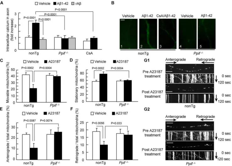
effect of CypD blockade. As shown infigure 3, Ab-treated nonTg axons had a 2.3-fold higher Fluo-4 intensity than vehicle-treated nonTg axons (Fig. 3A–B1–2), while axons treated with cyclosporine A (CsA), a pharmacological inhibitor of CypD, (Fig. 3A–B3) or genetic CypD-deficient axons (Fig. 3A–B4-5) showed no significant change in calcium levels in the presence of Ab. These results demonstrate that blockade of CypD attenuates Ab-induced neuronal calcium elevation and maintains intra-neuronal calcium homeostasis.
To further validate the role of CypD blockage on Ab-induced calcium elevation as shown in Fig. 3A–B on abnormal axonal mitochondrial transport, we evaluated the direct effect of calcium overload on mitochondrial transport. Because CypD deficiency protects cell death from A23187-induced Ca2+
overload, a strong inducer of calcium elevation in intact cells [27], we assessed the effect of CypD depletion on A23187-mediated alterations in axonal mitochondrial transport. Neurons were exposed to 5mM Calcium Ionophore (A23187). Axonal mitochondrial transport was recorded pre- and post-treatment with A23187 in the same neurons. Thirty minutes after A23187 treatment, the total of movable nonTg axonal mitochondria decreased by ,50% (Fig. 3C, 3G1; 20.0265.12% in post-treatment vs. 42.5463.31% in pre-treatment). As a result, the percentage of stationary mitochondria was significantly increased (Fig. 3D, 3G1;57.4663.31% in pre-treatment vs. 79.9865.12% in post-treatment). Similarly, A23187 treatment reduced anterograde and retrograde movement of mitochondria by,54% (Fig. 3E, 3G1; 23.7361.56% in pre-treatment vs. 10.7964.02% in post-treat-ment) and,50% (Fig. 3F, 3G1;18.8162.18% in pre-treatment vs. 9.2362.39% in post-treatment), respectively. Notably,Ppif2/2
axonal mitochondria are resistant to A23187-altered mitochon-drial movement when evaluated for the percentage of movable vs. stationary mitochondria, anterograde or retrograde movement
(Fig. 3C–G). These results indicate that calcium imbalance plays a role in axonal mitochondrial trafficking and that CypD depletion protects against calcium-mediated disruption of axonal mitochon-drial transport and motility.
Effect of CypD Deficiency on ROS-instigated Alterations in Axonal Mitochondrial Transport
Because mitochondria are a major site of reactive oxygen species (ROS) production and because the formation of CypD-mediated mPTP triggers mitochondrial ROS generation, we next
examined whether increased ROS generation contributes to impaired axonal mitochondrial transport. We first measured intra-axonal ROS levels using 2979-dichlorofluorescein diacetate (DCF-DA) fluorescent probe as an indicator of intracellular ROS in Ab-treated axons for the comparison with vehicle-treated axons. Ab-treated nonTg neurons revealed significantly higher DCF-DA intensity than the vehicle-treated group (Fig. 4A, 4B1–2), while Cyp D blockade produced by the addition of CsA (Fig. 4A, 4B3) or genetic deletion of CypD (Fig. 4A, 4B4–5) significantly blunted Ab-induced increase in axonal DCF-DA intensity. These results indicate that blockade of CypD attenuates axonal ROS production or accumulation following Abinsults.
To further evaluate whether increased axonal ROS production contributes to Ab- mediated alterations observed in axonal mitochondrial trafficking, we examined the effects of the antioxidant Probucol on Ab-impaired mitochondrial movement. Treatment with Probucol completely rescued the reduced percentage of movable mitochondria following Ab treatment (Fig. 4C–D;from 26.8761.59% to 41.5362.86%). Accordingly, probucol treatment protected against Ab-induced disruption of anterograde (from 14.4461.40% to 21.8361.28%) and retrograde mitochondrial movement (from 12.2361.61% to 17.3661.23%) (Fig. 4C–D).
CypD-dependent Activation of p38 MAP Kinase Underlies the Axonal Mitochondrial Injury
It is known that Abactivates a variety of kinases including p38/ MAP kinase [39,40] and that impaired mPTP leads to activation of p38 [41]. ROS and calcium are inducers for activation of p38/ MAP kinase [42–44]. P38 activation inhibits fast axonal transport (FAT) by phosphorylation of kinesin (motor protein associated with FAT), which are respectively responsible for axonal mitochondrial transport [45,46]. We therefore examined the relatively unexplored role of CypD-dependent mPTP in activation of p38 on Ab-mediated axonal mitochondrial damage. To do so, we first analyzed the effect of CypD on Ab-induced phosphory-lation of p38 by immunoblotting. As shown infigure 5A–B, Ab -treated nonTg neurons exhibited significantly increased levels of p38 phosphorylation compared to vehicle-treated nonTg neurons
(Fig. 5A). Addition of a specific p38 inhibitor (SB203580) to neurons completely suppressed p38 phosphorylation (Fig. 5A). Interestingly, CypD-deficient neurons were resistant to Ab -in-duced p38 phosphorylation (Fig. 5A–B). It was noted that the
Figure 2. Effect of CypD on Ab-induced changes in axonal mitochondrial morphology.(A) The average length of axonal mitochondria decreased in Ab-treated nonTg neurons, but was largely preserved inPpif2/2neurons. Data were collected from 3 independent experiments. (B, C)
Cumulative distribution data showed that Abtreatment caused a remarkable increase in fragmentation of small mitochondria and a decrease in the numbers of long mitochondria in nonTg neurons; this was partially attenuated inPpif2/2neurons.
baseline of phospho-p38 or total p38 in Ppif 2/2 neurons was comparable to that in nonTg neurons, suggesting no effect of CypD deficiency on p38 signal transduction under physiological condition. Based on the observations that CypD depletion significantly stabilized Ab-induced intracellular calcium and ROS perturbations in neurons and elevated intracellular calcium and ROS are associated with axonal mitochondrial transport and dynamics defects, we then tested whether CypD deficiency inhibits A23187-induced p38 activation by stabilizing intracellular calcium levels and whether Ab-induced p38 activation is suppressed by antioxidant. We first exposed nonTg and CypD deficient neurons to 5mM A23187. Indeed, A23187 treatment significantly in-creased phospho-p38 compared to vehicle-treated nonTg neurons (Fig. S2). No significant elevation of p38 phosphorylation was detected in A23187-treated CypD deficient neurons (Fig. S2). Similarly, the co-incubation of Probucol with Ab significantly suppressed Ab-induced p38 activation in nonTg neurons (Fig. S3). Taken together, these data suggest the linkage of p38 activation
with mPTP associated intracellular calcium and Ab-induced ROS disturbances.
To determine if there is a direct link of p38 activation to mitochondrial transport, we assessed the effect of p38 inhibitor on axonal mitochondrial transport following Ab treatment. Treat-ment with a specific p38 inhibitor (SB203580) resulted in a significantly higher percentage of movable mitochondria in Ab-insulted neurons than in neurons without SB20358 treatment (Fig. 5C; 35.3462.74% with SB203580 vs. 21.9462.95% without SB203580). Similarly, SB203580 treatment protected against Ab -induced alterations in both anterograde and retrograde mitochon-dria (Fig. 5C). As the result, SB203580 treatment attenuated Ab -induced reduction in axonal mitochondrial density (Fig. 5D; 0.19960.01/mm with SB203580 vs. 0.28360.02/mm without SB203580). These results demonstrate that CypD depletion reduces Ab-mediated activation of p38 contributing to the impairment of axonal mitochondrial transport.
Figure 3. Effect of CypD depletion on Ab-induced intra-axonal calcium elevation.(A) Ab-treated nonTg hippocampal neurons displayed an increase in axonal calcium levels. CypD-deficient or CsA-treated (500 nM for 24 hours) neurons diminished elevated levels of calcium. rAbhad no effect on axonal calcium levels. Data were derived from 3 independent experiments. (B) Representative images of axonal calcium staining in nonTg andPpif2/2hippocampal neurons at indicated treatment. Scale bar represents 2mm. (C–G2) Effect of CypD depletion on calcium ionophore
(A23187)-impaired axonal mitochondrial motility. NonTg and Ppif2/2hippocampal neurons were exposed to A23187 (5
mM for 30 min) and
subjected to recording of axonal mitochondrial movements including movable (C), stationary (D), anterograde (E) and retrograde (F) mitochondria. *P,0.05 vs. other groups of neurons. (G1–G2) The kymograph of axonal mitochondrial movement in nonTg (G1) andPpif2/2(G2) neurons before
and after A23187 treatment. A23187 treatment resulted in less movement than the vehicle-treated group.Ppif2/2 neurons revealed increased moving traces compared to nonTg neurons in the presence of A23187. Scale bar represents 10mm.
doi:10.1371/journal.pone.0054914.g003
CypD Depletion Protects Against Ab-induced Synaptic Damage
To analyze the contribution of abnormal axonal mitochondrial transport and/or its directionality to synaptic dysfunction and loss of synapses in Ab-rich environment, we measured synaptic activity by recording the spontaneous miniature excitatory post-synaptic currents (mEPSCs) and also quantified synaptic density. To determine the effect of CypD on synaptic activity, nonTg andPpif
2/2neurons were treated with Aband then subjected to whole-cell
patch-clamp recording of mEPSCs. The frequency of mEPSCs is largely associated with the probability of presynaptic release and the amplitude of mEPSCs at certain levels relies on the size of the vesicle-releasing pool in presynaptic regions [47–49]. Vehicle-treated nonTg andPpif 2/2neurons showed similar patterns of mEPSCs frequency and amplitude, suggesting no effect of CypD deficiency on the spontaneous nonaction potential-dependent activation of synapses under physiological condition. However, Ab-insulted nonTg neurons showed a 54.6% decrease in mEPSCs frequency, compared to 16.4% reduction in Ppif 2/2 neurons (Fig. 6A). As a result, Ab-superimposed Ppif 2/2 neurons significantly preserved mEPSCs frequency (Fig. 6A, 6C; 1.8460.24 Hz inPpif 2/2 neurons vs. 1.0960.23 Hz in nonTg neurons). Similarly, the amplitude of mEPSC was significantly increased in Ab-treatedPpif2/2neurons compared to Ab-treated nonTg neurons (Fig. 6B–C; 61.8863.05 pA in Ppif 2/2 vs. 47.0363.28 pA in nonTg neurons).
To examine the protective effect of CypD depletion on Ab -induced loss of synapses, we quantified synaptophysin-positive clusters attaching to dendrites in cultured hippocampal neurons
derived from nonTg and CypD-deficient mice. Synapses were recognized as synaptophysin-positive clusters attaching to den-drites and denden-drites were determined by MAP2 (microtubule-associated protein 2) staining. Ab-treated nonTg neurons exhibited significantly decreased presynaptic density compared to vehicle-treated control (Fig. 6D–E;vehicle: 0.49260.029/mm vs Ab: 0.27360.02/mm), whereas CypD depletion completely reversed the loss of presynaptic density (Fig. 6D–E;
0.52760.026/mm). The rAb did not affect synaptic density (Fig. 6D–E; 0.50660.019/mm). There was no difference in presynaptic density between nonTg andPpif2/2neurons in the vehicle-treated groups (Fig. 6D–E). To determine effect of p38 activation on loss of synapses, neurons were treated with specific p38 inhibitor (SB203580) for 30 min prior to Ab. A shown in Fig. 6C–D, the addition of SB203580 to culture increased synaptic density (Fig. 6F–G; 0.44260.033% with SB203580 vs. 0.27360.020% without SB203580). Taken together, our results indicate that lack of CypD protects neuron from Ab-insulted synaptic injury with involvement of CypD/Ab-associated P38 MAPK signaling, which is associated with compromised mito-chondrial transport in axon.
Discussion
Abnormal axonal mitochondrial transport is a recently recog-nized mitochondrial pathology induced by Ab [20–26,50]. The precise mechanisms underlying impairments in axonal mitochon-drial transport and the link of mitochonmitochon-drial dysfunction to synaptic damage in AD are not well understood. In this study, we
Figure 4. CypD depletion attenuates Ab-induced intra-axonal ROS elevation.(A) Quantification of DCF intensity in nonTg- orPpif2/2
hippocampal neurons treated with vehicle or Ab. Addition of CsA (500 nM) to cells for 24 hours reduced the DCF intensity. Data were derived from 3 independent experiments. (B) Representative images of axonal DCF staining in nonTg andPpif2/2hippocampal neurons for the indicated treatment.
Scale bar is 10mm. (C–D) Effect of antioxidant (Probucol) on Ab-induced axonal mitochondrial motility. (C) Administration of Probucol (5mM, 24
hours) ameliorated changes in Ab-induced axonal mitochondrial motility. (D) Kymograph images show the protected effects of axonal mitochondrial moving traces following Probucol treatment. Scale bar is 10mm.
analyzed the effect of CypD on Ab-mediated mitochondrial motility and distribution in hippocampal neurons using mice with genetic depletion of CypD. Our results show that CypD depletion protects against Ab-induced alterations in axonal mitochondrial transport as shown by increased mitochondrial motility and distribution, and improved anterograde and retrograde move-ment. The possible mechanisms underlying the protective effect of lacking CypD are suppressed mPTP opening, reduced ROS production, and increased calcium buffering capacity in axonal mitochondria. Furthermore, we also demonstrated that CypD-mediated p38 activation contributes to Ab-impaired axonal mitochondrial transport and synaptic injury. We focus our attention on the protective effect of CypD deficiency on axonal mitochondrial movement in view of the essential role of normal axonal mitochondrial trafficking in supporting synaptic plasticity. Our current study uncovers the role of CypD in Ab-mediated alterations in axonal mitochondrial motility and dynamics contributing to synaptic degeneration in AD.
An increasing body of evidence suggests that oligomeric Ab inhibits axonal mitochondrial transport and breaks the mitochon-drial fusion/fission balance. Ab-disrupted axonal mitochondrial trafficking is a mechanism underlying synaptic degeneration in AD
[20,23–26,50]. In the present of study, we examined the effect of relatively low concentration of Ab(200 nM) that did not alter cell viability on axonal mitochondrial transport to mimic lowin vivo
levels and chronic Abinsults in AD brain. Similar to what have been reported [20,23–26,50], under our experimental condition, 200 nM oligomeric Absignificantly reduced mitochondrial density and movement in axon by 30–40% (Fig. 1A–D and 2A) without significant changes in the cell viability. This suggests an early change in axonal mitochondrial trafficking is prior to neuronal death. A relatively low concentration of Ab(200 nM) used in our study may account for the modest effects on mitochondrial movement without significant neurotoxicity. Indeed, a study has shown that the acute treatment of monomeric Abdemonstrated significant inhibitory effect on neuronal mitochondrial movement [51], suggesting that both Ab species (monomeric or oligomeric forms) are toxic to neuronal mitochondrial transport. In consid-eration of the significance of oligomeric Ab-induced mitochondrial and synaptic dysfunction relevant to the AD pathogenesis [52] and our experimental condition (chronic treatment of low concentra-tion of 200 nM Ab for 24 hours) in which condition that monomeric Abis prone to form oligomers during incubation time [53], we used oligomeric Abfor all our experiments. In addition,
Figure 5. Effect of CypD on Ab-induced activation of p38 MAP kinase and axonal mitochondrial motility.(A) Quantification of phospho-p38 immunoreactive bands (pT180/pY182) in hippocampal neurons treated with vehicle, Ab, or SB203580 (SB, 1mM) plus Ab, respectively, which was normalized for the total p38. (B) Representative immunoblots for phospho- and total-p38. (C–E) Administration of p38 inhibitor, SB203580 (1mM, 24
hours) to cells ameliorated Ab-induced axonal mitochondrial motility changes (C) and mitochondrial density (D). (E) Kymographs showed the protected effects of axonal mitochondrial movement after SB203580 treatment. Scale bar is 10mm.
doi:10.1371/journal.pone.0054914.g005
reversed Abpeptide (rAb) that has the same molecular weight and composition of amino acids with Abbut without biological effects was used as a widely accepted control to verify the specific effects of Ab[54,55].
To elucidate the protective mechanisms of CypD depletion, we focused on the major consequences of mPTP formation on axonal mitochondrial motility and morphology: impaired mitochondrial calcium handling capacity and ROS generation. Ab has been reported to increase intracellular Ca2+
, which could have more targets than mitochondrial trafficking. In view of the role of CypD-dependent mPTP on maintaining intracellular Ca2+homeostasis, significance of Ab-impaired mitochondrial transport on synaptic degeneration, and unexplored role of CypD on mitochondrial transport, it is essential and logical to investigate the involvement of CypD on Ab-induced abnormal axonal mitochondrial trans-port. CypD is a key component for the formation of mPTP
contributing to maintaining calcium homeostasis. CypD deficiency inhibits opening of mPTP, subsequently, increases mitochondrial calcium buffering capacity in response to changes in intracellular calcium levels such as calcium overloading [12,27,29,56]. There-fore, CypD-dependent mPTP is an important regulating mecha-nism of intracellular Ca2+
homeostasis.
We have presented data showing that blockade of CypD by genetic depletion of CypD or pharmacological CypD inhibitor significantly suppressed Ab-induced elevation of the intracellular calcium in axon (Fig. 3A), which are consistent with our [12] and other [27] published studies. These results suggest that an inhibitory effect of CypD deficiency on Ab-mediated changes in intracellular Ca2+
levels is important for maintaining normal mitochondrial transport. To test this hypothesis, we examined a direct effect of CypD deficiency on ionomycin (A23187)-induced Ca2+
overload, a strong inducer of Ca2+
elevation in intact cells,
Figure 6. Effect of CypD on Ab-induced synaptic damage.(A–C) Electrophysiological recording of mEPSCs for Ab-treated nonTg andPpif2/2
neurons. CypD deficiency alleviated Ab-induced decrease in mEPSCs frequency (A) and amplitude (B). Data were derived from 16–19 neurons for each group. (C) Representative traces of mEPSCs in the indicated group. Scale bar represents 100 pA in amplitude and 25 seconds in time. (D–E) Effect of CypD deficiency on synaptic density. The results were derived from 20–30 neurons of each group. Dendrites were visualized by the staining of MAP2 and synapses were recognized as synaptophysin-positive clusters overlapping with dendrites. (E) Representative images for double staining of synaptophysin and MAP-2 in the indicated groups. MAP2 is shown in green color and synaptophysin is labeled by red fluorescence. (F–G) Effect of Ab-induced activation of p38 MAP kinase on synaptic density. (F) Administration of p38 inhibitor, SB203580 (1mM, 24 hours) to cells ameliorated Ab -induced synaptic loss. (G) Representative images showed the protected effects of synaptic density after SB203580 treatment.
and alterations in axonal mitochondrial transport [27]. As expected, CypD-deficient neurons blocked A23187-induced ele-vation of intracellular Ca2+(Fig. S4) and p38 activation (Fig. S2). This could be a mechanism of the protective effect of CypD deficiency on A23187-altered axonal mitochondrial trafficking (Fig. 3C–G). Compared to the effect of Ab, A23187 treatment had a greater effect on mitochondrial transport (50% decline in A23187 treatment vs. 30–40% in Ab-treated cells). A strong induction of calcium elevation in intact cells (high levels of Ca2+ ) by A23187 could be the explanation for a more dramatic effect of A23187 (50% in Fig. 3C, E–F) than Ab treatment (30% in
Fig. 1A–B, C2, D1and 40% in Fig. 2A–C) in which elevated levels of Ca2+are expected to be lower than A23187-treated cells. A direct role of intracellular calcium in controlling axonal mitochondrial motility and dynamics is also supported by the recent study. For example, calcium-induced mitochondrial disso-ciation has been postulated as a potential mechanism for modulation of mitochondrial docking under physiological condi-tions [57]. Increased calcium levels are reported to decrease mitochondrial movement/transport by interrupting Miro and kinesin complexes [57,58]. At pathological states with significant and sustained calcium elevation achieved by the activation ofN
-Methyl-D-aspartate (NMDA) receptors [37] or the application of A23187 [59], mitochondrial morphology and movement are substantially disrupted, suggesting the impact of pathological intra-calcium perturbations. In Ab-rich environment where calcium levels are abnormally high [60–63], increased mitochondrial detachments occur in Ab-treated axons (represented by an increased percentage of stationary mitochondria). Thus, axonal mitochondria are crucial to the calcium buffering process. Maintenance of axonal calcium homeostasis by CypD depletion is an underlying mechanism for controlling axonal mitochondrial
calcium in the face of Abinsults. Ab-mediated elevation of calcium is a potential mechanism at the nexus of Ab toxicity and alterations in mitochondrial motility. The dramatic protection of lacking CypD against A23187-disturbed calcium balance as well as mitochondrial motility and dynamics changes provides substantial evidence that the blockade of CypD-mediated mPTP counteracts calcium-instigated axonal mitochondrial alterations in trafficking and morphology.
Another major consequence of CypD-mediated mPTP forma-tion is increased ROS producforma-tion/accumulaforma-tion leading to release of ROS from mitochondrial to cytosol. As Ca2+
metabolism and oxidative stress are intertwined, especially in mitochondrial processes, these organelles can become severely dysfunctional during the permeability transition in combination with effects of oxidative stress and dysregulation of cytosolic free Ca2+
. Indeed, in the present study, we report reduced mitochondrial calcium buffering capacity, increased membrane permeability transition, and accumulation of ROS in axons in the presence of Ab. ROS has been implicated in disruption of mitochondrial movement. For example, zinc-induced ROS generation is associated with phosphatidylinositol (PI) 3-kinase activation, which in turn disrupts mitochondrial transport in neurons [64]. However, to our knowledge, the contribution of axonal ROS dysregulation to Ab-induced defects in mitochondrial transport has not yet been documented. We showed here that the addition of probucol, an antioxidant drug to suppress ROS generation, or genetic deletion of CypD to blunt oxidative stress and to enhance mitochondrial calcium buffer capability significantly rescues mitochondrial movement against Ab toxicity, indicating the significance of oxidative stress on Ab-altered axonal mitochondrial trafficking. These data support that Ab-induced intra-axonal ROS has deleterious effects on transport.
Activation of P38 mitogen-activated protein kinase (MAPK) is associated with increased intracellular calcium, ROS production/ accumulation, Ab stimulation, and mitochondrial stress [43,65– 71]. We demonstrated that levels of P38 phosphorylation were significantly increased in Ab-treated neurons. Antioxidant Probu-col blocked Ab-induced p38 activation, indicating a role of Ab -induced oxidative stress in disruption of signal transduction such as p38 MAP kinase contributing to abnormal axonal mitochondrial transport. Notably, Ab-induced p38 phosphorylation was blunted in neurons lacking CypD. The addition of a specific p38 inhibitor (SB203580) resulted in pronounced preservation of mitochondrial motility and morphology even in the face of Abinsults, indicating the involvement of CypD/Ab-associated p38 MAPK signaling in disruption of axonal mitochondrial trafficking. The application of p38 inhibitor did not interfere with Ab-induced calcium elevation (data not shown). These results suggest that p38 is a downstream target of Ab. Thus, we propose that CypD-dependent impaired calcium homeostasis and ROS production/accumulation in axons are responsible for p38 MAPK activation, which leads to further mitochondrial injury including abnormal axonal mitochondrial transport and loss of synapse. The detailed mechanisms of P38 activation in injuring axonal mitochondrial transport need further investigation. For example, P38 activation is connected with changes in mitochondrial movement via phosphorylation of kinesin [46] and dynein [72], which dissociates mitochondria from the motor proteins. In addition to p38, perturbations of several other signaling cascades including PKA [51] and GSK-3b [23] are also reported to be involved in Ab-induced disruption in mitochondrial transport. Given the tight interaction of these signaling cascades [73], they may work together in keeping axonal mitochondrial movement in normal fashion while their indepen-dent effects on mitochondrial trafficking remain unclear. In view
Figure 7. Working hypothesis. Ab-Cyclphilin D mediates impair-ments in axonal mitochondrial transport. In the present of Ab, there is an increase in the opening of CypD-mediated mitochondrial perme-ability transition pore (mPTP), leading to disruption of Ca2+balance and
increase in reactive oxygen species (ROS) production/accumulation. Consequently, elevation of Ca2+and oxidative activates downstream
signal pathway p38 MAP Kinase contributing to mitochondrial dysfunction, deficits in axonal mitochondrial trafficking, eventually, synaptic damage.
doi:10.1371/journal.pone.0054914.g007
of potential involvement of motor proteins in mitochondrial movement, we will further examine whether mPTP-associated axonal mitochondrial transport changes are related to changes of motor proteins such as hyperphosphorylation of dynein and kinesin and altered Miro activity state in near future. Nevertheless, our study suggests that CypD-involved activation of p38 signaling plays a role, at least in part, in Ab-insulted abnormal mitochon-drial transport in axon.
Axonal mitochondria are dynamic organelles and their traffick-ing and docktraffick-ing are critical for synaptic plasticity and function. Synaptic loss and deactivation are biological basis of AD. Increasing evidence emphasizes the importance of mitochondria for the maintenance of synaptic function. Defects in dendritic mitochondria lead to dendritic degeneration [74] and injured mitochondria in the presynapse region are associated with compromised presynaptic function [75]. Mitochondrial transport maintains functional mitochondria around synapses [76] Pre-viously, we and other groups showed that Ab insults results in impaired mitochondrial distribution and trafficking in axons [20,23–26], while in the present study, we demonstrate the protective effects of CypD depletion on Ab-mediated deficits in axonal mitochondrial transport and synaptic injury including synaptic activity and loss of synapses. Notably, blockade of p38 activation significantly rescue synaptic loss insulted by Ab(Fig. 6F– G), supporting a connection of CypD/Ab-involved signal trans-duction (p38) with mitochondrial and synaptic degeneration.
In summary, our data offer new insights into the mechanism of mitochondrial perturbation in the pathogenesis of AD, specifically the role of CypD in axonal mitochondrial transport. Ab-CypD interaction promotes opening of mitochondrial permeability transition pore, consequently, disrupts calcium balance and enhances production/accumulation of ROS, thereby further activating P38 MAPK signal transduction pathway. All these events disrupt mitochondrial trafficking and dynamics, ultimately causing synaptic damage (Fig. 7). We have clearly demonstrated that CypD depletion protects axonal mitochondrial transport from Abinsults along with suppressing Ab-induced elevation of calcium and accumulation of oxidative stress. Importantly, CypD depletion also suppressed Ab-induced activation of p38/MAPK and this inhibition rescued axonal mitochondrial movement and pre-synaptic density. Thus, our results provide evidence that CypD/ Ab-mediated mitochondrial dysfunction is correlated with disrup-tion of axonal mitochondrial transport and synaptic injury. These findings significantly enhance our understanding of the patholog-ical role of CypD in axonal pathology in AD.
Supporting Information
Figure S1 Cultured hippocampal neurons were transfected with pDsRedmito and observed under microscope. The figure showed the image of a transfected neuron. Middle part of the axon (in the frame) was selected for the experiment to detect mitochondrial movement.
(TIF)
Figure S2 CypD depletion suppresses A23187-induced p38 phosphorylation. NonTg and CypD deficient hippocampal neurons were exposed to 5mM A23187 for 15 and 30 min, respectively. Cell lysates were subjected to immunoblots for phospho- and total-p38. The treatment of A23187 on nonTg neurons significantly increased p38 phosphorylation level as compared to the vehicle-treated neurons (vehicle: 160.021 vs. A2318715 min: 1.8960.047; vehicle vs. A23187 30 min: 2.0660.18). CypD depletion significantly suppressed A23187-induced elevation of phospho-p38. Data were derived from 4 independent experiments.
(TIF)
Figure S3 Addition of Probucol attenuates Ab-induced p38 phosphorylation in nonTg neurons. nonTg neurons were treated with Ab co-incubated in the presence or absence of Probucol (5mM, 24 hours). Abtreatment resulted in a significant elevation of p38 phosphorylation level as compared to vehicle treatment (vehicle: 160.077 vs. Ab: 3.0660.27), while Ab-induced p38 phosphorylation was inhibited by the addition of Probucol (Ab: 3.0660.27 vs. Ab+Probucol: 1.1560.46). Data were derived from 3 independent experiments.
(TIF)
Figure S4 Effect of CypD depletion on A23187-induced intra-axonal calcium elevation. NonTg and Ppif 2/2 hippocampal neurons were exposed to A23187 (5mM for 30 min) and subjected to recording of intra-axonal calcium before and after A23187 treatment. A23187 treatment resulted in increased axonal calcium level in nonTg neurons. Ppif 2/2 neurons abolished A23187-induced calcium elevation.
(TIF)
Author Contributions
Conceived and designed the experiments: SSY JXC LG HD. Performed the experiments: LG HD SQY XPW. Analyzed the data: LG HD SQY XPW. Contributed reagents/materials/analysis tools: LG HD SQY XPW GMM. Wrote the paper: SSY LG HD.
References
1. Kang JS, Tian JH, Pan PY, Zald P, Li C, et al. (2008) Docking of axonal mitochondria by syntaphilin controls their mobility and affects short-term facilitation. Cell 132: 137–148.
2. Miller KE, Sheetz MP (2004) Axonal mitochondrial transport and potential are correlated. Journal of cell science 117: 2791–2804.
3. Billups B, Forsythe ID (2002) Presynaptic mitochondrial calcium sequestration influences transmission at mammalian central synapses. The Journal of neuroscience : the official journal of the Society for Neuroscience 22: 5840– 5847.
4. David G, Barrett EF (2000) Stimulation-evoked increases in cytosolic [Ca(2+)] in mouse motor nerve terminals are limited by mitochondrial uptake and are temperature-dependent. The Journal of neuroscience : the official journal of the Society for Neuroscience 20: 7290–7296.
5. Talbot JD, David G, Barrett EF (2003) Inhibition of mitochondrial Ca2+uptake affects phasic release from motor terminals differently depending on external [Ca2+]. Journal of neurophysiology 90: 491–502.
6. Chada SR, Hollenbeck PJ (2003) Mitochondrial movement and positioning in axons: the role of growth factor signaling. The Journal of experimental biology 206: 1985–1992.
7. Chada SR, Hollenbeck PJ (2004) Nerve growth factor signaling regulates motility and docking of axonal mitochondria. Current biology : CB 14: 1272– 1276.
8. Morris RL, Hollenbeck PJ (1993) The regulation of bidirectional mitochondrial transport is coordinated with axonal outgrowth. Journal of cell science 104 (Pt 3): 917–927.
9. Chang DT, Honick AS, Reynolds IJ (2006) Mitochondrial trafficking to synapses in cultured primary cortical neurons. The Journal of neuroscience : the official journal of the Society for Neuroscience 26: 7035–7045.
10. Baloh RH, Schmidt RE, Pestronk A, Milbrandt J (2007) Altered axonal mitochondrial transport in the pathogenesis of Charcot-Marie-Tooth disease from mitofusin 2 mutations. The Journal of neuroscience : the official journal of the Society for Neuroscience 27: 422–430.
11. Caspersen C, Wang N, Yao J, Sosunov A, Chen X, et al. (2005) Mitochondrial Abeta: a potential focal point for neuronal metabolic dysfunction in Alzheimer’s disease. FASEB journal : official publication of the Federation of American Societies for Experimental Biology 19: 2040–2041.
13. Du H, Yan SS (2010) Mitochondrial permeability transition pore in Alzheimer’s disease: cyclophilin D and amyloid beta. Biochimica et biophysica acta 1802: 198–204.
14. Hansson MJ, Mansson R, Morota S, Uchino H, Kallur T, et al. (2008) Calcium-induced generation of reactive oxygen species in brain mitochondria is mediated by permeability transition. Free radical biology & medicine 45: 284–294. 15. Lustbader JW, Cirilli M, Lin C, Xu HW, Takuma K, et al. (2004) ABAD
directly links Abeta to mitochondrial toxicity in Alzheimer’s disease. Science 304: 448–452.
16. Manczak M, Anekonda TS, Henson E, Park BS, Quinn J, et al. (2006) Mitochondria are a direct site of A beta accumulation in Alzheimer’s disease neurons: implications for free radical generation and oxidative damage in disease progression. Human molecular genetics 15: 1437–1449.
17. Takuma K, Fang F, Zhang W, Yan S, Fukuzaki E, et al. (2009) RAGE-mediated signaling contributes to intraneuronal transport of amyloid-beta and neuronal dysfunction. Proceedings of the National Academy of Sciences of the United States of America 106: 20021–20026.
18. Yao J, Irwin RW, Zhao L, Nilsen J, Hamilton RT, et al. (2009) Mitochondrial bioenergetic deficit precedes Alzheimer’s pathology in female mouse model of Alzheimer’s disease. Proceedings of the National Academy of Sciences of the United States of America 106: 14670–14675.
19. Mao P, Manczak M, Calkins MJ, Truong Q, Reddy TP, et al. (2012) Mitochondria-targeted catalase reduces abnormal APP processing, amyloid beta production and BACE1 in a mouse model of Alzheimer’s disease: implications for neuroprotection and lifespan extension. Hum Mol Genet.
20. Du H, Guo L, Yan S, Sosunov AA, McKhann GM, et al. (2010) Early deficits in synaptic mitochondria in an Alzheimer’s disease mouse model. Proceedings of the National Academy of Sciences of the United States of America 107: 18670– 18675.
21. Du H, Guo L, Yan SS (2012) Synaptic mitochondrial pathology in Alzheimer’s disease. Antioxid Redox Signal 16: 1467–1475.
22. Reddy PH, Tripathi R, Troung Q, Tirumala K, Reddy TP, et al. (2012) Abnormal mitochondrial dynamics and synaptic degeneration as early events in Alzheimer’s disease: Implications to mitochondria-targeted antioxidant thera-peutics. Biochim Biophys Acta 1822: 639–649.
23. Decker H, Lo KY, Unger SM, Ferreira ST, Silverman MA (2010) Amyloid-beta peptide oligomers disrupt axonal transport through an NMDA receptor-dependent mechanism that is mediated by glycogen synthase kinase 3beta in primary cultured hippocampal neurons. The Journal of neuroscience : the official journal of the Society for Neuroscience 30: 9166–9171.
24. Calkins MJ, Manczak M, Mao P, Shirendeb U, Reddy PH (2011) Impaired mitochondrial biogenesis, defective axonal transport of mitochondria, abnormal mitochondrial dynamics and synaptic degeneration in a mouse model of Alzheimer’s disease. Human molecular genetics 20: 4515–4529.
25. Calkins MJ, Reddy PH (2011) Amyloid beta impairs mitochondrial anterograde transport and degenerates synapses in Alzheimer’s disease neurons. Biochimica et biophysica acta 1812: 507–513.
26. Wang X, Perry G, Smith MA, Zhu X (2010) Amyloid-beta-derived diffusible ligands cause impaired axonal transport of mitochondria in neurons. Neuro-degenerative diseases 7: 56–59.
27. Baines CP, Kaiser RA, Purcell NH, Blair NS, Osinska H, et al. (2005) Loss of cyclophilin D reveals a critical role for mitochondrial permeability transition in cell death. Nature 434: 658–662.
28. Baines CP (2007) The mitochondrial permeability transition pore as a target of cardioprotective signaling. American journal of physiology Heart and circulatory physiology 293: H903–904.
29. Du H, Guo L, Zhang W, Rydzewska M, Yan S (2011) Cyclophilin D deficiency improves mitochondrial function and learning/memory in aging Alzheimer disease mouse model. Neurobiology of aging 32: 398–406.
30. Banker GA, Cowan WM (1979) Further observations on hippocampal neurons in dispersed cell culture. J Comp Neurol 187: 469–493.
31. Cai Q, Gerwin C, Sheng ZH (2005) Syntabulin-mediated anterograde transport of mitochondria along neuronal processes. The Journal of cell biology 170: 959– 969.
32. Oliva AA, Jr., Atkins CM, Copenagle L, Banker GA (2006) Activated c-Jun N-terminal kinase is required for axon formation. The Journal of neuroscience : the official journal of the Society for Neuroscience 26: 9462–9470.
33. Sharma G, Vijayaraghavan S (2003) Modulation of presynaptic store calcium induces release of glutamate and postsynaptic firing. Neuron 38: 929–939. 34. Trinchese F, Liu S, Ninan I, Puzzo D, Jacob JP, et al. (2004) Cell cultures from
animal models of Alzheimer’s disease as a tool for faster screening and testing of drug efficacy. J Mol Neurosci 24: 15–21.
35. Mattson MP, Cheng B, Davis D, Bryant K, Lieberburg I, et al. (1992) beta-Amyloid peptides destabilize calcium homeostasis and render human cortical neurons vulnerable to excitotoxicity. The Journal of neuroscience : the official journal of the Society for Neuroscience 12: 376–389.
36. Ferreira IL, Bajouco LM, Mota SI, Auberson YP, Oliveira CR, et al. (2011) Amyloid beta peptide 1–42 disturbs intracellular calcium homeostasis through activation of GluN2B-containing N-methyl-d-aspartate receptors in cortical cultures. Cell calcium.
37. Rintoul GL, Filiano AJ, Brocard JB, Kress GJ, Reynolds IJ (2003) Glutamate decreases mitochondrial size and movement in primary forebrain neurons. The Journal of neuroscience : the official journal of the Society for Neuroscience 23: 7881–7888.
38. Yi M, Weaver D, Hajnoczky G (2004) Control of mitochondrial motility and distribution by the calcium signal: a homeostatic circuit. The Journal of cell biology 167: 661–672.
39. Origlia N, Bonadonna C, Rosellini A, Leznik E, Arancio O, et al. (2010) Microglial receptor for advanced glycation end product-dependent signal pathway drives beta-amyloid-induced synaptic depression and long-term depression impairment in entorhinal cortex. The Journal of neuroscience : the official journal of the Society for Neuroscience 30: 11414–11425.
40. Zhu X, Mei M, Lee HG, Wang Y, Han J, et al. (2005) P38 activation mediates amyloid-beta cytotoxicity. Neurochemical research 30: 791–796.
41. Tomasello F, Messina A, Lartigue L, Schembri L, Medina C, et al. (2009) Outer membrane VDAC1 controls permeability transition of the inner mitochondrial membrane in cellulo during stress-induced apoptosis. Cell research 19: 1363– 1376.
42. Matsuyama D, Kawahara K (2011) Oxidative stress-induced formation of a positive-feedback loop for the sustained activation of p38 MAPK leading to the loss of cell division in cardiomyocytes soon after birth. Basic research in cardiology 106: 815–828.
43. Lee MW, Park SC, Yang YG, Yim SO, Chae HS, et al. (2002) The involvement of reactive oxygen species (ROS) and p38 mitogen-activated protein (MAP) kinase in TRAIL/Apo2L-induced apoptosis. FEBS letters 512: 313–318. 44. Wright DC, Geiger PC, Han DH, Jones TE, Holloszy JO (2007) Calcium
induces increases in peroxisome proliferator-activated receptor gamma coacti-vator-1alpha and mitochondrial biogenesis by a pathway leading to p38 mitogen-activated protein kinase activation. The Journal of biological chemistry 282: 18793–18799.
45. Morfini G, Pigino G, Szebenyi G, You Y, Pollema S, et al. (2006) JNK mediates pathogenic effects of polyglutamine-expanded androgen receptor on fast axonal transport. Nature neuroscience 9: 907–916.
46. De Vos K, Severin F, Van Herreweghe F, Vancompernolle K, Goossens V, et al. (2000) Tumor necrosis factor induces hyperphosphorylation of kinesin light chain and inhibits kinesin-mediated transport of mitochondria. The Journal of cell biology 149: 1207–1214.
47. Kimura A, Pavlides C (2000) Long-term potentiation/depotentiation are accompanied by complex changes in spontaneous unit activity in the hippocampus. J Neurophysiol 84: 1894–1906.
48. Zhang J, Yang Y, Li H, Cao J, Xu L (2005) Amplitude/frequency of spontaneous mEPSC correlates to the degree of long-term depression in the CA1 region of the hippocampal slice. Brain Res 1050: 110–117.
49. Rohrbough J, Spitzer NC (1999) Ca(2+)-permeable AMPA receptors and spontaneous presynaptic transmitter release at developing excitatory spinal synapses. J Neurosci 19: 8528–8541.
50. Pigino G, Morfini G, Atagi Y, Deshpande A, Yu C, et al. (2009) Disruption of fast axonal transport is a pathogenic mechanism for intraneuronal amyloid beta. Proceedings of the National Academy of Sciences of the United States of America 106: 5907–5912.
51. Rui Y, Tiwari P, Xie Z, Zheng JQ (2006) Acute impairment of mitochondrial trafficking by beta-amyloid peptides in hippocampal neurons. The Journal of neuroscience : the official journal of the Society for Neuroscience 26: 10480– 10487.
52. Benilova I, Karran E, De Strooper B (2012) The toxic Abeta oligomer and Alzheimer’s disease: an emperor in need of clothes. Nature neuroscience 15: 349–357.
53. Stine WB, Jr., Dahlgren KN, Krafft GA, LaDu MJ (2003) In vitro characterization of conditions for amyloid-beta peptide oligomerization and fibrillogenesis. The Journal of biological chemistry 278: 11612–11622. 54. Parameshwaran K, Sims C, Kanju P, Vaithianathan T, Shonesy BC, et al.
(2007) Amyloid beta-peptide Abeta(1–42) but not Abeta(1–40) attenuates synaptic AMPA receptor function. Synapse 61: 367–374.
55. Perez JL, Carrero I, Gonzalo P, Arevalo-Serrano J, Sanz-Anquela JM, et al. (2010) Soluble oligomeric forms of beta-amyloid (Abeta) peptide stimulate Abeta production via astrogliosis in the rat brain. Experimental neurology 223: 410– 421.
56. Nakagawa T, Shimizu S, Watanabe T, Yamaguchi O, Otsu K, et al. (2005) Cyclophilin D-dependent mitochondrial permeability transition regulates some necrotic but not apoptotic cell death. Nature 434: 652–658.
57. Macaskill AF, Rinholm JE, Twelvetrees AE, Arancibia-Carcamo IL, Muir J, et al. (2009) Miro1 is a calcium sensor for glutamate receptor-dependent localization of mitochondria at synapses. Neuron 61: 541–555.
58. Wang X, Schwarz TL (2009) The mechanism of Ca2+-dependent regulation of kinesin-mediated mitochondrial motility. Cell 136: 163–174.
59. Dubinsky JM, Levi Y (1998) Calcium-induced activation of the mitochondrial permeability transition in hippocampal neurons. Journal of neuroscience research 53: 728–741.
60. Hartmann H, Eckert A, Muller WE (1993) beta-Amyloid protein amplifies calcium signalling in central neurons from the adult mouse. Biochemical and biophysical research communications 194: 1216–1220.
61. Sberna G, Saez-Valero J, Beyreuther K, Masters CL, Small DH (1997) The amyloid beta-protein of Alzheimer’s disease increases acetylcholinesterase expression by increasing intracellular calcium in embryonal carcinoma P19 cells. Journal of neurochemistry 69: 1177–1184.
62. Lin F, Wang Y, Hosford DA (1999) Age-related relationship between mRNA expression of GABA(B) receptors and calcium channel beta4 subunits in cacnb4lh mice. Brain research Molecular brain research 71: 131–135.
63. Canevari L, Abramov AY, Duchen MR (2004) Toxicity of amyloid beta peptide: tales of calcium, mitochondria, and oxidative stress. Neurochemical research 29: 637–650.
64. Malaiyandi LM, Honick AS, Rintoul GL, Wang QJ, Reynolds IJ (2005) Zn2+ inhibits mitochondrial movement in neurons by phosphatidylinositol 3-kinase activation. The Journal of neuroscience : the official journal of the Society for Neuroscience 25: 9507–9514.
65. Mattson MP (1994) Calcium and neuronal injury in Alzheimer’s disease. Contributions of beta-amyloid precursor protein mismetabolism, free radicals, and metabolic compromise. Annals of the New York Academy of Sciences 747: 50–76.
66. Mattson MP (1995) Free radicals and disruption of neuronal ion homeostasis in AD: a role for amyloid beta-peptide? Neurobiology of aging 16: 679–682; discussion 683.
67. Bus AI, Huang X, Fairlie DP (1999) The possible origin of free radicals from amyloid beta peptides in Alzheimer’s disease. Neurobiology of aging 20: 335– 337; discussion 339–342.
68. Kulisz A, Chen N, Chandel NS, Shao Z, Schumacker PT (2002) Mitochondrial ROS initiate phosphorylation of p38 MAP kinase during hypoxia in cardiomyocytes. American journal of physiology Lung cellular and molecular physiology 282: L1324–1329.
69. Semenova MM, Maki-Hokkonen AM, Cao J, Komarovski V, Forsberg KM, et al. (2007) Rho mediates calcium-dependent activation of p38alpha and subsequent excitotoxic cell death. Nature neuroscience 10: 436–443. 70. Arancio O, Zhang HP, Chen X, Lin C, Trinchese F, et al. (2004) RAGE
potentiates Abeta-induced perturbation of neuronal function in transgenic mice. The EMBO journal 23: 4096–4105.
71. Chen KC, Kao PH, Lin SR, Chang LS (2008) p38 MAPK activation and mitochondrial depolarization mediate the cytotoxicity of Taiwan cobra phospholipase A2 on human neuroblastoma SK-N-SH cells. Toxicology letters
180: 53–58.
72. Kim S, Kim HY, Lee S, Kim SW, Sohn S, et al. (2007) Hepatitis B virus x protein induces perinuclear mitochondrial clustering in microtubule- and Dynein-dependent manners. Journal of virology 81: 1714–1726.
73. Thornton TM, Pedraza-Alva G, Deng B, Wood CD, Aronshtam A, et al. (2008) Phosphorylation by p38 MAPK as an alternative pathway for GSK3beta inactivation. Science 320: 667–670.
74. Li Z, Okamoto K, Hayashi Y, Sheng M (2004) The importance of dendritic mitochondria in the morphogenesis and plasticity of spines and synapses. Cell 119: 873–887.
75. Zenisek D, Matthews G (2000) The role of mitochondria in presynaptic calcium handling at a ribbon synapse. Neuron 25: 229–237.