Q uantitative studie s o f the vasculature
o f the caro tid bo dy in the chro nically
hypo xic rat
1Department of Physiology, The Royal Free and University College Medical School,
London, UK
2Department of Physiology, the Medical School, University of Birmingham,
Birmingham, UK
3Department of Environmental and Preventive Medicine, Wolfson
Institute of Preventive Medicine, Q ueen Mary and Westfield College, London, UK J.A. Clarke1,
M. de B. Daly1,
J.M. Marshall2,
H.W. Ead† and
E.M. Hennessy3
Abstract
The carotid bodies of rats made chronically hypoxic by breathing 12% O2 in a normobaric chamber (inspired PO2 91 mmHg) were compared
with those of controls. Serial 5-µm sections of the organs were examined using an interactive image analysis system. The total vol-ume of the carotid bodies was increased by 64%. The total vascular volume rose by 103% and was likely due to an increase in size of the large vessels (>12 µm lumen diameter) because the small vessel (5-12 µm lumen diameter) volume did not increase significantly while the small vessel density tended to decrease. The extravascular volume was increased by 57%. Expressed as a percentage of the total volume of the organ, the total vascular volume did not change, but the small vessel volume was significantly decreased from 7.83 to 6.06%. The large vessel volume must therefore have been increased. The proportion occupied by the extravascular volume was virtually unchanged (84 vs
82%). In accordance with these findings, the small vessel endothelial surface area per unit carotid body volume was diminished from 95.2 to 76.5 mm-1, while the extravascular area per small vessel was increased
from 493 to 641 µm2 or by 30%. In conclusion, the enlargement of the
carotid body in chronic hypoxia is most likely due to an increase in total vascular volume, mainly involving the large vessels, and to an increase in extravascular volume. This is in contrast to our previously published findings indicating that in the spontaneous insulin-depend-ent diabetic rat the enlargeminsulin-depend-ent of the carotid body is due solely to an increase in extravascular volume.
Co rre spo nde nce
M. de B. Daly Royal Free and University College Medical School Royal Free Campus Rowland Hill Street London NW3 2PF UK
Fax: + 44-171-433-1921
Research supported by grants from British Telecommunications plc to J.A. Clarke and from the British Heart Foundation to M. de B. Daly and to J.M. Marshall. †Deceased.
Some of these data were previously reported in abstract form in Ref. 10 (Journal of Physiology (1996) 495: 29P).
Received January 29, 1999 Accepted January 3, 2000
Ke y wo rds
·Rat carotid body
·Morphology
·Chronic hypoxia
Intro ductio n
Chronic hypoxia causes enlargement of the carotid bodies as first shown by Arias-Stella (1) and confirmed since in several studies in humans (2) and cattle living at high altitude (3). It also occurs in animals exposed
to an environment of lowered PO2 at sea
level and this enlargement is reversed by
raising the PO2 to normal (3-6).
organ or to the appearance of the systemic arteries, arterioles and the microvasculature in the organ under these conditions.
In this paper we report the results of an investigation to establish the role that vascu-lar structures might play in the envascu-largement of the carotid body in the chronically hy-poxic rat. The various compartments of the carotid body were analysed quantitatively and particular attention was paid to the vol-ume and proportion of the small vessels (5-12 µm in diameter) in the vascular compart-ment together with the density of small ves-sels. The term small vessels has been de-fined and used previously (8,9).
This study also constitutes a further in-vestigation by us of the pathophysiological conditions affecting the microvasculature of the carotid body, thereby permitting a com-parison of the effects of chronic hypoxia in the organ with those occurring in the sponta-neous insulin-dependent diabetic rat (9,10).
Mate rial and Me tho ds
Carotid bifurcation regions were exam-ined bilaterally in 5 control adult male rats (Wistar strain), age 77 days, 3 of which had been prepared at an earlier date as part of another study, and in 3 male rats age 80 days and of similar weight (Table 1), made chroni-cally hypoxic by breathing a mixture of 12% O2 in 88% N2 in a normobaric chamber for
31 days (inspired PO2 approximately 91
mmHg) (Supplier: Biomedical Services Unit, University of Birmingham, Birmingham, UK). The carotid bodies were analysed quan-titatively, particular attention being paid to the volume and proportion of the small vessels (5-12 µm lumen diameter) and larger vessels (>12 µm lumen diameter) in the vascular compartment, as defined by us previously (8,9). A complete description of the perfusion-fixation technique used to ex-amine the carotid bifurcation regions in rats has been given previously (9,11,12). Briefly, the animals were anaesthetized with
pento-barbitone sodium (Sagatal, Rhöne Mérieux; 40 mg/kg, intraperitoneally), heparinized, submitted to median sternotomy, and quickly bled to death via a large incision in the right atrium. Immediately, perfusion of the ca-rotid bifurcations was begun via the ascend-ing aorta with sodium chloride solution (154 mmol/l) at a pressure of 100 mmHg and temperature of 37o
C, the arterial pressure in the chronically hypoxic group being the same as in the control group breathing room air (13). Perfusion was followed by 3% glutar-aldehyde in isotonic phosphate buffer, pH 7.3, at the same pressure and temperature for 5 min. Each animal was stored overnight with the level of the carotid bifurcation re-gions 1-2 cm above that of the heart to maintain a normal venous pressure in these regions and to prevent retrograde movement of blood-stained fluid and occasional red cells from the trunk of the animal into the carotid body vasculature. Each carotid bifur-cation region was prepared routinely for light microscopy and ribbons of transverse serial 5-µm sections from paraffin wax blocks were cut and stained using a modification of the Martius Scarlet Blue method for fibrin (9,12). The various compartments of the carotid body were determined quantitatively using an interactive image analysis system. From an analysis of the histological sections taken at sample intervals of 25 µm, the following information was obtained by using Simpsons rule (5): 1) carotid body area and volume; 2) total vascular area and volume; 3) extravas-cular area and volume by subtraction (7); 4) small vessel endothelial surface area, i.e., a measurement of the surface area actually based on the external surface of the endothe-lial cells; 5) large vessel endotheendothe-lial surface area, and 6) ratio of small vessel endothelial surface area to the carotid body volume. Values for extravascular area of the carotid body and small vessel area were obtained by summing the respective areas of individual sections of the total organ (9).
val-ues ± SD. For each measure the outcome variable was the mean for the left and right carotid body. Students unpaired t-test was used to compare the means for hypoxic and control rats. We do not know whether as-sumptions regarding normality are appropri-ate and therefore the statistical significance of the results should be interpreted with caution, but nonparametric tests with such small numbers would not have had the power to detect statistical significance. An assump-tion of similar variances gives more conser-vative results than the method for different variances and was therefore used. Stata (14) was used for the statistical analysis. Values were taken as significant if P<0.05.
Re sults
Micro vasculature o f the caro tid bo dy
We have described this previously in de-tail (9), but we shall briefly review the rel-evant points which are applicable to this paper. Classically, the carotid body artery, having originated from the external carotid artery, approaches the caudal pole of the organ and branches at its caudal margin into several arterioles. Thereafter, two distinct circulatory routes through the organ are dis-cernible. Firstly, there is the straight through circulation which consists of three or four small arterioles confined to the central con-nective tissue framework of the carotid body. Secondly, there is the parenchymal circula-tion which consists of a dense capillary network which fans out from the caudal pole of the carotid body and travels rostrally to supply the type 1 and type 2 cells. This arrangement was clearly observed by us in a computer reconstruction of the vasculature of the rat carotid body (15; Clarke JA, Duff MJB and Ip HH-S, unpublished observa-tions).
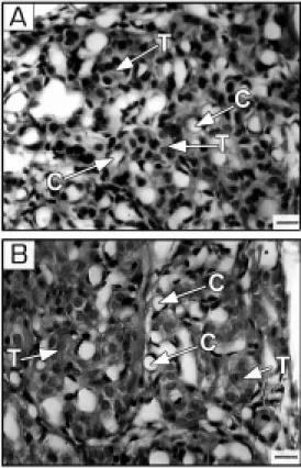
The general topographical description of the carotid bodies agreed well with that given previously (6) and is shown in Figure 1 taken
from a normal animal (A) and a hypoxic animal (B). The type 1 cells of the carotid bodies of the chronically hypoxic rats showed no evidence of recent hyperplasia. The nu-clei of the type 1 cells and type 2 cells, which could be unequivocally identified, were uni-formly devoid of metaphase spindles or other features of chromosome aggregation. Meas-urements of type 1 cells in randomly se-lected sections indicated that the size of the cells fell within the accepted normal range of 15-30 µm (16), but this variable has not been made the subject of a special study. The endothelial cells of the small vessels were prominent in contrast to those of the control organs, being more intensely stained. This resulted in many of the vessels appearing to have distinct cellular crescents adjacent to the lumen. These features were absent in the controls (Figure 1A,B). The nuclei of the endothelial cells showed no evidence of re-cent mitosis, as metaphase chromosome con-figuration was absent. No evidence of sys-temic arterial disease was noted in the
ca-Figure 1 - Photomicrographs of the carotid body from a control rat (A) and chronically hypoxic animal (B). Section thickness, 5 µm. M artius Scarlet Blue meth-od of staining. Bar, 10 µm. T, Type 1 cells; C, capillary. Note that the endothelial nuclei are not particularly conspicuous in
A, but are prominently stained in
rotid stem arteries or in their major branches in either the control or hypoxic group of animals. The capillary bed was patent in all parts of the carotid bodies, although in occa-sional sections, small areas of closely ar-ranged type 1 and 2 cells, constituting less than 5% of the field, were seen which had no apparent accompanying capillaries.
The computerized analysis of some hun-dreds of sections of the carotid bodies re-vealed the following findings, although they are not all apparent in a comparison of just two selected sections shown in Figure 1A,B. Although the rostral-caudal length of the carotid bodies was unchanged in the hypoxic group, the total volume was increased by 64% (Table 1). There was a significant in-crease in total vascular volume of 103% which must have been due to an increase in size of the large vessels >12 µm in diameter because the small vessel volume (5-12 µm in diameter) did not change (Table 1). In the chronically hypoxic animals, the venules situ-ated at the periphery of the organ, but within
the definable irregular border of the carotid body, were more conspicuous than in the controls and presumably contributed to the increase in total vascular volume. By sub-traction, extravascular volume was found to be increased significantly by 57% (Table 1). The sizes of the total vascular, small vessel and extravascular volumes expressed in pro-portion to the total volume of the carotid body are also shown in Table 1. The value for total vascular volume, expressed as a percentage of the total volume of the organ, tended to increase from 15.3% in the con-trols to 18.0% in the hypoxic group, that is, by 17.6%, but this was not statistically sig-nificant. However, the percent small vessel volume was significantly reduced, whereas the proportion of the carotid body occupied by the extravascular cells and tissues was unaffected (Table 1).
The values for the endothelial surface areas are shown in Table 2. In keeping with the reduced proportion of the small vessel compartment in the carotid bodies of the hypoxic animals, small vessel endothelial surface area per unit carotid volume was significantly diminished. On the other hand, the increased volume of the large vessels in the hypoxic carotid bodies tended to be asso-ciated with an augmented large vessel endo-thelial surface area both in absolute units and when expressed per unit carotid body volume, but the differences were not signifi-cant (Table 2). Table 2 also provides mean data for variables based on a sampling inter-val of 25 µm. In the hypoxic animals there was a significant increase of 73% in ex-travascular area and of 30% in exex-travascular area per small vessel. Correspondingly, the density of the small vessels was significantly reduced by about the same proportion.
D iscussio n
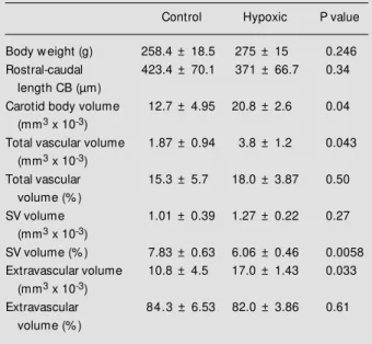
We have confirmed that in the chroni-cally hypoxic rat there is an increase in total volume of the carotid body (for references, Table 1 - Dimensions of control and hypoxic rat carotid bodies (CB).
Total vascular, small vessel (SV) and extravascular volumes are also expressed as a percentage of the total volume of the organ. Values are reported as means ± SD for carotid bodies of 5 control and 3 hypoxic rats. P values w ere determined by Student’s un-paired t-test.
Control Hypoxic P value
Body w eight (g) 258.4 ± 18.5 275 ± 15 0.246
Rostral-caudal 423.4 ± 70.1 371 ± 66.7 0.34
length CB (µm)
Carotid body volume 12.7 ± 4.95 20.8 ± 2.6 0.04
(mm3 x 10-3)
Total vascular volume 1.87 ± 0.94 3.8 ± 1.2 0.043
(mm3 x 10-3)
Total vascular 15.3 ± 5.7 18.0 ± 3.87 0.50
volume (% )
SV volume 1.01 ± 0.39 1.27 ± 0.22 0.27
(mm3 x 10-3)
SV volume (% ) 7.83 ± 0.63 6.06 ± 0.46 0.0058
Extravascular volume 10.8 ± 4.5 17.0 ± 1.43 0.033
(mm3 x 10-3)
Extravascular 84.3 ± 6.53 82.0 ± 3.86 0.61
see Introduction). The new information is that, by using image analysis techniques, this increase is found to be due to changes in the morphology of two compartments in the or-gan: a) an augmented total vascular volume, and b) an increase in extravascular volume. Other investigators (17) observed a generalised increase in vascularity of the hypoxic carotid body, but gave no indication of the luminal diameter of the affected ves-sels. In particular, they drew attention to dilated vessels and blood sinuses, a cat-egory of blood vessel which does not occur in the organ. A critical analysis of their technique indicates that their observations may be due in part to the method of prepara-tion of the carotid bodies, which were fixed in situ in the anaesthetised animal by drip-ping 8% glutaraldehyde on to the carotid bifurcation regions until the animals died or were killed by thoracotomy. As a conse-quence of this fixation method, venous con-gestion of cardiac origin must have occurred due to toxicity and/or asphyxia, so that a rise in venous pressure would have affected the carotid body vasculature passively. In an-other investigation (18) concerning the ef-fect of sympathectomy on long-term hypoxic rats, the authors gave insufficient informa-tion on their methods to allow us to comment on their findings of different sizes of blood vessels within the organ. We emphasise that the changes observed in the two compart-ments of the carotid body in our expericompart-ments occurred in the absence of an increase in venous pressure in vivo (19) and that during the perfusion-fixation procedure we used, retrograde filling and artificial distension of the organs venous microvasculature after death were prevented by raising the animals head just above the level of the thorax.
Our results showed that the total vascular volume and extravascular volume increased
pari passu with the total volume of the
or-gan, so that when these variables are ex-pressed as a percentage of the total volume of the organ there is no significant difference
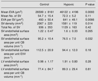
Table 2 - Vascular endothelial surface areas of control and hypoxic rat carotid bodies (CB), mean extravascular area (EVA), number of small vessels, extravascular area per small vessel, small vessel density, and total number of small vessels, all based on a sampling interval of 25 µm.
Values are reported as means ± SD for carotid bodies of 5 control and 3 hypoxic rats. P values w ere determined by Student’s unpaired t-test. EVV, Extravascular volume; LV, large vessel; SV, small vessel.
Control Hypoxic P value
M ean EVA (µm2) 26599 ± 8161 46122 ± 4196 0.0093
M ean No. of SV 54.7 ± 15.4 72.3 ± 5.2 0.11
EVA per SV (µm2) 493 ± 50.4 641 ± 49.1 0.0066
SV density (mm2) 2067 ± 220 1581 ± 119 0.014
Total No. of SV 936 ± 353 1129 ± 163 0.42
SV endothelial surface 1.22 ± 0.47 1.6 ± 0.33 0.265
area (mm2)
SV endothelial surface 95.2 ± 10.4 76.5 ± 7.0 0.032
area per unit CB volume (mm-1)
SV endothelial surface 112.5 ± 20.9 94.4 ± 12.0 0.18
area per unit EVV (mm-1)
LV endothelial surface 0.98 ± 1.17 1.91 ± 0.80 0.28
area (mm2)
LV endothelial surface 77.4 ± 84.7 89.3 ± 29.4 0.81
area per unit CB volume (mm-1)
group of animals, so that when expressed as a percentage of the total volume of the organ, the percentage value actually decreased sig-nificantly. In accordance with this, the small vessel endothelial surface area expressed per unit carotid body volume, and the den-sity of the small vessels in the organ were both diminished.
A question arises as to the possible mech-anisms underlying these observations. The finding that the size of the small vessel volume did not change in the hypoxic ca-rotid body and that there was a reduction in the volume of this compartment when ex-pressed as a percentage of the total volume of the organ can be explained solely in terms of the absolute increase in total volume. We have therefore concentrated below on the explanation for the changes in the large ves-sels (>12 µm in diameter) and in the ex-travascular extracellular compartments.
Firstly, we considered whether the ob-served changes in the carotid bodies of chronically hypoxic rats might reflect angio-genesis. Tissue hypoxia is known to stimu-late capillary angiogenesis in tissues other than the carotid body (20) and in the carotid body itself mitotic figures have been re-ported in endothelial cells of the hypoxic organ (21). Particular attention was, there-fore, paid to this issue in the present study. No evidence of mitoses was seen in any part of the extensive capillary network within the organ although endothelial cell nuclei were prominent and their deeply stained crescen-tic appearances were striking. However, we have to acknowledge that the absence of observable mitotic spindle does not exclude the possibility that mitosis had occurred prior to 31 days when the hypoxic animals were examined. As noted above, however, the size of the small vessel compartment was actually decreased in the hypoxic carotid bodies. Therefore it is very unlikely that angiogenesis occurred at the level of capil-laries.
Secondly, changes in the vascular
com-partment of the hypoxic carotid body might be due to remodelling of the vasculature. Arteriolar remodelling has been shown to occur in the skeletal muscles of rats exposed to chronic hypoxia such that capillaries gain smooth muscle and become arterioles (22). The arrangement of the arteriolar vessels in the carotid body is unusual in that those supplying capillaries to groups of type 1 and type 2 cells are largely confined to the caudal pole of the organ. Furthermore, the distribu-tion of blood flow within the organ is uneven and only a small proportion, up to 8%, of the total blood flow through the carotid body supplies type 1 and type 2 cells (9,23). The remainder bypasses the parenchymal tissue via straight through channels which are largely arteriolar and are confined to the more central parts of the connective tissue framework of the carotid body. Changes in arteriolar arrangements in the central part of the organ would therefore be apparent im-mediately in serial sections, but this was not observed. Furthermore, careful examination of all the serial sections from the peripheral parts of the carotid bodies from hypoxic animals did not reveal smooth muscle cells surrounding the endothelial cells of capillar-ies adjacent to type 1 and type 2 cells which would have been indicative of the formation of new arterioles. Such an arrangement would have resulted in a striking alteration in the appearance of the carotid body microvascu-lature, and again this was not observed.
body has such a variable morphology that, if remodelling had occurred prior to 31 days it would have been very difficult to detect in our sections. However, our feeling is that it was the diameter of the venous vessels rather than their number that increased. Certainly, in our histological sections the venous net-work of the cervical striated muscle and connective tissue surrounding the carotid body was more prominent in the chronically hypoxic animals, and this was apparently due to the fact that the venous vessels were larger than in the control animals. We have already discounted (see above) the possibil-ity that in our preparations a rise in venous pressure could have contributed to enlarge-ment of the large vessel compartenlarge-ment. How-ever, an explanation may be found in the changed morphology of the endothelial cells of the hypoxic carotid bodies, i.e., the promi-nent nuclei (see above). In keeping with current understanding of the effects of hy-poxia on endothelial cells (24,25), we sug-gest that substances such as nitric oxide, adenosine and prostaglandins are released during chronic hypoxia to cause the smooth muscle of the arterioles and venous vessels to relax. Furthermore, if this is the case, then as a consequence of the vasodilatation, and perhaps as a consequence of the release of substances that increase vascular permeabil-ity, we suggest there is a greater filtration of fluid out into the interstitium which would in turn have contributed to the augmented ex-travascular volume of the organ (see below). These hypotheses are in keeping with the finding that enlargement of the organ is al-most completely reversed when the arterial PO2 is restored to normal (26).
An increase in the number of type 1 cells might have contributed to the increase in the extravascular volume of the carotid body. We are aware of studies (6,21) reporting changes in the morphology of type 1 cells. Even so, in random selected sections of the carotid bodies from hypoxic animals, we found that the diameter of type 1 cells fell
within the normal range (16) and we did not see any evidence of mitotic spindles in type 1 cells. Various recent studies on the devel-opment of neural crest cells (27,28) have shown that cells from neural crest-derived ectomesenchyme have the ability to divide and undergo specialised development. Pre-sumably, type 1 cells of the carotid body originate from neural crest cells which have migrated through the 3rd pharyngeal arch, thus providing an explanation for the inner-vation of the organ via the carotid sinus branch from the 1X cranial nerve. Whether type 1 cells which have reached their final destination still have the capacity to divide is unknown. We can say only that type 1 cells may have undergone a brief period of mito-sis prior to 31 days of chronic hypoxia. However, we suggest that a brief wave of cellular proliferation would have made a minimal contribution to the overall increase in size of the organ reported under condi-tions of chronic hypoxia.
The reversal of the size of the carotid
body when the arterial PO2 is returned to
inter-stitial space. Therefore, in summary, we pro-pose that in the chronically hypoxic carotid body, dilatation of arterioles and particularly of the venous vessels of >12 µm together with increased filtration of fluid into the interstitium are mainly responsible for in-creasing the size of large vessel and ex-travascular compartments. Clearly, further investigations will be required to test these hypotheses.
It is of interest to compare our results with previous reports (10,11) on changes observed in the carotid body of the spontane-ously insulin-dependent diabetic rat. The cause of the enlargement of the carotid body in this condition differed from that seen in the hypoxic animals. In the diabetic rat, the total vascular volume did not change and was even diminished when expressed as a percentage of the total volume of the organ. Rather, the increased total volume of the diabetic rat carotid body was entirely attrib-utable to an increase in the extravascular volume, in contrast to the hypoxic state where the increased extravascular volume was only a partial contributor.
Po ssible significance o f mo rpho lo gical
change s in the caro tid bo dy to
acclimatiza-tio n to chro nic hypo xia
Exposure to a steady-state chronic hy-poxic environment in humans and other spe-cies leads to an increase in pulmonary venti-lation which is characterised by an immedi-ate increase followed by a time-dependent progressive rise in minute volume and a fall
in alveolar and arterial PCO2, termed the
ventilatory acclimatization to hypoxia (VAH; 29,30). Although the time course of VAH varies between species, the evidence indi-cates that the carotid body chemoreceptors play an important role in the genesis of VAH (29,30). The discharge in the chemoreceptor fibres of the carotid sinus nerve increases with a time course similar to that of pulmo-nary ventilation (30-32), and there is a
time-dependent increased sensitivity of the oxy-gen-responsive mechanism (33). In animals with denervated carotid bodies, VAH is sig-nificantly attenuated (29,34). There is still doubt about the mechanism underlying the increased hypoxic sensitivity of the carotid bodies in VAH. Hypoxia augments the ac-tivity of the efferent fibres in the carotid sinus nerve (35), but such activity is unlikely to be a cause of the progressive increase in the chemoreceptor discharge in the VAH, since it is predominately inhibitory to the carotid body (36). Other mechanisms have been considered, but the evidence has been somewhat equivocal, e.g., down-regulation of the inhibitory action of dopamine and a noradrenergic mechanism (30). The results of the present study suggest an alternative explanation based on the observed morpho-logical changes in the carotid body. It has been established that under normoxic condi-tions the mean carotid body PO2 is about 25 mmHg in the cat (37). It must be assumed that whatever the mechanism of signal trans-duction, the activity of the organ, as indi-cated by the discharge of impulses in affer-ent fibres in the carotid sinus nerve, will be dependent upon the level of tissue PO2.
Tis-sue PO2 depends upon a number of factors:
the interstitial space, and possibly of the type 1 and 2 cells as well, this would further increase the mean capillary-tissue distance. It is well recognised that on return to sea-level following a stay at high altitude, some hyperventilation persists temporarily (venti-latory deacclimatization; 39). It appears that this phenomenon is not simply a manifesta-tion of the same mechanism that accounts for VAH, but is in part secondary to respira-tory alkalosis that develops during the phase of hyperventilation (40). Nevertheless, the sensitivity of the carotid bodies to acute hypoxia presumably remains increased, at least during the immediate period of
deaccli-matization (31) and in time gradually returns to normal. On the basis of the observations reported here, we suggest that this phenom-enon could be attributed to initial persis-tence, followed by reversal, of the morpho-logical changes evoked in the first place by chronic hypoxia.
Ackno wle dgm e nts
We wish to acknowledge the technical expertise of Barbara A. Jackson, FIBMS, Department of Histopathology, St. Marga-rets Hospital, Epping, Essex, in the prepara-tion of the material.
Re fe re nce s
1. Arias-Stella J (1969). Human carotid body at high altitudes. In: 69th Program and Abstracts of the American Association of Pathologists and Bacteriologists, San Francisco. Item 150.
2. Heath D & Smith P (1985). The Pathology of the Carotid Body and Sinus. Arnold, London.
3. Edw ards C, Heath D, Harris P, Castillo Y, Krüger H & Arias-Stella J (1971). The ca-rotid body in animals at high altitude. Jour-nal of Pathology, 104: 231-238.
4. Arias-Stella J & Bustos F (1976). Chronic hypoxia and chemodectomas in bovines at high altitudes. Archives of Pathology and Laboratory M edicine, 100: 633-639. 5. Barer GR, Edw ards CW & Jolly AI (1976).
Changes in the carotid body and the ven-tilatory response to hypoxia in chronically hypoxic rats. Clinical Science and M olecu-lar M edicine, 50: 311-313.
6. Dhillon DP, Barer GR & Walsh M (1984). The enlarged carotid body of the chroni-cally hypoxic and hypercapnic rat: a mor-phometric analysis. Quarterly Journal of Experimental Physiology, 69: 301-317. 7. Pallot DJ (1987). The mammalian carotid
body. Advances in Anatomy, Embryology and Cell Biology, 102: 1-91.
8. Clarke JA, Daly M deB & Ead HW (1990). Comparison of the size of the vascular compartment of the carotid body of the fetal, neonatal and adult cat. Acta Anato-mica, 138: 166-174.
9. Clarke JA, Daly M deB, Ead HW & Kreclovic' G (1993). A morphological study
of the size of the vascular compartment of the carotid body in a non-human pri-mate (Cercopithicus ethiopus), and a com-parison w ith the cat and rat. Acta Anato-mica, 147: 240-247.
10. Clarke JA, Daly M deB, M arshall JM & Ead HW (1996). A comparison of the size of the vascular compartment of the carotid body in normal, chronically hypoxic and diabetic rats. Journal of Physiology, 495: 29P (Abstract).
11. Clarke JA, Daly M deB, Ead HW & Hennessy EM (1999). The carotid body in the spontaneous insulin-dependent dia-betic rat. Brazilian Journal of M edical and Biological Research, 32: 85-91.
12. Clarke JA & Daly M deB (1981). A com-parative study of the distribution of ca-rotid body type-1 cells and periadventitial type-1 cells in the carotid bifurcation re-gions of the rabbit, rat, guinea-pig and mouse. Cell and Tissue Research, 220: 753-772.
13. M ian R & M arshall JM (1996). The behav-iour of muscle microcirculation in chroni-cally hypoxic rats: the role of adenosine.
Journal of Physiology, 491: 489-498. 14. Stata Corp. (1997). Stata Statistical
Soft-w are: Release 50. Stata Corporation, Col-lege Station, TX.
15. Clarke JA, Duff MJB & Ip HH-S (1983). Re-construction of the vascular system of the carotid body of the rat using an array pro-cessor. Journal of Physiology, 341: 6P-7P. 16. Adams WE (1958). The Comparative M or-phology of the Carotid Body and Carotid
Sinus. C.C. Thomas, Springfield, IL, 140-142.
17. M cGregor KH, Gil J & Lahiri S (1984). A morphometric study of the carotid body in chronically hypoxic rats. Journal of Ap-plied Physiology, 57: 1430-1438. 18. Péquignot J-M & Hellström S (1983).
In-tact and sympathectomized carotid bod-ies of long-term hypoxic rats. Virchow s Archives A, 400: 235-243.
19. Neylon M , M arshall JM & Johns EJ (1997). The effects of chronic hypoxia on renal function in the rat. Journal of Physi-ology, 501.1: 243-250.
20. Adair TH, Gay WJ & M ontani JP (1990). Grow th regulation of the vascular system: evidence for a m etabolic hypothesis.
American Journal of Physiology, 259: R393-R404.
21. Bee D, Pallott DJ & Barer GR (1986). Divi-sion of type 1 and endothelial cells in the hypoxic rat carotid body. Acta Anatomica, 126: 226-229.
22. Price RJ & Skalak TC (1998). Arteriolar remodelling in skeletal muscle of rats ex-posed to chronic hypoxia. Journal of Vas-cular Research, 35: 238-244.
23. Degner F & Acker H (1986). M athematical analysis of tissue PO2 distribution in the cat carotid body. Pflügers Archiv, 407: 305-311.
25. Burnstock G & Ralevic V (1994). New in-sights into the local regulation of blood flow by perivascular nerves and endothe-lium. British Journal of Plastic Surgery, 47: 527-543.
26. Heath D, Edw ards C, Winson M & Smith P (1973). Effects on the right ventricle, pulmonary vasculature, and carotid bod-ies of the rat of exposure to, and recovery from, simulated high altitude. Thorax, 28: 24-28.
27. Gilbert SF (1997). Developmental Biology. 5th edn. Sinauer Association, Inc., Sun-derland, M A, 284-297.
28. Creazzo TL, Godt RE, Leatherburg L, Conw ay SJ & Kirby M L (1998). Role of cardiac neural crest cells in cardiovascular development. Annual Review of Physiolo-gy, 60: 267-286.
29. Sm it h CA, Bisgard GE, Nielsen AM , Daristotle L, Kressin NA, Forster HV & Dempsey JA (1986). Carotid bodies are required for ventilatory acclimatization to chronic hypoxia. Journal of Applied Physi-ology, 60: 1003-1010.
30. Bisgard GE (1994). The role of arterial chemoreceptors in ventilatory
acclimati-zation to hypoxia. Advances in Experimen-tal M edicine and Biology, 360: 109-122. 31. Vizek M , Pickett CK & Weil JV (1987).
Increased carotid body hypoxic sensitivity during acclimatization to hypobaric hy-poxia. Journal of Applied Physiology, 63: 2403-2410.
32. Nielsen AM , Bisgard GE & Vidruk EH (1988). Carotid chemoreceptor activity during acute and sustained hypoxia in goats. Journal of Applied Physiology, 65: 1796-1802.
33. Barnard P, Andronikou S, Pokorski M , Smatresk N, M okashi A & Lahiri S (1987). Time-dependent effect of hypoxia on ca-rotid body chemosensory function. Jour-nal of Applied Physiology, 63: 685-691. 34. Bouverot P & Bureau M (1975).
Ventila-tory acclimatization and CSF acid-base bal-ance on carotid chemodenervated dogs at 3,550 m. Pflügers Archiv, 361: 17-23. 35. Lahiri S, Smatresk N, Pokorski M , Barnard
P & M okashi A (1983). Efferent inhibition of carotid body chemoreception in chroni-cally hypoxic cats. American Journal of Physiology, 245: R678-R683.
36. O’Regan RG & M ajcherczyk S (1983).
Control of peripheral chemoreceptors by efferent nerves. In: Acker H & O’Regan RG (Editors), Physiology of the Peripheral Arterial Chemoreceptors. Elsevier, Am-sterdam, 257-298.
37. Acker H, Lübbers DW & Purves M J (1971). Local oxygen tension field in the glom us-carot icum of t he cat and it s change at changing arterial PO2. Pflügers
Archiv, 329: 136-155.
38. Krogh A (1919). The number and distribu-tion of capillaries in muscles w ith calcula-tions of the oxygen pressure head neces-sary for supplying the tissue. Journal of Physiology, 52: 409-415.
39. Dempsey JA, Forster HV, Bisgard GE, Chosy LW, Hanson PG, Kiorpes AL & Pelligrino DA (1979). Role of cerebrospi-nal fluid (H+) in ventilatory deacclimatiza-tion from chronic hypoxia. Journal of Clini-cal Investigation, 64: 199-205.