Synthesis of a new class of naphthoquinone
glycoconjugates and evaluation of their potential
as antitumoral agents†
Vinicius R. Campos,aAnna C. Cunha,*aWanderson A. Silva,aVitor F. Ferreira,a Carla Santos de Sousa,bPatr´
ıcia D. Fernandes,bVin´ıcius N. Moreira,aDavid R. da Rocha,aFlaviana R. F. Dias,aRaquel C. Montenegro,cMaria C. B. V. de Souza,a Fernanda da C. S. Boechat,aCaroline F. J. Francoaand Jackson A. L. C. Resended
A novel series of carbohydrate-based naphthoquinones was synthesized and evaluated for cytotoxicity against different cancer cell lines. The compounds derived from 5-hydroxy-1,4-naphthoquinone (juglone) showed better cytotoxicity profiles against HCT-116, A-549 and MDA-MB 435 human cancer cells than the parent compound. The results suggest that the hydroxyl group on the aromatic ring increased the pro-oxidant activity of these new naphthoquinone derivatives. Furthermore, two derivatives were found to be more active against melanoma cells (MDA-MB435) than the clinically useful anticancer agent doxorubicin, and none of the compounds caused mouse erythrocyte lysis.
1
Introduction
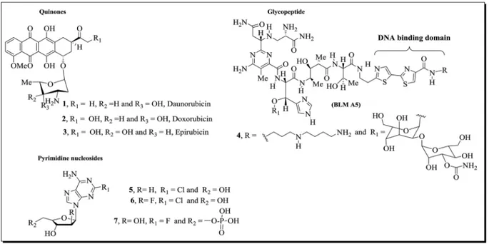
Natural glycoconjugated compounds,1–31
–7(Fig. 1), are useful
drugs that are commonly used clinically in the treatment of cancer. The importance of the carbohydrate residues in these structures has been well established: these moieties are important for solubilization and also decrease the toxicity and improve the pharmacokinetics of the compounds.4 Indeed,
several cases in the literature5 show the bene
t to biological activity of a carbohydrate moiety in the structure of a molecule. For example, the sugar moieties in antibiotic anthracycline derivatives1–3(Fig. 1) participate in the molecular recognition
of the main cellular targets, DNA and topoisomerase II, forming a DNA–drug–topoisomerase II ternary complex in which the enzyme is covalently linked to a broken DNA strand, which is critical for the induction of apoptosis and cell death.6,7
Other mechanisms can be involved in quinone toxicity of such as redox cycles, which result in the production of semi-quinone radicals and reactive oxygen species (ROS). These
events lead to the depletion of glutathione in cells as well as damage to other intracellular components.3
The importance of exploring the synthesis of sugar-conjugated compounds to increase the selectivity of drug uptake is based on the Warburg effect,8which is related to the large consumption of
glucose by cancerous cells compared to normal cells due to the high rate of aerobic glycolysis in the former.8
Recently, our group described the synthesis and pharmaco-logical evaluation of a series of carbohydrate-based quinones. In this study,5compounds8aand8b(Fig. 2) (IC
50below 3.0mM)
were found to exhibit signicant activity against a melanoma cell line (MDA-MB 435).
Quinones containing heterocyclic aromatic rings in their structures have been extensively explored by many research groups.9,10Structure
–activity relationship studies have revealed
that the number and position of nitrogen (N) atoms in the ve-or six-membered heterocyclic ring are critical factve-ors fve-or enhancing antitumor activity.9,10
Among nitrogen-containing heterocycles, 1,2,3-triazole derivatives exhibit a broad spectrum of biological properties11–15
and may serve as scaffold structures for the development of new pharmaceutical compounds. This heterocyclic system is resis-tant to metabolic degradation and can interact with biological targets through hydrogen bonds and dipole interactions, properties that may be responsible for the enhanced biological activities of many synthetically versatile molecules.16
To extend our investigation into the synthesis and biological evaluation of quinone glycoconjugate compounds5,17and
1,2,3-triazole derivatives,11–15we prepared a new series of
naphtho-triazoles,9a–c (Scheme 1), and assessed their activity against
three types of cancer cells: human colorectal carcinoma
aUniversidade Federal Fluminense, Departamento de Qu´ımica Orgˆanica, Programa de P´os-Graduaç˜ao em Qu´ımica, Outeiro de S˜ao Jo˜ao Batista, 24020-141 Niter´oi, RJ,
Brazil. E-mail: [email protected]ff.br; Fax: +55 21 26292145; Tel: +55 21 26292148
bUniversidade Federal do Rio de Janeiro, Instituto de Ciˆencias Biom´edicas, Laborat´orio de Farmacologia da Dor e da Inamaç˜ao, CCS, Bloco J, sala 10. Cidade Universit´aria, RJ, Brazil
cUniversidade Federal do Par´a, Instituto de Ciˆencias Biol´ogicas, Bel´em, PA, Brazil dUniversidade Federal Fluminense, Departamento de Qu´ımica Inorgˆanica, Laborat´orio
Regional de Difraç˜ao de Raios X (LDRX), 24020-141, Niter´oi, RJ, Brazil
†Electronic supplementary information (ESI) available. CCDC 1055524 and 1055525. For ESI and crystallographic data in CIF or other electronic format see DOI: 10.1039/c5ra19192k
Cite this:RSC Adv., 2015,5, 96222
Received 17th September 2015 Accepted 2nd November 2015
DOI: 10.1039/c5ra19192k
www.rsc.org/advances
RSC Advances
PAPER
Published on 09 November 2015. Downloaded by Federal University of Ceará on 15/06/2016 13:56:10.
View Article Online
(HCT-116), human lung adenocarcinoma (A-549) and human melanoma (MDA-MB435).
The synthesis of derivatives1710a
–c, which possess
amino-carbohydrate chains at the C-2 position of the quinone moiety (Scheme 1), was also performed to verify the possible pharma-cophoric effects of the 1,2,3-triazole moiety fused to naphthoquinone.
Naturally occurring hydroxy naphthoquinone (juglone, 12) exhibits a toxicological effect through its ability to undergo redox cycling to damage macromolecules, such as DNA, lipids and proteins.18Several studies have shown that the introduction
of a hydroxyl group at the carbon of the benzene moiety improves the pro-oxidant properties of naphthoquinone deriv-atives, stimulating lipid peroxidation and adduct formation with macromolecules.18
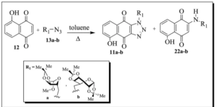
Based on these reports, we also decided to prepare a new series of hydroxylated naphthoquinones, 11a–c, exploring
cycloaddition between juglone (12) and azide-functionalized carbohydrates13a–c (Scheme 1), and to investigate the in
u-ence on biological activity of a hydroxyl group at position 5 of the benzene ring.
The compounds 10a–c and 11a–c were also evaluated for
their cytotoxic activities against cancer cell lines HCT-116, A-549 and MDA-MB435.
Fig. 1 Examples of natural glycoconjugated compounds (1–7) with antitumoral activity.
Fig. 2 Examples of synthetic molecules (8aand8b) with antitumoral activities.
Scheme 1 Strategy for the synthesis of naphthoquinone derivatives9a–c,10a–cand11a–c.
2
Results and discussion
2.1 ChemistryNaphthotriazole derivatives9a–c(Scheme 2) were obtained in
moderate yields via a [3 + 2] cycloaddition reaction between glycosyl azides13a–cand 1,4-naphthoquinone (14) (Scheme 2).
The probable mechanism for the formation of these compounds involves an initial 1,3-dipolar cycloaddition reac-tion between the azide carbohydrates13a–cand
1,4-benzoqui-none (14), producing the corresponding triazoline intermediates15a–c, which are tautomerized and oxidized to form the desired glycotriazole derivatives9a–c.
Glycosyl azides13a–cwere prepared from their
correspond-ing commercially available reagents D-ribose, D-xylose and D
-galactose using previously described methods for carbohydrate derivatization19,20(Scheme 3).
The aminonaphthoquinone analogs10a–c(Scheme 2) were
synthesized in moderate yieldsviaultrasound-accelerated 1,4-cycloaddition between 1,4-naphthoquinone (14) and different aminocarbohydrates (16a–c), according to our previous report.17
Reduction of the azide derivatives 13a–c in the presence of
a catalytic amount of 10% Pd/C (Scheme 3) led to the corre-sponding amines16a–c.
Juglone 12, prepared in 85% yield via the methodology described by Ferreira and coworkers,21was reacted with azide
compounds 13a and 13b. Although it is possible to form a mixture of the 5- and 8-hydroxy-naphthotriazole regioisomers (Scheme 4)viathis thermal 1,3-dipolar cycloaddition reaction, only 5-hydroxy-1-substituted-1H-naphtho[2,3-d ][1,2,3-]triazole-4,9-dione derivatives11a and11bwere isolated, in moderate yields. Furthermore, unexpected products 22a and 22b, pos-sessing a carbohydrate chain at the C-2 position of the quinone ring, were also isolated.
The reaction of compound13cwith juglone (12) gave a crude product which upon TLC analysis showed to be a mixture of two major compounds with retention factors (Rf) very similar. The 1H NMR spectrum of this mixture led to the conclusion that one
of the substances corresponded to11c, the desired compound, and that the other substance was the amine22c. Attempts to separate the products by chromatography on silica gel column were unsuccessful (Scheme 5).
Scheme 2 Synthesis of the novel naphthotriazole derivatives9a–cand 2-aminonaphthoquinones10a–c.
Scheme 3 Preparation of derivatives of carbohydrates13a–cand16a–c.
RSC Advances Paper
Published on 09 November 2015. Downloaded by Federal University of Ceará on 15/06/2016 13:56:10.
The structures of the new compounds9a–c,11a,11b,22a
and 22b were established by spectral data: one- and two-dimensional 1H and13C NMR spectra [1H,13C-APT, COSY-1H
1H, HSQC and HMBC], IR and elemental analysis.
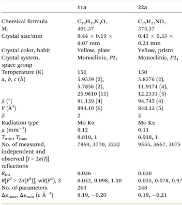
The1H NMR spectra of11a,11b,22aand22bindicate that only one of the two possible isomers was formed. The regio-chemistry of the reaction was investigated based on X-ray crys-tallographic analysis of substances11aand22a(Fig. 3). Fig. 3 shows the ORTEP diagrams of these compounds, and crystal data and renements are provided in Table 2. The diagram shows that the carbohydrate chains are in theanti-position to the hydroxyl group attached to the quinone ring of11aand22a. The carbohydrate moieties of11aand22aadopt envelope and twist conformations, respectively.
2.2 Biological assay
Compounds9a–c,10a–c,11a,11b,12,14, 22aand22b were
testedin vitroagainst three cancer cell lines and normal cells, human blood peripheral leukocytes, using the MTT assay. Doxorubicin was used as the positive control. Erythrocyte lysis was evaluated at a concentration of 200mM. The concentrations inducing 50% inhibition of cell growth (IC50) are reported in
Table 2. The compounds were classied according to their activity as highly active (IC50< 2mM), moderately active (2mM <
IC50< 10mM), or inactive (IC50> 10mM).22
The naphthoquinone derivatives9a–c,10a–c,11a,11b,12,
14,22aand22b(Table 2) did not exhibit a lytic effect against either erythrocytes or normal human leukocytes.
Among naphthotriazoles 9a–c, only derivative 9b showed
signicant activity against HCT-116 cells, with an IC50value of
4.85mM. This result indicates that the presence of the glycol 1,2,3-triazole subunit determines the biological activity assessed. Aminoquinones10a–cshowed no activity against the
three cell lines, allowing us to speculate that the presence of the amino group attached to carbon 2 has no effect on the biolog-ical activity of the compounds.
Aminoquinone analogs22aand22bshowed higher cytotoxic activities than compounds10aand10b. This can be attributed to the presence of the hydroxyl group connected to the benzene ring in these derivatives, possibly contributing to their pro-oxidant activities.18
In addition, the new glycoconjugate compounds22aand22b
were also more active than juglone (12). We speculate that cellular permeability of these 5-hydroxy-1,4-naphthoquinonic derivatives could be improved by the presence of the carbohy-drate chain at the C-2 position of the quinone moiety. As22a
and22bwere active against different cell lines, it appears that they can bind to different targets in a specic background.
The series of quinones11a–cdisplayed greater activity in all
tested cancer cell lines than naphthoquinone derivatives9aand
9b, possibly indicating that these quinones have increased pro-oxidant capacity. One can speculate that the hydroxyl attached to the C5 carbon in these derivatives is responsible for their increased anti-tumor activity.
Scheme 4 Preparation of isomeric glycotriazole derivatives11a and band aminonaphthoquinone derivatives22a and b.
Scheme 5 Synthesis of naphthoquinone compounds11cand22c.
Fig. 3 ORTEP diagrams of compounds11a(a) and22a(b), showing 50% probability ellipsoids and the atom-numbering scheme.
Derivatives 11a and 11b were also more active against melanoma cancer cells (MDA-MB435) than the clinically useful anticancer agent doxorubicin. Although doxorubicin is consid-ered an important drug for cancer chemotherapy, it has several clinical limitations, such as cardiotoxic effects and a high incidence of multi-drug resistance. Therefore, glycoconjugate compounds11a,11b,22aand22bcan be considered promising prototypes in drug development for cancer therapy.
3
Conclusion
The thermal cycloaddition reaction of glycosyl azides13a–cwith
1,4-naphthoquinone (14) produced the new glyconaphtho-triazoles9a–cin moderate yields. Under the same conditions,
parent compound 21 afforded the corresponding glyconaph-thotriazoles, 11a and11b, in moderate yields as well as the unexpected aminonaphthoquinones 22a and 22b. These carbohydrate-based naphthoquinones,11a,11b,22aand22b, derived from juglone (12) showed the best cytotoxicity prole against human cell lines HCT-116, A-549 and MDA-MB 435 when compared with the parent compounds,9a,9b,10aand
10b, respectively. These data show the importance of the hydroxyl group to the pro-oxidant action of these naph-thoquinone compounds. Indeed, derivatives11aand11bwere more active than the clinically useful anticancer agent doxoru-bicin against melanoma cancer cells (MDA-MB435 cells). However, none of the compounds exhibited lytic effects against mouse erythrocytes.
Molecules11a,11b,22aand22bcan be considered prom-ising prototypes for cancer therapy.
4
Experimental
4.1 Materials and measurements
Melting points were determined with a Fisher-Johns–Melting
Point Apparatus instrument, and the values are uncorrected. Infrared (IR) spectra were recorded on a Perkin-Elmer FT-IR 1600 spectrophotometer using KBr pellets. NMR spectra were recorded on a Varian Unity Plus 300 MHz or 500 MHz spec-trometer using the specied solvents. Chemical shis (d) are reported in ppm, and the coupling constants (J) are expressed in Hz. Column chromatography was performed with silica gel
ash from Acros. The reactions were routinely monitored by thin layer chromatography (TLC) on silica gel pre-coated F254
Table 1 X-ray crystallographic data for compounds11aand22a
11a 22a
Chemical formula C19H19N3O7 C19H21NO7
Mr 401.37 375.37
Crystal size/mm 0.440.19
0.07 mm
0.430.33
0.21 mm Crystal color, habit Yellow, plate Yellow, prism Crystal system,
space group
Monoclinic,P21 Monoclinic,P21
Temperature (K) 150 150
a,b,c(˚A) 5.9539 (2), 5.7856 (2), 25.9610 (11)
5.8376 (2), 11.9174 (4), 12.2331 (5)
b() 91.139 (4) 94.745 (4) V(˚A3) 894.10 (6) 848.13 (5)
Z 2 2
Radiation type Mo Ka Mo Ka
m(mm 1) 0.12 0.11
Tmin,Tmax 0.810, 1 0.918, 1
No. of measured, independent and observed [I> 2s(I)] reections
7869, 3770, 3232 9555, 3667, 3075
Rint 0.036 0.030
R[F2> 2s(F2)], wR(F2),S 0.042, 0.098, 1.10 0.035, 0.078, 0.97 No. of parameters 263 248
Drmax,Drmin(eA˚ 3) 0.19, 0.20 0.19, 0.21
Table 2 Cytotoxic activity expressed as the IC50(mM) of the compounds against different cell linesd
Compound
Cancer cell linea
Human blood leukocytes Erythrocytesc HCT-116 A-549 MDA-MB 435
9a >10 >10 >10 >100 >200
9b 4.85 >10 >10 >100 >200
9c >10 >10 >10 >100 >200
10a >10 >10 8.9 >100 >200
10b >10 >10 >10 >100 >200
10c >10 >10 >10 >100 >200
11a 0.67 1.22 0.47 >100 >200
11b 0.52 0.99 0.29 >100 >200
12 1.60 3.7 NT >100 >200
14 >10 NT NT >100 >200
22a 1.26 0.77 5.0 >100 >200
22b 0.43 1.76 NT >100 >200 Doxorubicinb 0.10 0.47 0.88 3.41 >25 aData are presented as IC
50values (mM), and 95% condence intervals were obtained by nonlinear regression for all cell lines from three
independent experiments.bDoxorubicin was used as a positive control. Only compounds with an IC50value lower than 5 mg mL 1for at least
one cell line were considered active.cConcentration of compound that induced erythrocyte lysis.dNT
–not tested.
RSC Advances Paper
Published on 09 November 2015. Downloaded by Federal University of Ceará on 15/06/2016 13:56:10.
Merck plates. Microanalyses were performed using a Perkin-Elmer Model 2400 instrument, and all values were within
0.4% of the calculated compositions.
4.2 General procedure for the preparation of new naphthotriazoles 9a–c
The glycosyl azides13a–c(1.0 mmol) and 1,4-naphthoquinone
(14) (0.158 mg, 1.0 mmol) were dissolved in toluene and heated at 100C for 24 hours. Aer completion of the reaction, which was monitored using TLC (eluted with hexane/EtOAc¼1/1), the mixture was cooled, and the solvent was removed. The residual solid product was puried by column chromatography on silica gel and eluted with an increasing polarity gradient mixture of hexane and ethyl acetate (100% to 50%) to afford compounds
9a–c.
(Methyl-50-deoxy-20,30-O-isopropylidene-b-D-ribofuranosid-50 -yl)-1H-naphtho[2,3-d][1,2,3]triazole-4,9-dione (9a). The 1,3-dipolar cycloaddition reaction between naphthoquinone12and glycosyl azide13ayielded glyconaphthotriazole9a(121.0 mg, 63%) as an orange solid: mp 225–227C; IR (KBr)n(cm 1): 1588
and 1686 (C]O).
1H NMR (500.00 MHz, CDCl
3)d: 1.30 (s, 3H, CH3), 1.44 (s, 3H,
CH3), 3.41 (s, 3H, OCH3), 4.72–4.73 (m, 1H, H-40), 4.74 (d, 1H,J ¼6.0 Hz, H-20), 4.86 (d, 1H,J¼6.0 Hz, H-30), 5.01 (dd, 2H,J¼
7.5 and 1.0 Hz, H-50a and H-50b), 5.02 (s, 1H, H-10), 7.82 (td, 1H,J ¼7.5 and 1.5 Hz, H-7), 7.86 (td, 1H,J¼7.5 and 1.5 Hz, H-6), 8.24 (dd, 1H,J¼7.5 and 1.5 Hz, H-8), 8.34 (dd, 1H,J¼7.5 and 1.5 Hz, H-5) ppm.
13C NMR (125.0 MHz, CDCl
3)d: 25.1 (CH3), 26.5 (CH3), 53.2
(C-50), 55.9 (OCH
3), 81.8 (C-30), 84.6 (C-40), 85.3 (C-20), 110.4
(C-10), 113.1 (
–OCO–), 127.5 (C-8), 128.5 (C-5), 132.8 (C-8a), 133.5
(C-4a), 133.6 (C-9a), 134.5 (C-7), 135.4 (C-6), 145.7 (C-3a), 175.6 (C-9), 176.7 (C-4) ppm. Anal. calc. for C19H19N3O6: C, 59.22; H,
4.97; N, 10.90. Found: C, 59.05; H, 5.29; N, 10.79%.
1-(60-Deoxy-10,20:30,40-di-O-isopropylidene-D -galactopyranos-60-yl)-1H-naphtho[2,3-d][1,2,3]triazole-4,9-dione (9b). The
1,3-dipolar cycloaddition reaction between naphthoquinone12and glycosyl azide13byielded glyconaphthotriazole 9b(126.0 mg, 57%) as a light yellow solid: mp 160–162C; IR (KBr)n(cm 1):
1689 and 1593 (C]O).1H NMR (300.00 MHz, CDCl3)d: 1.22 (s,
3H, CH3), 1.35 (s, 3H, CH3), 1.38 (s, 3H, CH3), 1.57 (s, 3H, CH3),
4.31 (dd, 1H,J¼5.0 and 2.7 Hz, H-20), 4.36 (dd, 1H,J¼7.8 and
2.0 Hz, H-30), 4.46
–4.49 (m, 1H, H-50), 4.68 (dd, 1H,J¼7.8 and
2.6 Hz, H-40), 4.82 (dd, 1H,J¼13.9 and 3.4 Hz, H-60a), 5.32 (dd,
1H,J¼13.9 and 9.6 Hz, H-60b), 5.40 (d, 1H,J¼5.0 Hz, H-10),
7.80 (td, 1H,J¼7.5 and 1.5 Hz, H-7), 7.85 (td, 1H,J¼7.5 and 1.5 Hz, H-6), 8.24 (dd, 1H,J¼7.6 and 1.2 Hz, H-8), 8.34 (dd, 1H,J¼
7.6 and 1.2 Hz, H-5) ppm.
13C NMR (75.0 MHz, CDCl
3)d: 24.8 (CH3), 24.9 (CH3), 25.8
(CH3), 26.2 (CH3), 50.7 (C-60), 67.2 (C-50), 70.4 (C-20), 71.2 (C-40),
71.4 (C-30), 96.3 (C-10), 109.1 (
–OCO–), 110.4 (–OCO–), 127.4
5), 127.9 8), 133.0 8a), 133.6 4a), 134.3 7), 134.4 (C-9a), 135.2 (C-6), 145.6 (C-3a), 175.5 (C-9), 176.9 (C-4) ppm. Anal. calc. for C22H23N3O7: C, 59.86; H, 5.25; N, 9.52. Found: C,
59.32; H, 5.63; N, 9.25%.
1-(50-Deoxy-10,20-O-isopropylidene-D-xylofuranos-50-yl)-1H -naphtho[2,3-d][1,2,3]triazole-4,9-dione (9c). The 1,3-dipolar
cycloaddition reaction between naphthoquinone 12 and glycosyl azide 13c yielded glyconaphthotriazole 9c (90.0 mg, 48%) as a light yellow solid: mp 240–243C; IR (KBr)n(cm 1):
1687 and 1592 (C]O).1H NMR (500.00 MHz, DMSO)d: 1.21 (s, 3H, CH3), 1.31 (s, 3H, CH3), 4.18 (d, 1H,J¼2.7 Hz, H-30), 4.50 (d,
1H,J¼3.6 Hz, H-20), 4.61
–4.64 (m, 1H, H-40), 5.00–5.01 (m, 2H,
H-50a and H-50b), 5.88 (d, 1H,J¼3.6 Hz, H-10), 7.92 (td, 1H,J¼
7.5 and 1.5 Hz, H-7), 7.95 (td, 1H,J¼7.5 and 1.5 Hz, H-6), 8.17 (dd, 1H,J¼7.5 and 1.5 Hz, H-8), 8.19 (dd, 1H,J¼6.5 and 1.5 Hz, H-5) ppm.13C NMR (125.0 MHz, DMSO)d: 26.1 (CH
3), 26.7
(CH3), 49.4 (C-50), 73.8 (C-30), 78.5 (C-40), 85.1 (C-20), 104.5 (C-10),
110.9 (–OCO–), 126.8 5), 126.9 8), 132.9 8a), 133.1
(C-4a), 134.3 (C-9a), 134.5 (C-7), 135.1 (C-6), 144.8 (C-3a), 175.1 (C-9), 176.8 (C-4) ppm. Anal. calc. for C18H17N3O6: C, 58.22; H,
4.61; N, 11.32. Found: C, 58.61; H, 4.35; N, 11.52%.
4.3 General procedure for the preparation of new naphthoquinone derivatives 11a, 11b, 22a and 22b
The glycosyl azides 13a and13b (1.0 mmol) and juglone 12
(0.174 mg, 1.0 mmol) were dissolved in toluene and heated at 100 C for 24 hours. The reaction mixture was concentrated
under reduced pressure and the resulting residue was puried by column chromatography using silica gel and eluted with an increasing polarity gradient mixture of hexane and ethyl acetate (100% to 70%) to give the desired compounds11aand11band the unexpected aminoquinones22aand22b.
5-Hydroxy-(methyl-50-deoxy-20,30-O-isopropylidene-b-D -ribo-furanosid-50-yl)-1H-nao[2,3-d][1,2,3]triazole-4,9-dione (11a) and 5-hydroxy-2-(methyl-50-deoxy-20,30-O-isopropylidene-b-D -ribofuranosid-50-yl)-amino-1,4-naphthoquinone (22a).The
1,3-dipolar cycloaddition reaction between juglone12and glycosyl azide13agave glyconaphthotriazole11a(136.0 mg, 68%) and amino compound22a(5.6 mg, 3%).
The compound11awas obtained as a yellow solid: mp 144–
147C; IR (KBr)n(cm 1): 3434 (OH), 1649 (C
]O), 1449 (C]C).
1H NMR (500.00 MHz, CDCl
3)d: 1.31 (s, 3H, CH3), 1.45 (s, 3H,
CH3), 3.41 (s, 3H, OCH3), 4.70–4.72 (m, 1H, H-40), 4.72 (d, 1H,J ¼6.0 Hz, H-20), 4.85 (d, 1H,J¼6.0 Hz, H-30), 4.98 (dd, 2H,J¼
7.5 and 2.5 Hz, H-50a and H-50b), 5.02 (s, 1H, H-10), 7.38 (dd, 1H, J¼8.5 and 1.0 Hz, H-6), 7.68 (dd, 1H,J¼8.5 and 7.5 Hz, H-7), 7.80 (dd, 1H,J¼7.5 and 1.0 Hz, H-8), 12.31 (s, 1H, OH) ppm.
13C/APT NMR (125.0 MHz, CDCl
3)d: 25.1 (CH3), 26.5 (CH3),
53.3 (C-50), 56.0 (OCH
3), 81.8 (C-40), 84.6 (C-20), 85.3 (C-30), 110.4
(C-10), 113.1 (
–OCO–), 115.5 (C-4a), 120.8 (C-8), 126.9 (C-6), 133.2
(C-8a), 133.9 (C-9a), 136.9 (C-7), 145.5 (C-3a), 163.6 (C-5), 174.8 (C-9), 182.7 (C-4) ppm. Anal. calc. for C19H19N3O7: C, 56.86; H,
4.77; N, 10.47. Found: C, 58.04; H, 5.25; N, 9.05%.
The compound22awas obtained as a red solid: mp 128–130 C; IR (KBr)n(cm 1): 3401 (OH), 3263 (N
–H); 1623, 1592 (C]O), 1468 (C]C).1H NMR (500.00 MHz, CDCl3)d: 1.32 (s, 3H, CH3),
1.50 (s, 3H, CH3), 3.29–3.34 (m, 2H, H-50a and H-50b), 3.41 (s,
3H, OCH3), 4.49–4.51 (m, 1H, H-40), 4.63 (dd, 1H,J¼6.0 and 1.0
Hz, H-30), 4.66 (d, 1H,J¼6.0 Hz, H-20), 5.05 (s, 1H, H-10), 5.64 (s,
1H, H-3), 6.66–6.68 (m, 1H, N–H), 7.25 (dd, 1H,J¼8.5 and 1.0
Hz, H-6), 7.47 (dd, 1H,J¼8.5 and 7.5 Hz, H-7), 7.59 (dd, 1H,J¼
7.5 and 1.0 Hz, H-8), 12.98 (s, 1H, OH) ppm. 13C/APT NMR (125.0 MHz, CDCl3)dppm: 25.1 (CH3), 26.6 (CH3), 45.6 (C-50),
55.7 (OCH3), 82.3 (C-30), 84.5 (C-40), 85.4 (C-20), 100.5 (C-3), 109.8
(C-10), 113.1 (
–OCO–), 115.0 (C-4a), 119.2 (C-8), 126.1 (C-6), 130.7
8a), 134.2 7), 149.0 2), 161.3 5), 181.1 1), 189.2 (C-4) ppm. Anal. calc. for C19H21NO7: C, 60.79; H, 5.64; N, 3.73.
Found: C, 61.61; H, 6.00; N, 3.21%.
5-Hydroxy-1-(60-deoxy-10,20:30,40-di-O-isopropylidene-D -gal-actopyranos-60-yl)-1H-nao[2,3-d][1,2,3]triazole-4,9-dione (11b)
and 5-hydroxy-2-(60-deoxy-10,20:30,40-di-O-isopropylidene-D -gal-actopyranos-60-yl)-amino-1,4-naphthoquinone (22b). The
1,3-dipolar cycloaddition reaction between juglone12and glycosyl azide13b gave glyconaphthotriazole 11b(64.0 mg, 28%) and amino compound22b(4.3 mg, 2%).
The compound 11bwas obtained a yellow solid: mp 160–
162C; IR (KBr)n(cm 1): 3416 (OH), 1654 (C
]O), 1453 (C]C).
1H NMR (300.00 MHz, CDCl
3)d: 1.23 (s, 3H, CH3), 1.36 (s, 3H,
CH3), 1.38 (s, 3H, CH3), 1.57 (s, 3H, CH3), 4.32 (dd, 1H,J¼5.0
and 2.5 Hz, H-20), 4.36 (dd, 1H,J¼7.5 and 2.0 Hz, H-40), 4.44 –
4.47 (m, 1H, H-50), 4.69 (dd, 1H,J¼7.5 and 2.5 Hz, H-30), 4.81
(dd, 1H,J¼14.0 and 3.5 Hz, H-60a), 5.31 (dd, 1H,J¼14.0 and
10.0 Hz, H-60b), 5.39 (d, 1H,J¼5.0 Hz, H-10), 7.36 (dd, 1H,J¼
8.5 and 1.5 Hz, H-6), 7.66 (dd, 1H,J¼8.5 and 7.5, H-7), 7.77 (dd, 1H,J¼7.5 and 1.0 Hz, H-8), 12.34 (s, 1H, OH) ppm.13C/APT NMR (75.0 MHz, CDCl3)d: 24.9 (CH3), 25.0 (CH3), 25.8 (CH3),
26.2 (CH3), 50.8 (C-60), 67.1 (C-50), 70.4 (C-20), 71.2 (C-30), 71.4
(C-40), 96.3 (C-10), 109.1 (
–OCO–), 110.5 (–OCO–), 115.6 (C-4a), 120.5
8), 126.6 6), 133.4 8a), 134.4 9a), 136.7 7), 145.2 (C-3a), 163.5 (C-5), 174.7 (C-9), 183.0 (C-4) ppm. Anal. calc. for C22H23N3O8: C, 57.76; H, 5.07; N, 9.19. Found: C, 57.99; H, 5.34;
N, 8.78%.
The compound22bwas obtained as a red solid: mp 203–205 C; IR (KBr)n(cm 1): 3400 (OH), 3341 (N
–H), 1615 (C]O), 1469 (C]C).1H NMR (300.00 MHz, CDCl3)d: 1.33 (s, 3H, CH3), 1.37
(s, 3H, CH3), 1.49 (s, 3H, CH3), 1.52 (s, 3H, CH3), 3.44–3.48 (m,
2H, H-60a and H-60b), 4.00
–4.05 (m, 1H, H-50), 4.25 (dd, 1H,J¼
7.8 and 1.8 Hz, H-40), 4.35 (dd, 1H,J¼5.1 and 2.4 Hz, H-20), 4.65
(dd, 1H,J¼7.8 and 2.4 Hz, H-30), 5.55 (d, 1H,J¼5.1 Hz, H-10),
5.67 (s, 1H, H-3), 6.52–6.56 (m, 1H, N–H), 7.23 (dd, 1H,J¼8.4
and 1.2 Hz, H-6), 7.46 (dd, 1H,J¼8.4 and 7.5 Hz, H-7), 7.59 (dd, 1H,J¼7.5 and 1.2 Hz, H-8), 13.05 (s, 1H, OH) ppm.13C/APT
NMR (75.0 MHz, CDCl3)d: 24.5 (CH3), 25.0 (CH3), 26.1 (CH3),
26.2 (CH3), 43.3 (C-60), 64.9 (C-50), 70.5 (C-30), 70.6 (C-40), 71.9
(C-20), 96.5 (C-10), 99.9 (C-3), 109.0 (
–OCO–), 110.0 (–OCO–), 115.1
4a), 119.2 8), 125.9 6), 130.7 8a), 134.0 7), 149.1 (C-2), 161, 2 (C-5), 181.0 (C-1), 189.1 (C-4) ppm. Anal. calc. for C22H25NO8: C, 61.25; H, 5.84; N, 3.25. Found: C, 61.97; H, 6.18;
N, 3.31%.
5
X-ray determination
X-ray diffraction was performed using an Oxford Diffraction Xcalibur Atlas Gemini ultra Diffractometer at 150 K with Mo-Ka
radiation. The collection and reduction of the data were per-formed using the CrysAlisPro soware. The structures (Table 1) were solved by direct methods and rened using full-matrix
least squares onF2with the SHELX-97 soware package.23The
CCDC reference numbers for 11a and 22a are 1055525 and 1055524, respectively.†
6
Biology
6.1 Cytotoxicity against cancer cell lines
The compounds (0.15–20mM) were tested for cytotoxic activity against cell lines HCT-116 (human colorectal carcinoma, ATCC # 247), A-549 (human lung adenocarcinoma, ATCC # CCL-185) and MDA-MB435 (human melanoma, ATCC # HTB-129) as well as against freshly prepared human blood leukocytes and erythrocytes. All cell lines were maintained in DMEM medium supplemented with 10% fetal bovine serum, 2 mM glutamine, 100 U mL 1penicillin, and 100 mg mL 1streptomycin at 37C
in an atmosphere containing 5% CO2. Each compound was
dissolved with DMSO and diluted with the cell culture medium to obtain a concentration of 100mM, which was then incubated with the cells for 72 hours. The negative control received the same amount of DMSO (0.005% at the highest concentration). Doxorubicin was used as a positive control. Cell viability was determined by the reduction of the yellow dye 3-(4,5-dimethyl-2-thiazol)-2,5-diphenyl-2H-tetrazolium bromide (MTT) to a blue formazan product aer 3 hours of incubation, as described by Denizot (1986).24
6.2 Cell membrane disruption
Cytotoxicity testing was performed as described previously25in
96-well plates using a 2% mouse erythrocyte suspension in 0.85% NaCl containing 10 mM CaCl2. The compounds, which
were diluted as mentioned above, were tested at a concentration of 200mM. Aer incubation at room temperature for 30 min and centrifugation, the supernatant was removed, and the liberated hemoglobin was measured spectrophotometrically at 540 nm. DMSO was used as a negative control and Triton X-100 (1%) was used as a positive control.
Acknowledgements
This work was supported by the Brazilian agency FAPERJ-PRONEX. Fellowships granted to UFF, by FAPERJ, CAPES, and CNPq-PIBIC are gratefully acknowledged. We would like to thank the LabCri/UFMG for the use of the X-ray facilities.
References
1 U. Galm, M. H. Hager, S. G. V. Lanen, J. J. J. S. Thorson and B. Shen,Chem. Rev., 2005,105, 739.
2 J.-L. Yu, Q.-P. Wu, Q.-S. Zhang, Y.-H. Liu, Y.-Z. Li and Z.-M. Zhou,Bioorg. Med. Chem. Lett., 2010,20, 240.
3 M. N. da Silva, V. F. Ferreira and M. C. B. V. de Souza,Quim. Nova, 2003,26, 407.
4 V. K. Tiwaria, R. C. Mishra, A. Sharma and R. P. Tripathi,
Mini-Rev. Med. Chem., 2012,12, 1497.
5 V. R. Campos, E. A. dos Santos, V. F. Ferreira, R. C. Montenegro, M. C. B. V. de Souza, L. V. Costa-Lotufo,
RSC Advances Paper
Published on 09 November 2015. Downloaded by Federal University of Ceará on 15/06/2016 13:56:10.
M. O. de Moraes, A. K. P. Regufe, A. K. Jord˜ao, A. C. Pinto, J. A. L. C. Resende and A. C. Cunha, RSC Adv., 2012, 2, 11438–11448.
6 C. Temperini, M. Cirilli, M. Aschi and G. Ughetto,Bioorg. Med. Chem., 2005,13, 1673.
7 Z. S. Saify, N. Mushtaq, F. Noor, S. Takween and M. Arif,Pak. J. Pharm. Sci., 1999,12, 21.
8 Y. Zhao, E. B. Butler and M. Tan,Cell Death Dis., 2013,4, e532.
9 M. A. Castro, A. M. Gamito, V. Tangarife-Casta˜no, V. Roa-Linares, J. Ma, M. del Corral, A. C. Mesa-Arango, L. Betancur-Galvis, A. M. Francesch and A. San Feliciano,
RSC Adv., 2015,5, 1244.
10 J. S. Kim, H.-K. Rhee, H. J. Park, S. K. Lee, C.-O. Lee and H.-Y. P. Choo,Bioorg. Med. Chem., 2008,16, 4545–4550.
11 A. C. Cunha, J. M. Figueiredo, J. L. M. Tributino, A. L. P. Miranda, H. C. Castro, R. B. Zingali, C. A. M. Fraga, M. C. B. V. de Souza, V. F. Ferreira and E. J. Barreiro,Bioorg. Med. Chem., 2003,11, 2051.
12 A. K. Jord˜ao, V. F. Ferreira, E. S. Lima, M. C. B. V. de Souza, E. C. L. Carlos, H. C. Castro, R. B. Geraldo, C. R. Rodrigues, M. C. B. Almeida and A. C. Cunha,Bioorg. Med. Chem., 2009,
17, 3713.
13 R. Menegatti, A. C. Cunha, V. F. Ferreira, E. F. R. Perreira, A. El-Nabawi, A. T. Eldefrawi, E. X. Albuquerque, G. Neves, S. M. K. Rates, C. A. M. Fraga and E. J. Barreiro, Bioorg. Med. Chem., 2003,11, 4807.
14 M. S. Costa, N. Boechat, E. A. Rangel, F. C. da Silva, A. M. T. de Souza, C. R. Rodrigues, H. C. Castro, I. N. Junior, M. C. S. Lourenço, S. M. S. V. Wardell and V. F. Ferreira,Bioorg. Med. Chem., 2006,14, 8644.
15 V. R. Campos, P. A. Abreu, H. C. Castro, C. R. Rodrigues, A. K. Jord˜ao, V. F. Ferreira, M. C. B. V. de Souza, F. C. Santos, L. A. Moura, T. S. Domingos, C. Carvalho, E. F. Sanchez, A. L. Fuly and A. C. Cunha, Bioorg. Med. Chem., 2009,17, 7429.
16 S. G. Agalave, S. R. Maujan and V. S. Pore,Chem.–Asian J.,
2011,6, 2696–2718.
17 C. F. J. Franco, A. K. Jord˜ao, V. F. Ferreira, A. C. Pinto, M. C. B. V. de Souza, J. A. L. C. Resende and A. C. Cunha,
J. Braz. Chem. Soc., 2011,22, 187.
18 K. Murakami, M. Haneda, S. Iwata and M. Yoshino,Toxicol. In Vitro, 2010,24, 905.
19 B. S. Moon, A. Y. Shim, K. C. Lee, H. J. Lee, B. S. Lee, G. I. An, S. D. Yang, D. Y. Chi, C. W. Choi, S. M. Lim and K. S. Chun,
Bull. Korean Chem. Soc., 2005,26, 1865.
20 S. B. Ferreira, A. C. R. Sodero, M. F. C. Cardoso, E. S. Lima, C. R. Kaiser, F. P. Silva Jr and V. F. Ferreira,J. Med. Chem., 2010,53, 2364.
21 D. R. Rocha, A. C. G. Souza, J. A. L. C. Resende, W. C. Santos, E. A. Santos, C. Pessoa, M. O. Moraes, L. V. Costa-Lotufo, R. C. Montenegro and V. F. Ferreira, Org. Biomol. Chem., 2011,9, 4315.
22 E. P´erez-Sacau, R. G. D´ıaz-Penate, A. Est´˜ evez-Braun,
A. G. Ravelo, J. M. Garc´ıa-Castellano, L. Pardo and
M. Campillo,J. Med. Chem., 2007,50, 696.
23 G. M. Sheldrick,Acta Crystallogr., Sect. D: Biol. Crystallogr., 2010,66, 479.
24 F. Denizot and R. Lang,J. Immunol. Methods, 1986,89, 271. 25 P. Sharma and J. D. Sharma,J. Ethnopharmacol., 2001,74,
239.