336
Oliveira PCR et al. Gorham’s disease of scapula and clavicle
Radiol Bras. 2011 Set/Out;44(5):336–338
Gorham’s disease of scapula and clavicle: case report
and two-year follow-up of a rare disorder
*
Doença de Gorham da escápula e clavícula: relato de caso e evolução de dois anos de uma afecção rara
Paulo Cesar Rocha Oliveira1, Fabio Peixoto Alcantara1, Paola Lima Pasini Judice1, Giovani Rodrigues Batista1, Ilailson de Goes Teles1, Julio Salgado Antunes2
Gorham’s disease is a rare osteolytic disorder of still controversial etiology that may affect any bone. The histopathological substrate for such a condition is the replacement of normal bone by aggressive non-neoplastic expansile vascular tissue. The authors describe radiographic, computed tomography and magnetic resonance imaging findings in a case of this entity initially affecting the left scapula and, two years later, the ipsilateral clavicle.
Keywords: Gorham’s disease; Massive osteolysis; Computed tomography; Magnetic resonance imaging.
A doença de Gorham é uma rara desordem osteolítica, de etiologia controversa, que pode afetar qualquer osso. O substrato histopatológico é a substituição óssea por uma formação expansiva de natureza vascular agressiva, não neoplásica. Descrevemos os achados radiográficos, tomográficos e de ressonância magnética de um caso desta afec-ção acometendo a escápula esquerda e, dois anos depois, a clavícula ipsilateral.
Unitermos: Doença de Gorham; Osteólise maciça; Tomografia computadorizada; Imagem por ressonância
magné-tica.
Abstract
Resumo
* Study developed at SARAH Hospital – Brasília Center of SARAH Network of Rehabilitation Hospitals, Brasília, DF, Brazil.
1. MD, Diagnostic Imaging Department, SARAH Network of Rehabilitation Hospitals, Brasília, DF, Brazil.
2. MD, Surgical Pathology Laboratory, SARAH Network of Rehabilitation Hospitals, Brasília, DF, Brazil.
Mailing Address: Paulo Cesar Rocha Oliveira, MD. Diagnostic Imaging Department, Hospital SARAH-Brasilia, SARAH Network of Rehabilitation Hospitals, SMHS Qd 501, Bl A. Brasilia, DF, Brazil, 70335-901. E-mail: [email protected].
Received December 4, 2010. Accepted after revision April 18, 2011.
Rocha Oliveira PC, Alcantara FP, Judice PLP, Batista GR, Teles IG, Antunes JS. Gorham’s disease of scapula and clavicle: case report and two-year follow-up of a rare disorder. Radiol Bras. 2011 Set/Out;44(5):336–338.
0100-3984 © Colégio Brasileiro de Radiologia e Diagnóstico por Imagem CASE REPORT
male, 20-year-old patient. A brief literature review is carried out.
CASE REPORT
In July 2007, a female 18-year old pa-tient was admitted with complaints of in-termittent pain in her left shoulder for one year, with progressive incapacity of the ipsilateral upper limb. No history of fever, focal inflammatory signs, previous trauma, surgery or radiotherapy was reported. The clinical examination demonstrated pain at palpation and functional incapacity, simi-lar to a massive rotator cuff tear, without a palpable mass. Neurological tests were normal. Laboratory tests only demonstrated a subtle increase in C-reactive protein lev-els (0.9 mg/dl).
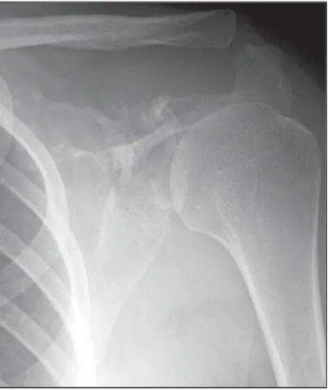
Shoulder radiographs revealed cortical rupture and bone fragmentation affecting the upper segment of the scapula, with no sign of bone expansion (Figure 1). Com-puted tomography (CT) clearly demon-strated the involvement of the whole scapula (Figure 2). Magnetic resonance imaging (MRI) revealed heterogeneous bone signal intensity, with areas of spon-taneous hyperintense signal intensity on INTRODUCTION
Gorham’s disease is a rare disorder characterized by extensive osteolysis and anomalous vascular proliferation, whose diagnosis is difficult and the prognosis, unpredictable. Such condition was firstly described by Jackson in 1838, and its clini-cal and pathologiclini-cal characteristics were established by Gorham and colleagues (1954) and Gorham & Stout (1955)(1–3). It
is also known by several terms such as Gorham’s syndrome, Gorham-Stout syn-drome, Morbus Gorham-Stout disease, massive osteolysis, and vanishing bone disease(2,4).
The authors report a case of Gorham’s disease affecting the right scapula and, two years after, the ipsilateral clavicle of a
fe-Figure 1. Frontal radiograph of left shoulder dem-onstrating cortical bone rupture and fragmentation of the upper half of the scapula, with a relative pres-ervation of the distal segment. Note the normal appearance of the distal extremity of the clavicle.
337
Oliveira PCR et al. Gorham’s disease of scapula and clavicle
Radiol Bras. 2011 Set/Out;44(5):336–338
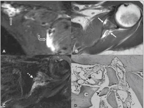
Chest CT did not demonstrate any other alteration. Gorham’s disease was the main radiological hypothesis in this case. Bone biopsy was performed and histopathology demonstrated trabecular bone destruction with prominent bone formation and resorp-tion by osteoblastic and osteoclastic activ-ity, respectively. The bone marrow was re-placed by loose fibrous tissue, with exten-sive vascular proliferation (Figure 3D).
Two years later, follow-up radiographs and CT demonstrated distal clavicular os-teolysis and progression of scapular resorp-tion (Figure 4). The patient has been con-servatively treated with oral analgesics and muscle strength exercises, and pain man-agement has been achieved.
DISCUSSION
Gorham’s disease may affect any bone, particularly the scapular and pelvic girdles. Unifocal involvement is usual, while mul-tifocal involvement is rarely observed(2,5).
Osteolysis may involve joints(4,6) – in the
present case, osteolysis extended from the
scapula towards the clavicle. In most of cases this disease occurs under the age of 40, with neither sex predilection nor he-reditary predisposition(2,5).
Clinical presentation depends on loca-tion, including sudden or insidious pain onset, pathological fractures, progressive functional limb disability – like in the present case – and soft tissue atrophy(2,7).
Laboratory tests are non-specific and gen-erally present normal results. The clinical disease course may be progressive, rela-tively stable or rapidly leading to death. Spontaneous resolution is extremely rare(2,5).
Severe complications such as chylothorax, pleural or pericardial effusion, osteomyeli-tis, septicemia and spinal cord compression are not frequently observed(2,7).
The Gorham’s disease etiopathogeny still remains obscure. The histopathologi-cal substrate for the disease is the replace-ment of healthy bone by an aggressively expansile, non-neoplastic vascular tissue similar to hemangioma and lymphangioma, which is progressively replaced by fibrous connective tissue. Also, fatty replacement of bone marrow may occur(1–3,8).
Specific and effective treatment is not available yet. Radiotherapy, surgery, anti-osteoclastic medication (bisphosphonates) and interferon alpha-2b have been utilized with diverging results(2,5,8).
Imaging methods, particularly radiogra-phy and CT, play a relevant role in the ini-Figure 2. Computed tomography with 3D
recon-struction demonstrating extensive areas of osteoly-sis in practically the whole scapula, with deformity of the choracoid process and glenoid neck (arrows). but without continuity, measuring 4.6 cm in the longest axis (Figure 3A), that was not biopsied because it was considered as a probable formation of lymphatic nature.
Figure 4. Frontal radiograph of left shoulder acquired two years after the patient admission, demonstrat-ing unequivocal progression of scapular resorption and appearance of osteolysis on the inferior aspect of the distal segment of the clavicle (arrows).
338
Oliveira PCR et al. Gorham’s disease of scapula and clavicle
Radiol Bras. 2011 Set/Out;44(5):336–338 tial evaluation of patients with this disease.
The initial suspicion, the clinical progres-sion and the post-therapy follow-up are based on findings of such imaging meth-ods. The radiographic findings demonstrate the sequential disease progression with a clear absence of new bone formation(1,6,7).
Initially multiple radiolucent foci are seen in a relatively normal bone, followed by progression of deformity and bone mass loss with later osteolysis dissemination to-ward adjacent tissues. Subsequently, corti-cal bone erosion and focorti-cal invasion of soft tissues by the angiomatous mass are ob-served, culminating in the disappearance of the remaining bone(1,6,7). CT can confirm
the radiological findings and, additionally evaluates soft tissues and guides the bi-opsy(7), better demonstrating the disease
extent in complex structures such as the scapular girdle. MRI is an adjuvant method with great variability in signal intensity and post-contrast enhancement, reflecting the
spectrum of neovascular and fibrous pro-gression(1,7).
Differential diagnoses for Gorham’s dis-ease include osteomyelitis, metastasis, os-teolysis secondary to rheumatoid arthritis and hyperparathyroidism, besides several osteolytic syndromes. The diagnosis is based on clinical, radiological and mainly histo-pathological findings(1,2,5,7,8). It is important
to highlight the relevance of imaging meth-ods in the setting of an initial diagnostic suspicion, as well as in the evaluation of the disease progression and in the post-therapy follow-up in patients with this disease.
Acknowledgements
The authors thank to the librarians of the institution for their usual efficiency, promptness and cordiality.
REFERENCES
1. Chung C, Yu JS, Resnick D, et al. Gorham syn-drome of the thorax and cervical spine: CT and MRI findings. Skeletal Radiol. 1997;26:55–9.
2. Patel DV. Gorham’s disease or massive osteolysis. Clin Med Res. 2005;3:65–74.
3. Gorham LW, Stout AP. Massive osteolysis (acute spontaneous absorption of bone, phantom bone, disappearing bone); its relation to hemangiomato-sis. J Bone Joint Surg Am. 1955;37:985–1004. 4. Glass-Royal M, Stull MA. Musculoskeletal case of
the day. Gorham syndrome of the right clavicle and scapula. AJR Am J Roentgenol. 1990;154:1335– 6.
5. Rubel IF, Carrer A, Gilles JJ, et al. Progressive Gorham disease of the forearm. Orthopedics. 2008; 31:284.
6. Torg JS, Steel HH. Sequential roentgenographic changes occurring in massive osteolysis. J Bone Joint Surg Am. 1969;51:1649–55.
7. Cano B, Insa S, Cifrián C, et al. Radiologic find-ings in Gorham-Stout syndrome. Radiologia. 2006; 48:33–6.