Tomographic findings of lobar consolidation in primary
pulmonary tuberculosis*
Aspectos tomográficos da consolidação lobar na tuberculose pulmonar primária
Bruno Alberto Falcão Pereira1, Solange Gonçalves David de Macêdo2, Renata do Amaral Nogueira3, Lola Celeste Pantoja Castiel4, Cláudia Renata Rezende Penna5
OBJECTIVE: To describe tomographic findings of lobar consolidation as early manifestation of primary pulmonary tuberculosis. MATERIALS AND METHODS: The present study was developed at Hospital Municipal Jesus, Rio de Janeiro, RJ, Brazil, in the period between 2002 and 2006, retrospectively evaluating tomographic findings in four children aged from 3 to 14 months with lobar consolidation as an early manifestation of primary pulmonary tuberculosis. RESULTS: The most frequently found radiological pattern was lobar consolidation with calcifications, cavitation and intermingle necrotic areas, associated with bulging fissure. Signs of bronchogenic dissemination and lymph node enlargement were observed in all of the four children. Consolidation with a pseudotumor aspect and masslike effect was observed in one case. CONCLUSION: The cases included in the present study have demonstrated that primary pulmonary tuberculosis manifested as lobar consolidation presents typical tomographic images such as cavitation, hypodense areas and calcifications intermingled with consolidation. The association with lymph node enlargement with central necrosis and signs of bronchogenic dissemination reinforce the diagnosis of tuberculosis.
Keywords: Pulmonary tuberculosis; Child; Computed tomography.
OBJETIVO: Descrever os aspectos tomográficos da tuberculose pulmonar primária manifestada inicialmente como consolidação lobar. MATERIAIS E MÉTODOS: O trabalho foi realizado no Hospital Municipal Jesus, Rio de Janeiro, RJ, no período de 2002 a 2006, avaliando-se retrospectivamente os aspectos tomográficos de quatro crianças de 3 a 14 meses de idade com tuberculose pulmonar primária manifestada inicialmente como consolidação lobar. RESULTADOS: O padrão radiológico mais frequente foi a consolidação lobar com calcificações, escavações e áreas de necrose de permeio, associada a abaulamento da cissura. Sinais de disseminação broncogênica e linfadenomegalia foram observados em todas elas. Consolidação de aspecto pseudotumoral, com efeito de massa, foi observada em um caso. CONCLUSÃO: Nos casos estudados ob-servou-se que a tuberculose pulmonar primária manifestada como consolidação lobar apresenta imagens ca-racterísticas à tomografia computadorizada, como escavações, áreas hipodensas e calcificações de permeio à consolidação. A associação com linfonodomegalias com centro necrótico e sinais de disseminação bron-cogênica reforçam o diagnóstico de tuberculose.
Unitermos: Tuberculose pulmonar; Criança; Tomografia computadorizada. Abstract
Resumo
* Study developed at Hospital Municipal Jesus, Rio de Janeiro, RJ, Brazil.
1. Post-graduate in Radiology and Imaging Diagnosis by Santa Casa da Misericórdia do Rio de Janeiro, Rio de Janeiro, RJ, Bra-zil, MD, Resident at Hôpital Européen Georges Pompidou, Paris, France.
2. Master in Pediatrics, MD, responsible by the Unit of Pneu-mology at Hospital Municipal Jesus, Rio de Janeiro, RJ, Brazil.
3. MD, Radiologist, Unit of Pediatrics at Clínica de Diagnóstico Por Imagem (CDPI), Rio de Janeiro, RJ, Brazil.
4. Post-graduation in Radiology and Imaging Diagnosis by Santa Casa da Misericórdia do Rio de Janeiro, Rio de Janeiro, RJ, Bra-zil, MD, Radiologist at Clínica Radiológica Dr. Samuel Castiel, Porto Velho, RO, Brasil.
5. MD, Radiologist responsible by the Service of Pediatric Radiology at Hospital Municipal Jesus, Rio de Janeiro, RJ, Bra-zil.
Mailing address: Dra. Cláudia Renata Rezende Penna. Rua Lauro Müller, 96/907, Botafogo. Rio de Janeiro, RJ, Brazil, 22260-190. E-mail: [email protected]
Received October 30, 2008. Accepted after revision Novem-ber 14, 2008.
a challenging task for several reasons. Most of times, the patients are asymptomatic at the moment of the diagnosis, bacteriologi-cal confirmation is hardly achieved be-cause of the difficulty the sputum collec-tion, and positive gastric lavages are found in a small number of cases. Other relevant aspects to be taken into consideration in the diagnostic investigation are the following: epidemiological history of recent exposure to the bacillus, tuberculin test (PPD) and chest radiography(4,6,7).
Chest computed tomography presents advantages over conventional radiography and can detect tuberculosis in patients with normal or equivocal chest radiographs(1).
Falcão Pereira BA, Macêdo SGD, Nogueira RA, Castiel LCP, Penna CRR. Tomographic findings of lobar consolidation in primary pulmonary tuberculosis. Radiol Bras. 2009;42(2):109–113.
INTRODUCTION
Tuberculosis still remains as a relevant cause of morbi-mortality worldwide, par-ticularly in developing countries. Children represent a risk group, especially those under the age of five(1–3).
In this age range, the most frequent pre-sentation of this condition is primary pul-monary tuberculosis, and most commonly radiological findings include: lymph node enlargement, parenchymal disease, atelectasis, pleural effusion and miliary disease(4,5).
The present study is aimed at describ-ing tomographic finddescrib-ings of primary pul-monary tuberculosis initially manifesting as lobar consolidation.
MATERIALS AND METHODS
The present study was developed in the Hospital Municipal Jesus, a reference in pediatrics in the State of Rio de Janeiro, Brazil. In the period between 2002 and 2006, tomographic findings of four chil-dren in the age range between 3 and 14 months with primary pulmonary tubercu-losis initially manifested as lobar consoli-dation were retrospectively evaluated.
The children were clinically evaluated, with investigation of possible previous contact with tuberculosis, tuberculin test, culture of gastric and bronchoalveolar as-pirates for tubercle bacillus and human immunodeficiency virus (HIV) serum test, besides imaging investigation.
Chest radiography and computed to-mography were requested as a diagnostic complementation for the clinical suspicion of pulmonary tuberculosis.
The following aspects were analyzed on the computed tomography images:
– Consolidation pattern (presence of cavitation, calcifications and hypodense areas compatible with caseous necrosis);
– pattern and distribution of nodular opacities;
– mediastinal or hilar lymphadenopa-thy, with or without the presence of central necrosis and/or peripheral calcification;
– complications such as masslike effect, airway stenosis, atelectasis, pleural effu-sion and miliary disease.
The pseudotumoral aspect was taken into consideration for consolidations with or without masslike effect with enhance-ment after contrast injection without the presence of air bronchogram(1).
Bronchogenic dissemination was re-ported for air-space nodules, centrilobular nodules and tree-in-bud pattern(8).
The terms utilized in the present paper follow the recommendations of the Con-senso Brasileiro sobre a Terminologia dos Descritores de Tomografia Computadori-zada do Tórax (Brazilian Consensus on Terminology Used to Describe Computed Tomography of the Chest)(9,10).
RESULTS
The study sample included three girls and one boy in the age range between 3 and 14 months, three of them with malnutrition. History of cough and fever for at least one month was present in all of the four chil-dren (Table 1). All of them had been vac-cinated with BCG at their first weeks of life and none was HIV-positive.
The diagnosis of tuberculosis was achieved with a positive culture of bron-choalveolar lavage in two of the children and positive culture of gastric aspirate in all of them; three patients presented with tu-berculin skin test with an area of induration of 14 mm or greater (strong reactor); two of them had previous contact with house-hold members diagnosed with pulmonary tuberculosis; initially, the four children had been unsuccessfully treated for pneumonia and, only after institution of specific therapy presented remission of symptoms and radiological aspects.
All of the children were initially evalu-ated by chest radiography presenting lobar consolidation, bulging fissure similar to Klebsiella pneumonia, with no significant respiratory symptom. The investigation was extended with chest computed tomog-raphy for evaluation of parenchymal le-sions characteristics (Tables 2 and 3).
Case 1 – M.I.C., three-month-old fe-male patient with chest radiograph demon-strating right upper lobe consolidation and nodular opacity in the pulmonary basis and
right paracardiac nodular opacity (Figure 1). Tomographic findings included consolida-tions with sparse air bronchograms and subtle calcifications, irregular hypodense areas and a single cavitation; confluent signs of bronchogenic dissemination at right in the lower lobes; centrally hypodense precarinal and infracarinal lymph nodes with no calcification (Figures 2 and 3).
Case 2 – N.O.C., 11-month-old male patient presented lobular consolidation (right upper lobe) on chest radiograph. Com-puted tomography demonstrated consolida-tion in the right upper lobe with air broncho-grams, hypodense areas, single cavitation and subtle, intermingled calcifications; signs of bronchogenic dissemination in the right lower lobe; cantrally hypodense infracarinal lymphadenopathy (Figure 4).
Case 3 – M.E.S., 14-month-old female patient with chest radiograph demonstrat-ing pulmonary hyperaeration and consoli-dation in the left upper lobe, with tracheal compression and deviation to the right. Tomographic findings demonstrated a large opacity with intermingled hypodense areas with a masslike effect, without air bron-chogram, calcifications or cavitations. Other findings were: mediastinal deviation to the right, reduction in the left source bronchus caliber and atelectasis, rare centrilobular opacities in the left lower lobe, small nodule in the left upper lobe and lymphadenomegaly with central hypodensity determining infracarinal wid-ening (Figure 5).
Table 2 Chest radiography findings in the patients evaluated by the present study.
Case 1 2 3 4 Consolidation (local) RUL RUL LUL RUL Fissure bulging + + + + Bronchogenic dissemination + – – + Mediastinal deviation – – + – Emphysema – – + +
RUL, right upper lobe; LUL, left upper lobe.
Table 1 Summary of clinical and laboratory findings.
Case 1 2 3 4 Age (months) 3 11 14 6 Sex Female Male Female Female Nutrition level Eutrophic Malnutrition Malnutrition Malnutrition Household member contact + – – + BCG history + + + + PPD + + + – Gastric lavage + + + + BAL – + + –
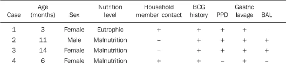
Figure 1. Chest radiograph showing consolidation in the right upper lobe bulging the fissure.
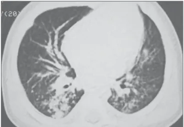
Figure 3. Chest computed tomography demonstrating centrilobular nodules confluent to the right in the lower lobes.
Figure 4. Non-contrast-enhanced chest computed tomography demonstrat-ing consolidation with air bronchograms and subtle intermdemonstrat-ingled calcifications in the right upper lobe.
Figure 2. Contrast-enhanced chest computed tomography demonstrating parenchymal consolidation in the right upper pulmonary lobe, with heteroge-neous contrast uptake, hypodense areas and sparse intermingled air bronchograms, precarinal lymphadenopathy with central necrosis.
L
Table 3 Chest computed tomography findings in the patients evaluated by the present study.
Case
1
2
3
4
Calcifications
+
+
–
+
Lymph nodes (with necrosis)
+
+
+
+*
Bronchogenic dissemination
+
+
+
+
Atelectasis
–
–
+
+
Emphysema
–
–
+
+
RUL, right upper lobe; LUL, left upper lobe. *With calcifications.
Consolidation
Site
RUL
RUL
LUL
RUL
Cavitation
+
+
+
+
Necrosis
+
+
+
+
Case 4 – K.V., six-month-old female patient. Chest radiograph with consolida-tion in the left upper lobe, hyperinsuflaconsolida-tion of the left lung and nodular opacities in the right lung basis and the whole left lung. Chest computed tomography
demon-strated extensive consolidation in the right upper lobe with air bronchograms inter-mingled with hypodense areas, consolida-tion in the lingula, middle lobe and right lower lobe with intermingled areas of cavi-tation, diffused centrilobular opacities in
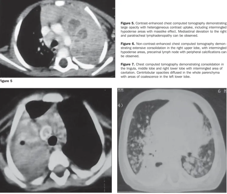
Figure 5. Contrast-enhanced chest computed tomography demonstrating large opacity with heterogeneous contrast uptake, including intermingled hypodense areas with masslike effect. Mediastinal deviation to the right and paratracheal lymphadenopathy can be observed.
Figure 6. Non-contrast-enhanced chest computed tomography demon-strating extensive consolidation in the right upper lobe, with intermingled hypodense areas, precarinal lymph node with peripheral calcifications can be observed.
Figure 7. Chest computed tomography demonstrating consolidation in the lingula, middle lobe and right lower lobe with intermingled area of cavitation. Centrilobular opacities diffused in the whole parenchyma with areas of coalescence in the left lower lobe.
Figure 5
Figure 6 Figure 7
DISCUSSION
Most pulmonary tuberculosis cases ob-served in children are primary tuberculo-sis. The primoinfection is initiated with ba-cilli deposition in the pulmonary alveoli. This process produces a focal alveolar consolidation corresponding to the pri-mary focus that is not always visualized on chest radiographs(1). This consolidation
focus may develop and affect a segment of the whole lobe. The infection may dis-seminate from the primary focus to the central lymph nodes through lymphatic pathways resulting in regional lymph-adenomegaly. The primary focus and the enlarged lymph nodes are called Ranke
complex. In most of cases, parenchymal lesions and lymphadenomegaly present spontaneous resolution(1,7,11,12).
In some cases, particularly involving in-fants, the lymph nodes continue to enlarge progressing up to caseous necrosis. This enlargement may result in regional bron-chus compression causing stenosis, ob-struction and emphysema. Additionally, inflamed lymph nodes may perforate adja-cent bronchus and discharge caseous ma-terial into the bronchial tree, causing bron-chogenic dissemination and/or focal or lo-bar pneumonia(1,7,12).
The most frequent radiological findings in primary pulmonary tuberculosis of chil-dren are lymph node enlargement,
paren-chymatous disease, atelectasis, pleural ef-fusion and military disease(4).
In a tomographic study involving 25 children with ages up to 12 months, Kim et al.(1) have observed consolidation in
100% of patients, 59% with masslike pre-sentation, 41% with necrosis, and 21% with cavitation intermingled with consoli-dation. Bronchogenic dissemination was found in 41% of cases. Parenchymal cal-cifications could be observed six months after, during the treatment(1,2).
In another study(7), evaluating
Leung et al.(4), analyzing chest
radio-graphs of 191 patients in the age range between 20 days to 15 years, observed that the prevalence of parenchymal infiltration has reached 51% for children aged under three, and that hilar or mediastinal lym-phadenopathy would be the most frequent abnormality in this age range(4),
represent-ing the second disease progression site(4,12).
Parenchymal lesions would be most fre-quently found in children aged between 4 and 15 (88%) and would be seen as inter-stitial, alveolar or mixed opacities(4).
Also, there are studies in the literature reporting masslike presentation without satellite lymphadenopathy(6) in children
aged under two, which would be even more uncommon.
In the cases described in the present study with four children aged under 14 months, the most frequent radiological pattern was lobar consolidation with calci-fications, cavitations with intermingled necrotic areas in association with fissure bulging. Signs of bronchogenic dissemina-tion and lymph node enlargement were observed in all of them. pseudotumoral consolidation with masslike effect was observed in one case.
Infracarinal was the preferential local-ization of lymphadenopathy. All of the lymph nodes presented central caseous necrosis and, in two cases peripheral cal-cification was observed. Atelectasis and emphysema were observed in two patients. Miliary disease and pleural effusion were
not observed in the cases included in the present study.
Parenchymal calcifications were ob-served in three of the four cases in the present study, before specific treatment in-stitution. In the studies reviewed(1,4,12), this
finding was observed only during the fol-low-up of the patients along a period of more than ten months. However, Marais et al.(12) report that this finding may be
ob-served at a short time interval in younger children, so the presence of calcifications may be related to the immunological re-sponse and to the time of the disease pro-gression associated with the institution of specific therapy, allowing the utilization of this finding as an additional positive crite-rion for tuberculosis.
CONCLUSION
In the present cases, the authors ob-served that primary pulmonary tuberculo-sis manifested with lobar consolidation is seen with characteristic images at com-puted tomography such as cavitations, hypodense areas and calcifications inter-mingled with consolidation. The associa-tion between lymphadenopathies with cen-tral necrosis and signs of bronchogenic dissemination reinforces the diagnosis of tuberculosis.
REFERENCES
1. Kim WS, Choi J, Cheon JE, et al. Pulmonary tu-berculosis in infants: radiographic and CT find-ings. AJR Am J Roentgenol. 2006;187:1024–33.
2. Amodio J, Abramson S, Berdon W. Primary pul-monary tuberculosis in infancy: a resurgent dis-ease in the urban United States. Pediatr Radiol. 1986;16:185–9.
3. Sant’Anna CC, Hijjar MA. Recente contribuição da Organização Mundial de Saúde para o controle da tuberculose na infância. Rev Saúde Pública. 2007;41(Supl. 1):117–20.
4. Leung AN, Müller NL, Pineda PR, et al. Primary tuberculosis in childhood: radiographic manifes-tations. Radiology. 1992;182:87–91.
5. Leung AN. Pulmonary tuberculosis: the essen-tials. Radiology. 1999;210:307–22.
6. Cherian MJ, Dahniya MH, al-Marzouk N, et al. Primary pulmonary tuberculosis presenting as mass lesions and simulating tumours in children. Australas Radiol. 1998;42:309–12.
7. Lamont AC, Cremin BJ, Pelteret RM. Radiologi-cal patterns of pulmonary tuberculosis in the pae-diatric age group. Pediatr Radiol. 1986;16:2–7. 8. Campos CA, Marchiori E, Rodrigues R. Tuber-culose pulmonar: achados na tomografia compu-tadorizada de alta resolução do tórax em pacien-tes com doença em atividade comprovada bacte-riologicamente. J Pneumol. 2002;28:23–9.
9. Pereira-Silva JL, Kavakama J, Terra Filho M, et al. Consenso brasileiro sobre a terminologia dos descritores de tomografia computadorizada do tórax. J Bras Pneumol. 2005;31:149–56.
10. Souza Jr AS, Araujo Neto C, Jasinovodolinsky D, et al. Terminologia para a descrição de tomogra-fia computadorizada de tórax (sugestões iniciais para um consenso brasileiro). Radiol Bras. 2002; 35:125–8.
11. Kim WS, Moon WK, Kim IO, et al. Pulmonary tuberculosis in children: evaluation with CT. AJR Am J Roentgenol. 1997;168:1005–9.