In vitro
evaluation of an alternative method to
bond molar tubes
Célia Regina Maio PINZAN-VERCELINO1, Arnaldo PINZAN2, Júlio de Araújo GURGEL3, Fausto Silva BRAMANTE1,
Luciana Maio PINZAN4
1- DDS, MSc, PHD, Assistant Professor, Department of Orthodontics, University Center of Maranhão (UNICEUMA), São Luís, MA, Brazil.
2- DDS, MSc, PhD, Associate Professor, Department of Pediatric Dentistry, Orthodontics and Community Health, Bauru School of Dentistry, University of São Paulo, Bauru, SP, Brazil.
3- DDS, MSc, PhD, Professor and Program Director of Master in Orthodontics, University Center of Maranhão (UNICEUMA), São Luís, MA, Brazil. 4- Undergraduate student, Clinical practice, Bauru, SP, Brazil.
Corresponding address: Dr. Célia Regina Maio Pinzan-Vercelino - Alameda dos Sabiás, 58 - Portal dos Pássaros - Boituva - SP - 18550-000 - Brazil - Phone: 55 15 33635958 - Fax: 55 14 32271282 - e-mail: [email protected]
!
ABSTRACT
D
espite the advances in bonding materials, many clinicians today still prefer to place bands on molar teeth. Molar bonding procedures need improvement to be widely accepted clinically. Objective: The purpose of this study was to evaluate the shear bond strength when an additional adhesive layer was applied on the occlusal tooth/tube interface to provide reinforcement to molar tubes. Material and methods: Sixty third molars were selected and allocated to the 3 groups: group 1 received a conventional direct bond followed by the application of an additional layer of adhesive on the occlusal tooth/tube interface, group 2 received a conventional direct bond, and group 3 received a conventional direct bond and an additional cure time of 10 s. The specimens were debonded in a universal testing machine. The results were analyzed statistically by ANOVA and Tukey’s test (D=0.05).groups 2 and 3. No difference was detected between groups 2 and 3 (p>0.05). Conclusions: The present in vitro ! ! "
on the tooth/tube interface increased the shear bond strength of the bonded molar tubes.
Key words: Molar. Shear strength. Dental bonding.
INTRODUCTION
Presently, clinicians are concerned about the clinical effectiveness of the orthodontic procedures. Orthodontists, patients and patients’ parents want to obtain the best results in the shortest time. There are many factors that influence treatment duration, including characteristics associated with the problem, such as amount of anteroposterior or vertical discrepancy; age; patient compliance; psychological implications ! 5,21,23,26,27. Besides,
rebonding of the attachments and recementation of the bands should have been considered, which are undesirable in a private clinic because they are
costly and time-consuming procedures18,20.
In contemporary orthodontic practice, direct bond in molars is a less frequently adopted practice, in spite of its advantages: it reduces chairside time to
# $ !" maintenance of periodontal health, eliminates the spaces caused by the bands, facilitate the detection
of caries and provides a better esthetics2,29. A study9
performed in the United States showed that only 22-30% of the orthodontists usually bond the molars. This fact is likely related to the clinical failure rate of this procedure.
It has been extensively demonstrated13,15,19,28 a
clinical failure rate of bonded molars of 14.8% at least. However, no emphasis is given in the literature
to the loose bands, except for one study2 that
compared bonded and banded molars. Boyd and
Baumrind2 (1992) evaluated banded and bonded
more frequently than banded ones.
In spite of advances in orthodontic bond materials, there is still a need to improve current
bonding procedures20, especially for teeth that
are subjected to higher occlusal forces, like the
molars22.
The aim of this study was to evaluate if the application of an additional adhesive layer on the occlusal teeth/tube interface provides reinforcement to molar tubes.
MATERIAL AND METHODS
Approval of this protocol was received from the institutional review board prior to the start of the study (Protocol 00951/08).
Sixty human mandibular third molars with indication for surgical removal were selected. The criteria for tooth selection included the following: intact buccal enamel not subjected to any pretreatment chemical agents, e.g., hydrogen, no cracks from the presence of the extraction forceps, no caries, and no abrasion or any other crown defect.
The teeth were obtained from a private practice, and were stored in a refrigerated chloramine-T solution for decontamination. Afterwards, the teeth were placed in PVC rings with self-curing acrylic resin; the long axis of each tooth was vertical, and only the crowns were exposed. Each tooth was mounted with the buccal surface of the crown positioned perpendicularly to the PVC ring base with the help of an acrylic squadron, to facilitate the mechanical tests. After resin polymerization, all specimens were immediately immersed in distilled water.
The specimens were randomly divided into 3 groups of 20 teeth each. Each group underwent a different bond protocol: group 1 (experimental) underwent conventional direct bonding followed by the application of an additional layer of adhesive on the occlusal tooth/tube interface, and 10 s of light-cure; group 2 (control 1) underwent conventional direct bonding only; and group 3 (control 2) underwent conventional direct bonding followed by an interval of 40 s, and additional 10 s of light-cure.
Before the direct bond, the buccal surface of each tooth was polished for 10 s with pumice and rubber prophylactic cups with a low-speed handpiece. The surface was then rinsed under water and dried with an oil-free air spray for 30 s. The prophylactic cups were changed after every 10 teeth to obtain a better procedure pattern. The teeth were then etched with 37% phosphoric acid, for 30 s, rinsed with an air-water spray for 30 more s and air-dried until a proper white frosted appearance was observed. The next step was the application of a thin coat of primer (Transbond XT Primer; 3M
Unitek Orthodontic Products, Monrovia, CA, USA), followed by the direct bonding of the single tube bracket convertible for bonding (Morelli Ortodontia,
Sorocaba, SP, Brazil, base area of 13.6 mm2), using
the Transbond XT adhesive. The tubes were kept in the manufacturer’s packaging until immediately prior to bonding and they were handled at all times with bonding tweezers to avoid any contamination of the bonding base, which could interfere with the results. The adhesive was placed on the tube base, & surface to secure a thin layer of adhesive and to
pattern the procedure15. After removal of excess
adhesive from around the attachment, the adhesive was light-cured at a distance of approximately 5 mm above the interproximal contact for 20 s (10 s on each side), according to the manufacturer’s instructions.
The described procedure was followed for group 2 (Figures 2A and 2B), but the specimens in the other groups were prepared with some technical differences.
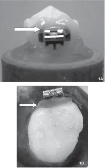
In group 1, immediately after the conventional
direct bond was performed, an additional layer of adhesive was applied on the occlusal tooth/tube interface, followed by 10 additional s of light-cure (Figures 1A and 1B). To standardize the quantity of the applied adhesive, a metal spatula with a demarcation line at 2 mm was used in order to avoid extreme adhesive plantation. The adhesive & ' " was applied using microbrush tips, only close to the bond interface, without extending beyond the attachment limits.
In group 3, the specimens were light cured for an additional 10 s from the occlusal side.
All steps were performed by a single person throughout the experiment, and all the intervals were controlled by use of a chronometer.
After bonding, the specimens were stored
in distilled water at 37°C for 24 h. At the end
of this period the specimens were debonded by using a universal testing machine (EMIC, line DL, series 385, São José dos Pinhais, PR, Brazil) at a crosshead speed of 0.5 mm/min. The specimens were debonded in a direction parallel to the buccal surface. An occlusogingival load was applied to the tubes to produce a shear force at the tooth/ tube interface until fracture occurred. A computer electronically connected to the test machine recorded the results of each test. To calculate bond strength, the debonding force values (N) were
converted to MPa (N/mm2) by taking into account
the surface area data provided by the manufacturer. Descriptive statistics including the mean, standard deviation, median and minimum and maximum values were calculated for each group of teeth tested.
ANOVA was used to compare the results of the three groups. Tukey’s pairwise comparison test was used to determine which groups were statistically different from each other. The results & ! " & than 0.05. These analyses were performed with Statistica software (Statistica for Windows version 6.0; Statsoft, Tulsa, OK, USA).
RESULTS
The descriptive statistics for the shear bond strength of each of the groups tested are presented in MPa in Table 1.
ANOVA indicates that the shear bond strength of group 1 (which received an additional adhesive layer on the occlusal tooth/tube interface) was significantly higher (p<0.05) than that of the other groups (Table 1). There was no statistically
Figure 2- Specimen preparation. Group 2 and Group 3: conventional direct bond (A: vestibular view; B: occlusal view)
Group 1 Group 2 Group 3 (experimental) (control 1) (control 2)
MPa Kg MPa Kg MPa Kg
Mean 17.08A 23.69 12.53B 17.37 12.60C 17.48
SD 3.28 4.55 2.09 2.9 1.97 2.74
Median 16.35 22.66 13.25 18.38 13.1 18.16
Minimum 11.68 16.2 8.65 12.01 8.38 11.63
Maximum 24.54 34.03 15.78 21.88 15.68 21.75
Table 1- Bond strength data [means, standart deviation (SD), median, minimum and maximum values], in MPa and in kilograms (Kg)
!! * & 2 and 3 in relation to the duration of light-cure (Table 1).
DISCUSSION
Advances in adhesives have helped reducing the failure rate of orthodontic procedures. Presently, it seems more important to improve clinical operating procedures than to increase the retentive
strength of the adhesives currently being used28.
Several studies have evaluated the failure rate of bonded molars, but only one study has tested an
alternative method to reduce the rate of failure8.
Johnston and McSherry8 (1999) evaluated the
effect of sandblasting on the bond strength of molar attachments and concluded that sandblasting foil mesh bases is likely to provide only a minimal improvement in clinical performance when bonding molar teeth. As a result, the purpose of the present study was to evaluate a possible alternative technique that could reduce the rate of failure of bonded molars.
Mandibular third molars were selected because, with the evolution of conservative dental treatment, # ! !+ The third molars were relatively easier to obtain.
Millet, et al16 (2001) and Retamoso, et al25 (2009)
also used human third molars to conduct in vitro
studies evaluating molar tubes. Despite the possible morphological variations that can be presented in this group of teeth, the selection criteria were applied with extreme precision to eliminate any possible source of undue variation.
The Transbond XT adhesive was selected to bond the tubes because this resin is commonly used in
shear bond strength evaluations3,4,14-17,19,20,25.
Initially, the tubes were bonded in all 3 groups according to the manufacturer’s instructions. In group 1, an additional layer of adhesive was applied on the occlusal tooth/tube interface immediately after bonding, followed by an additional 10 s of light-cure (Figures 1A and 1B). We opted for 10 s of light-cure since the light was applied directly to the adhesive; moreover, as outlined in the manufacturer’s instructions, this is the time recommended to bond esthetic brackets, since direct illumination of the adhesive throughout the bracket is possible.
To evaluate the results of this alternative method $ ? & ' control group was group 2, in which the tubes were bonded traditionally (Figures 2A and 2B). However, because the total time of light-cure for group 1 was 30 s, a second control group was included to keep the same light-cure time. Therefore, for group 3, 40 s after the conventional direct bond, an additional 10 s of light-cure was applied to the occlusal tooth
surface. This 40-s interval corresponds to the mean time that was spent for the application of the reinforcement in group 1. Group 3 was established to clarify whether the adhesive reinforcement or the longer illumination time caused the difference in the results. Since group 1 showed the highest shear bond strength and there was no difference between groups 2 and 3 (Table 1), the adhesive reinforcement really was the responsible by the highest values obtained in group 1.
The teeth were etched with 37% phosphoric
acid for 30 s based on preliminary studies6,7 that
demonstrated that an etch time of 30 s is required to produce a satisfactory etch pattern on molar teeth.
The bonding procedures were performed by the same operator using a standardized technique. This is an important consideration, based on data from
Millett, et al.15 @@@$ & &
difference in molar tube survival when the procedure was performed by different operators. According to
those authors15, this difference could be related to
the tube location and non-uniformity in the resin thickness between the enamel and bonding bases.
As in other studies2,3,6,12,14,16,18,24,25, the specimens
were stored in distilled water at 37°C until testing
24 h later. Hajrassie and Khier4 (2007) compared
the bond strengths of orthodontic brackets bonded to enamel and debonded at various times (10 min, ?Q $ &Y Q &Y " bond strength values were not time-dependent. In
addition, McCourty, et al14 (1991) did not observe
!! ?Q h and 30 days after a direct bond with Transbond adhesive.
To test shear bond strength, many investigators
have used a universal testing machine2-4,14,16,24,25
with a crosshead speed of 0.5 mm/min3,4,14,24,
which is the same method used in the present
study. A recent study10 showed that crosshead
speed variations between 0.1 and 5 mm/min do Z ! the mode of failure of brackets bonded to enamel with a composite adhesive.
The amount of adhesive remaining on the enamel surface was not evaluated since this study was testing the proposed reinforcement, not the adhesive system. However, in all specimens in which failure occurred, the failure occurred predominantly at the enamel/adhesive interface; this is commonly
observed when bond strengths are higher11,as
observed in this study.
Despite the controversy about i n v i t r o
evaluation13$ !
should be considered important because the application of an additional adhesive layer in specimens was previously tested than in humans,
validate this proposed method in vivo, it should
25.
The results clearly show that the application of an additional adhesive layer in the occlusal tooth/ tube interface provides a better bond strength than the conventional direct bond (Table 1). The values obtained in group 1 were higher than those observed in the other two groups. According to the theory of material resistance, when a force is # (tooth) using an adhesive material (resin), the tension (T) is calculated as the force applied (F) divided by the contact area (A) between the bodies bonded (T=F/A). Considering that the adhesive is a material with the lowest possible rupture tension, we should increase the surface area in order to increase the shear bond strength of the tube/ adhesive/tooth complex. With this purpose in mind, we applied an adhesive reinforcement. The results showed that the additional adhesive layer created an additional contact area between the tooth and the tube so that the applied force was distributed over a more extended surface area, yielding better results for group 1.
The results of the control groups (groups 2 and 3, Table 1) are in agreement with those obtained by
Knoll, Gwinnett and Wolff11 (1986), who observed
a shear bond strength of 11±4.0 MPa for bonded molars. These results are also close to those
observed by Bishara et al1 (1998) (11.8±4.1 MPa).
The increase in light-cure time exposure did Z ' $ since there was no difference between groups 2 and 3. This fact emphasizes the importance of the [ Z strength, the higher values obtained in group 1 were achieved by the proposed method of bonding molar tubes.
Clinical studies have demonstrated different
failure rates for bonded molar tubes15,19,28. These
failures can be related to several factors, including possible moisture contamination, heavy occlusal contacts, tube base adaptation to the curved buccal molar surface, non-uniform resin thickness and the
age of the patient at the initiation of treatment13,15.
\!$ ] ^#22 (1983) demonstrated
that, in normal jaws, the maximum biting force is around 30 kg. In the present study, the values achieved by group 1, despite not having attempted this measurement, are closer than those of the control groups (group 2 and group 3; Table 1). Since many of the factors described above cannot be changed by orthodontists, the alternative method to bond molars presented here seems to be the best option available to increase the quality of the direct bond.
_ $ & !
! 13 or in
placing the tubes in the desired position in posterior regions during direct bonding. However, it is important to emphasize that, although none of the three groups was affected by the above mentioned problems, group 1 showed the best results.
Many factors must be accounted for when deciding to bond or to band molars, such as the possibility of an enamel etch, the quality of the adhesive material available to bond, the retentive potential of the attachment surface, the substrate (amalgam, resin, porcelain, enamel, or other metallic alloys) and the clinical necessities (type of movement, height of the clinical crown and use
of headgear, lip bumpers, or palatal bars)2,28. After
critical analysis of these factors, if direct bonding is chosen, the alternative method proposed in this study seems to be effective.
Bonding may also be useful in mandibular ! to band, particularly when they are only partly
erupted28.
Most adhesives are rough enough to easily
attract plaque28, but the regions where the excess
adhesive was placed are easy to clean, and the adhesive was applied far from the gingival margin. Therefore, gingival health will probably not be any worse than when bands are used, and patients can also be told to reinforce oral hygiene in the region to
which the adhesive is applied. Boyd and Baumrind2
(1992) demonstrated that banded molars in both adolescents and adults had significantly more + " Z than bonded molars.
Another important clinical point when using this technique in the lower arch is the necessity to check the contact points in the areas where the reinforcement was applied over the molar tubes. This can be done during or after the bonding procedure.
An enamel fracture during debonding was
observed in only one specimen. The fracture
occurred in the tooth that had the highest debonding
value (24.54 MPa, Table 1). A study4 that evaluated
in vitro and in vivo bond strengths showed that the in vivo &
strength values compared to the in vitro group.
Care should be taken with the interpretation of
the results, as the experimental conditions in vitro
do not reproduce the oral environment. A long-term clinical evaluation of a larger specimen population is necessary to provide clinically valid data.
CONCLUSION
The present in vitro
ACKNOWLEDGEMENTS
We gratefully acknowledge Morelli Ortodontia, and 3M Unitek Orthodontic Products, for providing commercial products. We also acknowledge Inser Indústria, Comércio e Serviços Ltda, Sorocaba, SP, Brazil, for allowing us to use the quality control laboratory to complete this study.
REFERENCES
1- Bishara SE, Gordan VV, VonWald L, Olson ME. Effect of an acidic primer on shear bond strength of orthodontic brackets. Am J Orthod Dentofacial Orthop. 1998;114:234-7.
2- Boyd RL, Baumrind S. Periodontal considerations in the use of bonds or bands on molars in adolescents and adults. Angle Orthod. 1992;62:117-26.
3- Bradburn G, Pender N. An in vitro study of the bond strength of two light-cured composites used in the direct bonding of orthodontic brackets to molars. Am J Orthod Dentofacial Orthop. 1992;102:418-26.
4- Hajrassie MK, Khier SE. In-vivo and in-vitro comparison of bond strengths of orthodontic brackets bonded to enamel and debonded at various times. Am J Orthod Dentofacial Orthop. 2007;131:384-90.
5- Janson G, Graciano JT, Henriques JF, Freitas MR, Pinzan A, Pinzan-Vercelino CR. Occlusal and cephalometric Class II division 1 malocclusion severity in patients treated with and without extraction of 2 maxillary premolars. Am J Orthod Dentofacial Orthop. 2006;129:759-67.
6- Johnston CD, Burden DJ, Hussey DL, Mitchell CA. Bonding to molars - the effect of etch time (an in vitro study). Eur J Orthod. 1998;20:195-9.
7- Johnston CD, Hussey DL, Burden DJ. The effect of etch duration on the microstructure of molar enamel: an in vitro study. Am J Orthod Dentofacial Orthop. 1996;109:531-4.
8- Johnston CD, McSherry PF. The effects of sanblasting on the bond strength of molar attachments - an in vitro study. Eur J Orthod. 1999;21:311-7.
9- Keim RG, Gottlieb EL, Nelson AH, Vogels DS. 2002 JCO study of Orthodontic diagnosis and treatment procedures. Part 1. Results and trends. J Clin Orthod. 2002;36:553-68.
[ `Y {$ `[^Y | _Z ! [ orthodontic bond strength testing. Dent Mater. 2005;21:139-44. 11- Knoll M, Gwinnett AJ, Wolff MS. Shear strength of brackets bonded to anterior and posterior teeth. Am J Orthod. 1986;89:476-9.
12- Linklater RA, Gordon PH. An ex vivo study to investigate bond strengths of different tooth types. J Orthod. 2001;28:59-65.
13- Linklater RA, Gordon PH. Bond failure patterns in vivo. Am J Orthod Dentofacial Orthop. 2003;123:534-9.
14- McCourt JW, Cooley RL, Barnwell S. Bond strength of light-cure Z[ [ Y " Am J Orthod Dentofacial Orthop. 1991;100:47-52.
15- Millett DT, Hallgren A, Fornell AC, Robertson M. Bonded molar tubes: a retrospective evaluation of clinical performance. Am J Orthod Dentofacial Orthop. 1999;115:667-74.
16- Millett DT, Letters S, Roger S, Cummings A, Love J. Bonded molar tubes - an in vitro evaluation. Angle Orthod. 2001;71:380-5. 17- Öztoprak MO, Isik F, Sayinsu K, Arun T, Aydemir B. Effect of blood and saliva contamination on shear bond strength of brackets bonded with 4 adhesives. Am J Orthod Dentofacial Orthop. 2007;131:238-42.
}[ ~Y |$ Y $ `Y {$ |$ ~ ] _Z of different tooth types on the bond strength of two orthodontic adhesive systems. Eur J Orthod. 2008;30:407-12.
19- Pandis N, Christensen L, Eliades T. Long-term clinical failure rate of molar tubes bonded with a self-etching primer. Angle Orthod. 2005;75:1000-2.
20- Pasquale A, Weinstein M, Borislow AJ, Braitman LE. In-vivo
prospective comparison of bond failure rates of 2 self-etching primer/adhesive systems. Am J Orthod Dentofacial Orthop. 2007;132:671-4.
21- Petrone J, Fishell J, Berk NW, Kapur R, Scioti J, Weyant RJ. Relationship of malocclusion severity and treatment fee to # ! { ~ Dentofacial Orthop. 2003;124:41-5.
??[ \! $ ] $ ^# ~ ! and long-face adults. J Dent Res. 1983;62:566-70.
?[ \! $ ' ] \ __ now or wait? Am J Orthod Dentofacial Orthop. 2002;121:560-2. 24- Rêgo EB, Romano FL. Shear bond strength of metallic brackets photo-activated with light-emitting diode (LED) at different exposure times. J Appl Oral Sci. 2007;15:412-5.
25- Retamoso LB, Collares FM, Ferreira ES, Samuel SM. Shear bond ! Y Z ! " Appl Oral Sci. 2009;17:190-4.
?[ ' {$ `Y { \ Z of orthodontic treatment. Am J Orthod Dentofacial Orthop. 1998;113:29-39.
27- Turbill EA, Richmond S, Wright JL. The time-factor in orthodontics: what influences the duration of treatments in National Health Service practices? Community Dent Oral Epidemiol. 2001;29:62-72.
28- Zachrisson BU. A posttreatment evaluation of direct bonding in orthodontics. Am J Orthod. 1977;71:173-89.
![Table 1- Bond strength data [means, standart deviation (SD), median, minimum and maximum values], in MPa and in kilograms (Kg)](https://thumb-eu.123doks.com/thumbv2/123dok_br/14982062.511068/3.892.86.424.334.806/table-strength-standart-deviation-median-minimum-maximum-kilograms.webp)
