Anesthetic technique for inferior alveolar nerve
block: a new approach
Dafna Geller PALTI1, Cristiane Machado de ALMEIDA1, Antonio de Castro RODRIGUES2, Jesus Carlos ANDREO2,
José Eduardo Oliveira LIMA3
1- DDS, MSc, Department of Pediatric Dentistry, Orthodontics and Community Health, Bauru School of Dentistry, University of São Paulo, Bauru, SP, Brazil. 2- MSc, PhD, Associate Professor, Department of Biological Sciences, Bauru School of Dentistry, University of São Paulo, Bauru, SP, Brazil.
3- DDS, MSc, PhD, Department of Pediatric Dentistry, Orthodontics and Community Health, Bauru School of Dentistry, University of São Paulo, Bauru, SP, Brazil.
Corresponding address: Jesus Carlos Andreo - Faculdade de Odontologia de Bauru - USP - Departamento de Biologia Oral - Disciplina de Anatomia - Alameda Octavio Pinheiro Brisolla, 9-75 - Vila Universitária - 17012-901 - Phone: +55-14-3235-8226 - e-mail: jcandreo@usp.br
!
ABSTRACT
B
ackground: Effective pain control in Dentistry may be achieved by local anesthetic techniques. The success of the anesthetic technique in mandibular structures depends on the proximity of the needle tip to the mandibular foramen at the moment of anesthetic injection into the pterygomandibular region. Two techniques are available to reach the inferior alveolar nerve where it enters the mandibular canal, namely indirect and direct; these techniques differ in the number of movements required. Data demonstrate that the indirect technique is considered ineffective in 15% of cases and the direct technique in 13-29% of cases. Objective: The aim of this study was to describe an alternative technique for inferior alveolar nerve block using several anatomical points for reference, simplifying the procedure and enabling greater success and a more rapid learning curve. Materials and Methods: A total of 193 mandibles (146 with permanent dentition and 47 with primary dentition) from dry skulls were used to establish a relationship between the teeth andgroove and middle point of the mesial slope of the distolingual cusp of the primary second side), a line can be achieved whose projection coincides with the left mandibular foramen. !"!!# molar, and in 93.62% of cases using the primary second molar. Conclusion: This method is potentially effective for inferior alveolar nerve block, especially in Pediatric Dentistry.
Key words: Anesthesia, dental. Nerve block. Inferior alveolar nerve.
INTRODUCTION
Adequate anesthesia is fundamental for the accomplishment of most dental procedures5. Some investigators state that anesthesia is essential for both the patient and the dental professional; also, the opinion of patients on their dental treatment is strictly related to their experience with local anesthesia15. Other authors have reported that many patients select their dentists based on their ability to offer a painless dental treatment13.
The use of local anesthetic techniques is important in the different dental specialties, especially in Pediatric Dentistry. McDonald and Avery21 (1994) stated that pain control is one of
$ behavior during dental treatment.
Inferior alveolar nerve block is the technique most frequently used for local anesthesia when performing restorative and surgical procedures in the mandible8; however, the approximate failure rate of these procedures ranges from 5 to 15%31 or 15 to 20% according to Kaufman14 (1984), reaching even higher percentages in pulpal anesthesia6,32.
anesthetic techniques for mandibular structures present a lower success rate compared to those for maxillary structures, because of the greater density of the mandibular alveolar bone, limited access to the inferior alveolar nerve, marked anatomical variations, in addition to the need for deeper needle penetration into the soft tissue17.
For the achievement of effective anesthesia, the anesthetic solution should be injected as close as possible to the nerve31. For this reason, the technique must be based on precise anatomical knowledge regarding the correct location of the mandibular foramen, making use of procedures through which it may be reached even though it is surrounded by soft tissues. Therefore, the failures are related to several factors, such as lack of knowledge on the anatomical structures, technical ''$ infection and damaged anesthetic solutions.
Concerning anatomical variations, Desantis and Liebow4 (1996) described four anatomical variations of the mandibular nerve that may complicate the local anesthesia, namely the accessory mylohyoid nerve, bifid mandibular nerve, presence of retromolar foramen and contralateral innervation of anterior teeth4. The presence of the retro-molar foramen is also cited in other papers, with a frequency of 7.7%25.
Several authors have contributed to improve the && and creating new techniques in order to enhance the success rate of these procedures2,3,7,9,10,18,29.
Considering that anesthesia of the inferior alveolar nerve is often applied in dental treatments and that the achievement of a successful technique is a concern among professionals, primarily those with less experience or who work with children or anxious patients, an alternative method is suggested for inferior alveolar nerve block, using anatomical reference points that are easily observed during application of anesthesia, simplifying the learning process, the technique and consequently increasing the success rate.
MATERIAL AND METHODS
Measurements were performed on 193 mandibles (146 with permanent dentition and 47 with primary dentition) from dry skulls from the Anatomy Museums of the Federal University of São Paulo (UNIFESP) and the University of São Paulo (USP).
Inclusion Criteria
- Presence of the reference teeth permanent * second molars, contralateral to the side to be anesthetized.
- No mesial displacement of the reference teeth
due to early loss of the adjacent teeth.
- Presence of two grooves on the buccal surface primary mandibular second molars, one mesial and one distal, dividing the teeth in three unequal portions; and one groove on the lingual surface, & and two lingual. These grooves are usually observed in 95.5% of cases26.
- Integrity of these teeth to allow establishment of the proposed anatomical references.
Exclusion criteria
- Absence of reference teeth.
- Reference teeth with decayed or worn crowns. - Distal and mesial displacement of reference teeth.
- Absence of buccal grooves on the reference teeth.
Observations
The observations to establish a correlation between the grooves of the permanent mandibular mandibular foramen were always performed by the same operator. Two no. 9 straight orthodontic wires were used, positioned according to the following three steps:
Right Side
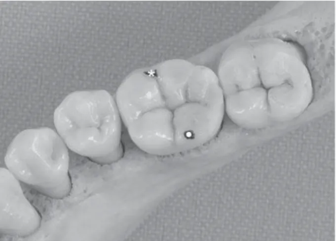
1. Locate the mesiobuccal groove and middle point of the mesial slope of the distolingual cusp of
+/" mesiobuccal groove and middle point of the mesial slope of the distolingual cusp of the primary second +"< second wire must follow the occlusal plane on the left side (Figure 3).
Left Side
The same reference points were used, yet changing the sides.
When the tip of the orthodontic wire coincided with the center of the mandibular foramen or a 3.5 mm radius from this point, the technique was & measuring 20 mandibular foramens in ten dry mandibles, in order to calculate the mean size of the foramen.
When there was no coincidence between the wire tip and the mandibular foramen, the distance from the wire tips to the center of the foramen was calculated using a digital pachymeter (Mitutoyo Corporation, Suzano, SP, Brazil, code number 700-113, model number SC-6, series number 0013491), considering this value as positive when the tip was located mesial to the foramen and negative when it was distally positioned.
Statistical analysis
These values were submitted to descriptive statistical analysis for achievement of the arithmetic mean and calculation of the percentage of coincidence.
RESULTS
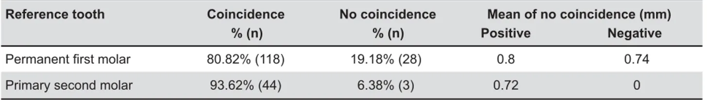
The obtained data are presented in Table 1. = were taken as reference, the percentage of coincidence was lower compared to the primary mandibular second molars (80.82% and 93.62%, respectively).
When there was no coincidence, positive distances were greater for both permanent and primary teeth (0.8 mm for the permanent mandibular first molars and 0.72 mm for the primary mandibular second molars).
Negative distances were not observed for the primary mandibular second molars.
DISCUSSION
As previously mentioned, local anesthesia is fundamental in dental treatment and is widely used in several dental specialties.
Two techniques are described in the literature to reach the inferior alveolar nerve where it enters the mandibular canal, namely indirect and direct; these techniques differ in the number of movements required17,24.
The approach most commonly used for anesthesia of the inferior alveolar nerve in the United States is the traditional Halstead method18, a direct technique
Reference tooth Coincidence No coincidence Mean of no coincidence (mm) % (n) % (n) Positive Negative
3HUPDQHQW¿UVWPRODU 80.82% (118) 19.18% (28) 0.8 0.74
Primary second molar 93.62% (44) 6.38% (3) 0.72 0
Table 1- Mean and percentage values obtained
Figure 2- 7KH ¿UVW ZLUH SDVVLQJ IURP WKH PHVLREXFFDO groove and the middle point of the slope of the distolingual cusp (or distolingual angle line) (note Figure 1) of the PDQGLEXODU¿UVWPRODUSHUPDQHQW
in which the inferior alveolar nerve is reached by an intraoral access before it penetrates the mandibular canal. This block method has success rates from 71 to 87%14 and incomplete anesthesia is not uncommon. Also, it has been shown that the indirect technique is ineffective in 15% of cases23.
More recently, Galdames, et al.7 (2008) suggested an anesthetic technique for the inferior alveolar, buccal and lingual nerves, taking the retromolar trigone as reference. This technique is safer for patients with blood dyscrasias, yet is less effective than the conventional technique of inferior alveolar nerve block.
The literature shows that failures in the anesthesia of the inferior alveolar nerve occur due to several factors, such as lack of knowledge on the anatomical structures, lack of experience, technical ''$ infection, damaged anesthetic solutions31, changes in anatomical structures4,30, inadequate mouth opening, inadequate positioning of the needle, hurry16 and needle deviation27.
Concerning the anatomical variations of the & & presence of retromolar foramen and contralateral innervation of anterior teeth5, the failure in the anesthesia of the inferior alveolar nerve should be solved using the supplementary anesthetic techniques described in the literature1,4,19,20.
Considering that the success of the anesthetic technique in mandibular structures depends on the proximity of the needle tip to the mandibular foramen at the moment of anesthetic injection, this anatomical structure was addressed in this study.
With regard to the anatomical variation of the mandibular foramen, the literature reports that the position of this foramen changes with skeletal growth both in craniocaudal and anteroposterior directions22. This should be taken into account when selecting the mandibular anesthetic technique11,12,28.
In the primary dentition, the mandibular foramen is positioned at or below the occlusal level28. Concerning its anteroposterior positioning, some investigators believe it is positioned in the third quadrant, starting on the anterior margin of the mandibular ramus11, while other authors believe it is found behind an imaginary line that divides the mandibular ramus in the half12.
Some studies using panoramic radiographs reveal variations in the position of the mandibular foramen in relation to other anatomical references2. Other studies have shown that the distance between the mandibular foramen and the anterior margin of the mandibular ramus was greater than the distance between the mandibular foramen and its posterior margin, both in the primary and permanent dentitions28.
In this study, the possible anatomical variations
were not taken into consideration, since the age of people whose mandibles were used was not available.
Studies in the literature attempt to determine the location of the mandibular foramen in craniocaudal and anteroposterior directions in the mandibular ramus. However, these measurements > anatomical reference in the oral cavity that may simplify the access to the mandibular foramen inside the soft tissues.
' the reference teeth, one point on the mesiobuccal groove and middle point of the mesial slope of the distolingual cusp of the primary second molar or
direction in the mandibular ramus, while the straight line passing through the occlusal plane on $ these two lines determines the point of penetration and the needle angulation for anesthesia of the inferior alveolar nerve. This study revealed that this technique may be used at any age, providing the inclusion and exclusion criteria are followed.
This work suggests an alternative technique for inferior alveolar nerve block, based on an anatomical ' a straight line, with possibility of success rate of 80.82% for the permanent dentition and 93.62% for the primary dentition, considering the coincidence of the points suggested with the mandibular foramen.
The cases of no coincidence or “failures” in this study were 19.18% for the permanent dentition and 6.38% for the primary dentition. The mean distance from the wire tip mesial to the center of the mandibular foramen was considered positive and revealed values of 0.8 mm and 0.72 mm for the permanent and primary dentitions, respectively. When the wire tip was positioned distal to the foramen, the distance was considered negative and revealed a mean value of 0.74 mm for the permanent dentition, without any case in the primary dentition. However, when 3.5 mm is subtracted from these measurements, which is the mean radius of the foramen, these distances present to be minimal and probably would not interfere with the effectiveness of the anesthetic technique suggested. Thus, the success rate of this technique might be even higher.
can be concluded that the method proposed in this study is effective to reach the inferior alveolar nerve, especially in pediatric dentistry. Clinical studies are
REFERENCES
1- Afsar A, Haas DA, Rossouw PE, Wood RE. Radiographic localization of mandibular anesthesia landmarks. Oral Surg Oral Med Oral Pathol Oral Radiol Endod. 1998;86(2):234-41. "?@JQ@VX@ inferior alveolar nerve block in patientes with irreversible pulpitis. J Endod. 2009;35(7):925-9.
3- Akinosi JO. A new approach to the mandibular nerve block. Br J Oral Surg. 1977;15(11):83-7.
4- DeSantis JL, Liebow C. Four common mandibular nerve anomalies that lead to local anesthesia failures. J Am Dent Assoc. 1996;127(7):1081-6.
5- Fan S, Chen W, Pan C, Huang Z, Xian M, Yang Z, et al. Anesthetic &&> periodontal liagament injection with articaine in patients with & ]^] Med Oral Pathol Oral Radiol Endod. 2009;108(5):e89-93. _?+=X` @>`@ alveolar nerve block in mandibular posterior theeth. Anesth Prog. 2007;54(4):163-9.
7- Suazo Galdames IC, Contín López MG, Zavando Matamala DA. Inferior alveolar nerve block anesthesia via the retromolar triangle, na alternative for patients with blood dyscrasias. Med Oral Patol Oral Cir Bucal. 2008;13(1):E43-7.
8- Goldberg S, Reader A, Drum M, Nusstein J, Beck B. {& alveolar, Gow-Gates, and Vazirani-Akinosi techniques. J Endod. 2008;34(11):1306-11.
9- Gow-Gates GA. Mandibular conduction anesthesia: a new technique using extraoral landmarks. Oral Surg Oral Med Oral Pathol. 1973;36(3):321-8.
10- Gustainis JF, Peterson LJ. An alternative method of mandibular nerve block. J Am Dent Assoc. 1981;103(1):33-6.
11- Hayward J, Richardson ER, Malhotra SK. The mandibular foramen: its anteroposterior position. Oral Surg Oral Med Oral Pathol. 1977;44(6):837-43.
12- Hetson G, Share J, Frommer J, Kronman JH. Statistical evaluation of the position of the mandibular foramen. Oral Surg Oral Med Oral Pathol. 1988;65(1):32-4.
13- Johnson TM, Badovinac R, Shaefer J. Teaching alternatives to the standard inferior alveolar nerve block in dental education: outomes in clinical practice. J Dental Educ. 2007;7(9):1145-52. /}?V~=`X& local anesthesia. J Am Dent Assoc. 1984;108(2):205-8.
15- Kohler BR, Castellón L, Laissle G. Gow-Gates technique: a pilot study for extraction procedures with clinical evaluation and review. Anesth Prog. 2008;55(1):2-8.
16- Madan GA, Madan SG, Madan AD. Failure of inferior alveolar nerve block. J Am Dent Assoc. 2002;133(7):843-6.
17- Malamed SF. Handbook of local anesthesia. 4th ed. St. Louis:
Mosby; 2001. p.193-219.
18- Malamed SF. The periodontal ligament (PDL) injection: an alternative to inferior alveolar nerve block. Oral Surg Oral Med Oral Pathol. 1982;53(2):117-121.
19- Matthews R, Drum M, Reader A, Nusstein J, Beck M. Articaine patients with irreversible pulpitis when the inferior alveolar nerve block fails. J Endod. 2009;35(3):343-6.
20- Meechan JG, Kanaa MD, Cobert IP, Steen IN, Whitworth JM. a double-blind randomized cross-over trial comparing buccal and &~ J. 2006;39(10):764-9.
21- McDonlad RE, Avery DR. Local anesthesia for the child and adolescent. In: McDonald RE, Avery DR, eds. Local anesthesia for the child and adolescent. 6th ed. St. Louis: Mosby; c1994. p.
294-306.
22- Nicholson ML. A study of the position of the mandibular foramen in the adult human mandible. Anat Rec. 1985;212(1):110-2. 23- Northrop PM. Practical technics in administration of local anesthetic agents: questions and answers. J Am Dent Assoc. 1949;38(4):444-8.
24- Ogle OE. Local anesthesia. In: Dym H. Atlas of Minor Oral Surgery. Philadelphia: Saunders; 2000. p. 30-40.
25- Sawyer DR, Kiely ML. Retromolar foramen: a mandibular variant important to dentistry. Ann Dent. 1991;50(1):16-8. 26- Serra DO. Observações morfológicas sobre os dentes jugais humanos. Revista APCD. 1952;5(3):21-6.
27- Steinkruger G, Nusstein J, Reader A, Beck M, Weaver J. The && inferior alveolar nerve block. J Am Dent Assoc. 2006;137(2):1685-91.
"!? foramen from primary to early permanent dentition. J Clin Pediatr Dent. 2004;28(3):215-9.
29- Watson JE, Gates GA. A clinical evaluation of the Gow-Gates mandibular block technique. N Z Dent J. 1976;72(330):220-3.
30- Wilson S, Johns P, Fuller PM. The inferior alveolar and mylohyoid nerves: an anatomic study and relationship to local anesthesia of the anterior mandibular teeth. J Am Dent Assoc. 1984;108(3):350-2.
31- Wong MK, Jacobsen PL. Reasons for local anesthesia failures. J Am Dent Assoc. 1992;123(1):69-73.