656
Srp Arh celok Lek. 2015 Nov-Dec;143(11-12):656-661 DOI: 10.2298/SArH1512656D
ОРИГИНАЛНИ РАД / OrIGINAL ArTIcLE UDc: 616.28-089.843
correspondence to: Dragan DANKUC Vase Stajića 32 21000 Novi Sad Serbia
dragan.dankuc@neobee.net
SUMMARY
Introduction The first modern cochlear implantation in Serbia was performed on November 26, 2002 at the Center for Cochlear Implantation of the Clinic for Ear, Nose and Throat Diseases, Clinical Center of Vojvodina.
Objective The aim of the paper is the analysis of intraoperative and postoperative complications. Major complications include those resulting in the necessity for revision surgery, explantation, reimplantation, severe disease or even lethal outcomes. Minor complications resolve spontaneously or can be managed by conservative therapy and do not require any prolonged hospitalization of the patient.
Methods In the 2002–2013 period, 99 patients underwent surgical procedures and 100 cochlear implants were placed. Both intraoperative and postoperative complications were analyzed in the investigated patient population.
Results The analysis encompassed 99 patients, the youngest and the oldest ones being one year old and 61 years old, respectively. The complications were noticed in 11 patients, i.e. in 10.5% of 105 surgical procedures. The majority of procedures (89.5%) were not accompanied by any post-surgical complications. Unsuccessful implantation in a single-step procedure (4.04%) and transient facial nerve paralysis can be considered most frequent among our patients, whereas cochlear ossification (1.01%) and transient ataxia (2.02%) occurred rarely. Stimulation of the facial nerve (1.01%), intraoperative perilymph liquid gusher (1.01%), device failure and late infections (1.01%) were recorded extremely rarely. Conclusion Complications such as electrode extrusion, skin necrosis over the implant or meningitis, which is considered the most severe postoperative complication, have not been recorded at our Center since the very beginning. Absence of postoperative meningitis in patients treated at the Center can be attributed to timely pneumococcal vaccination of children.
keyword: cochlear implantation; intraoperative complications; postoperative complications
Complications in Cochlear Implantation at
the Clinical Center of Vojvodina
Dragan Dankuc1,2, Ljiljana Vlaški1,2, Nemanja Pejaković1,2, Vladimir Mrdjanov3
1ENT Clinic, Clinical Center of Vojvodina, Novi Sad, Serbia; 2University of Novi Sad, Faculty of Medicine, Novi Sad, Serbia; 3Audio BM d.o.o., Novi Sad, Serbia
INTRODUCTION
The first successful cochlear implantation in Serbia has been performed at the Center for Cochlear Implantation of the Clinic for Ear, Nose and Throat Diseases, Clinical Center of Vojvodina. The first modern cochlear im-plant, Nucleus 24, was placed on November 26 of 2002 in a 40-year-old female patient with postlingual hearing impairment. The surgery was performed by Prof. Dr. J. Jori and Prof. J. G. Kiss from the ENT Clinic, Szeged, Hungary, and Prof. Dr. Dragan Dankuc from the ENT Clinic, Novi Sad, Serbia.
Subsequently, for the first time in Serbia, Dragan Dankuc, under the assistance of Prof. Dr. J. Jori, has performed the first implantation of an artificial inner ear – a cochlear implant Nucleus 24 [1]. Ever since, the cochlear implant surgery in Novi Sad has been exclusively per-formed by an experienced team led by eminent professors Zoran Komazec, Dragan Dankuc, Ljiljana Vlaški, Slobodanka Lemajić Komazec, specialized surdopedagogists Spomenka Ne-deljkov, Ivana Sokolovac and Oliver Vajs, as well as engineers Tibor Mendrei and Vladimir Mrdjanov [2].
OBjECTIVE
The paper’s objective is to analyze possible in-traoperative and postoperative complications. Major complications may result in the necessity for revision surgery, explantation, reimplanta-tion, severe disease or even lethal outcomes. Minor complications, on the other hand, can resolve spontaneously and do not require any prolonged hospitalization of the patient.
METHODS
657
Srp Arh Celok Lek. 2015 Nov-Dec;143(11-12):656-661
RESULTS
The analysis encompassed 99 patients, the youngest and the oldest ones being one year old and 61 years old, re-spectively. The patient population included 11 (11.1%) adults and 88 (88.9%) children as the majority of the pa-tient population.
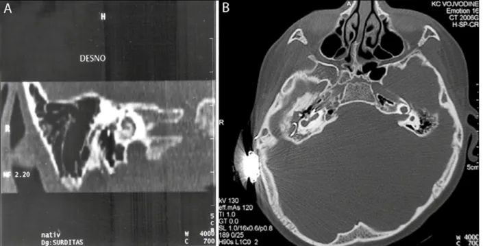
Both intraoperative and postoperative complications were analyzed in the investigated patient population. The complications were noticed in 11 patients, i.e. in 10.5% of 105 surgical procedures. Implant placement in a single-stage procedure was not possible in four cases because of acute otitis media in one patient, diagnosed during surgery, and the ossification of the cochlea that prevented electrode array placement in the remaining three patients (Figure 1A-B). The second surgery was successfully performed in all four patients, without any subsequent complications. Transient facial nerve paresis was recorded in four (4.04%) patients, which completely subsided two months post sur-gery after appropriate therapeutic treatment. Transient ataxia was observed in two (2.02%) patients [3].
Some rare complications, such as facial nerve stimu-lation associated with electro-stimustimu-lation of the cochlea, late complication occurring one year post surgery, device failure identified at fitting session and late infection, were observed in one (1.01%) patient each. All complications were successfully managed by incision and drainage, while preserving the functionality of the device (Table 1).
DISCUSSION
The youngest patient was only one year old (i.e. 14.7 months) at the moment of surgery. In 27 (30.7%) children, the surgery was performed at the age below three, whereas 50 (56.8%) children underwent implantation procedure at
the age under four years old. The aims of early implanta-tion are to reduce the hearing deprivaimplanta-tion period and to improve the development of auditory performance [4].
In adult patients with postlingual deafness, comprehen-sive evaluation of the ratio between potential benefit and risks associated with surgery itself and potential comor-bidities should be performed. However, age is not the cri-terion for excluding a patient from implantation procedure unless other risk factors are present [5, 6].
Complications associated with cochlear implantation can be categorized as major and minor ones. Major complica-tions include those resulting in the necessity for revision surgery, explantation, reimplantation, severe disease or even lethal outcomes. Minor complications resolve spontaneous-ly or can be managed by conservative therapy, and do not require any prolonged hospitalization of the patient [7, 8].
Cohen et al. [9] characterized implant-related complica-tions as major if they required revision surgery, and minor
if they resolved with minimal or no treatment. A survey reported 55 major (12%) and 32 minor (7%) complications.
Webb et al. [10] reporting their experience with 153 patients found 13.7% major and minor complications.
Table 1. Complications of cochlear implants at the Center for Cochlear Implantation of the Clinic for Ear, Nose and Throat Diseases, Clinical Center of Vojvodina
Complications Number
Without complications 94
Unsuccessful implantation 4
Transient paresis of n. VII 4
Cochlea ossification 3
Transient ataxia 2
Unwanted stimulation of n. VII 1
Perilymph gusher 1
Device failure 1
Late infection of incision site 1
658
Dankuc D. et al. Complications in Cochlear Implantation at the Clinical Center of Vojvodina
Hoffman and Cohen [11] noted that in later follow-up 220 (8%) major and 119 (4.3%) minor complications oc-curred among 2,751 implantations.
At our Center for Cochlear Implantation of the Clinical Center of Vojvodina, complications were observed in 11 patients, that is 10.05% of performed surgical procedures [12]. This incidence corresponds with the incidence rates reported from related centers worldwide, which is around 10%. In four (4.04%) patients treated at our Center, the single-stage surgery had not been initially possible, thus implantation was postponed and successfully accomplished in the second stage. In one patient, successful implantation using another type of electrode was performed on the same side. In three other patients, the second-stage surgery was performed on the other side with favorable outcome.
In cases of congenital malformations of the inner ear in two of our pediatric patients, the placement of the elec-trode into the altered cochlea could not be accomplished in spite of the surgical navigation system (Figure 2A-B).
Transient postoperative peripheral facial nerve paresis was observed in four (4.04 %) patients. This condition is considered a minor complication and is explained by tran-sient edema of the facial nerve in the fallopian canal in-duced by the heating of its immediate surrounding struc-tures during posterior tympanotomy. This impairment of nerve function was transient in all our patients. The symptoms resolved completely within the first month post surgery after conservative corticosteroid therapy without any need for subsequent surgical nerve decompression.
Major complications include facial nerve paralysis and implant exposure due to skin flap necrosis. Necrosis of the skin flap can lead to wound infection and device extru-sion, necessitating scalp flap reviextru-sion, and, when intrac-table infection is present, device removal with or without replacement.
Transient ataxia was observed in two (2.02%) patients, who presented with symptoms of postoperative instability and nausea. The patients responded well to symptomatic therapy and recovered rapidly without any consequences.
This complication might be explained by perilymph and endolymph leakage during the formation of the cochle-ostoma. After the surgery, upon reestablishment of the homeostasis of the semicircular canals, ataxia resolves spontaneously without need for any specific therapy.
Postoperative facial nerve stimulation was observed in one (1.01%) female patient. According to the available literature, this major complication of cochlear implanta-tion occurs in some 0.31–14% of cases. Switching off the electrodes that directly stimulate the nerve might be the potential solution in such cases; however, this can result in reduced sound perception, which was the case in our patient. Thus, implantation of the second ear was per-formed to accomplish satisfactory overall hearing perfor-mance through bilateral stimulation at sub-maximal level. Instead of electrode remapping, facial nerve stimulation can be managed by botulinum toxin injections; however, this therapeutic option requires repeated administration at three- to six-month intervals [13, 14].
Extensive intraoperative perilymph gusher was ob-served in one (1.01%) patient. This is considered a minor complication, and was successfully managed during surgi-cal procedure without affecting the outcome.
Function of external sound processor may be lost due to direct trauma, exposure to water and most frequently normal wear and tear of connecting lead-wires linking the sound processing unit with the magnetically retained antenna that relays information and power to the internal device. An internal device failure typically presents as ei-ther an immediate cessation of function or intermittency associated with reduced quality of sound and a period of diminishing function over days to weeks. Reports of pain-ful stimulation have been noted, bat are rare. Device failure is the most common indication for revision surgery and cochlear reimplantation.
Device failure at mapping session occurred in one (1.01%) patient. Such failures are considered major com-plications, as they inevitably require second surgery, which was successfully performed in our patient.
659
Srp Arh Celok Lek. 2015 Nov-Dec;143(11-12):656-661
Infection and flap breakdown require reimplantation less frequently [15]. Luetje and Jackson[16] reported a 9% rate of device failure in a review of 55 children.
Cochlear implants are rarely complicated by microbial infections. When such infections do occur, they can be difficult to treat with conventional antibiotic therapy, and may consequently require surgical removal of the implant [17, 18, 19]. Several studies have demonstrated that infec-tions of cochlear implants are due to biofilm formation [20, 21]. A biofilm-related infection is difficult to treat, as biofilm formation results in increased bacterial resistance both to the body’s immune response and to antibiotic ther-apy [22, 23]. Numerous studies have demonstrated that antimicrobial resistance is considerably increased when bacteria grow in biofilm mode [24, 25].
The increased antibiotic resistance of bacteria growing in a biofilm has been attributed to a number of factors, including decreased antibiotic penetration, altered me-tabolism of bacteria growing in biofilm and expression of biofilm-specific antibiotic resistance genes.
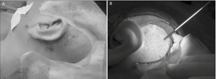
Skin infection in implant region was noticed as a late complication in one (1.01%) adult patient several months post surgery (Figure 3A-B). Prophylactic perioperative application of antibiotics can greatly reduce such
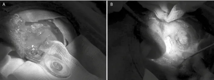
infec-tions, but other authors also reported the occurrence of this complication at the incidence of some 1%. In our pa-tient, the infection was successfully managed by drainage and antibiotic therapy while preserving the functionality and position of the implant. In our case, the patient will have the infected implant temporarily removed and bathed in 3% hydrogen peroxide solution for approximately 30 minutes, in an attempt to eradicate the bacterial biofilm (Figure 4A-B). After this period of disinfection, the im-plant is replaced as before and the patient is discharged on a high-dose course of appropriate antibiotics (Figures 5A-B and 6A-B).
The risk of bacterial infection of an implanted device producing labyrinthitis or meningitis and associated reac-tive fibrosis and destruction of neural elements appears to be low. Reefhuis et al. [26] conducted a study of 4,264 children implanted between 1997 and 2002 and found 29 cases of bacterial meningitis of all implanted children. This rate of meningitis caused by Streptococcus pneumoniae was 30 times the incidence in the general population.
Complications reported in the literature, such as elec-trode extrusion, skin necrosis over the implant, or men-ingitis, which is considered the most severe postoperative complication, have not been recorded at our Center since
Figure 3. Implant region: A) skin infection; B) skin incision
660
the very beginning. Absence of postoperative meningitis in patients treated at the Center can be attributed to timely pneumococcal vaccination of children.
CONCLUSION
The majority of our patients, i.e. 84 (84.9%), manifested prelingual hearing loss, whereas postlingual type of deaf-ness was observed in 15 (15.1%) cases.
At our Center for Cochlear Implantation of the Clinical Center of Vojvodina, the majority of procedures (89.5%) were not accompanied by any post-surgical complications.
The complications were observed in 11 patients, which is 10.5% of performed surgical procedures. Unsuccessful im-plantation in a single-step procedure and transient facial nerve paralysis can be considered to be the most frequent complications among our patients, whereas cochlear os-sification and transient ataxia occurred rarely. Stimulation of the facial nerve, intraoperative perilymph gusher, device failure and late infections were recorded extremely rarely.
Complications such as electrode extrusion, skin necro-sis over the implant, or meningitis, considered to be the most severe postoperative complication, have not been recorded at our Center, which can be attributed to timely pneumococcal vaccination of children.
1. Dankuc D. Kohlearni implant. Uvodnik. Med Pregl. 2005; 58(7-8):329-32. [PMID: 16296573]
2. Komazec Z, Dankuc D, Vlaški Lj, Lemajić-Komazec S, Nedeljkov S, Sokolovac I. Kohlearna implantacija na Klinici za bolesti uva, grla i nosa Kliničkog centra Vojvodine. Med Pregl. 2007; 60(11-12):643-8. [DOI: 10.2298/MPNS0712643K] [PMID: 18666611]
3. Komazec Z, Lemajić-Komazec S, Dankuc D, Vlaški Lj. Kohlearna implantacija – rizici i komplikacije. Med Pregl. 2008; 61(Suppl 2):27-30. [PMID: 18924587]
4. Valencia DM, Rimell FL, Friedman BJ, Oblander MR, Helmbrecht J. Cochlear implantation in infants less than 12 months of age. Int J Pediatr Otorhinolaryngol. 2008; 72(6):767-73.
[DOI: 10.1016/j.ijporl.2008.02.009] [PMID: 18403026] 5. Baumgartner WD, Pok SM, Egelierler B, Franz P, Gstoettner W,
Hamzavi J. The role of age in pediatric cochlear implantation. Int J Pediatr Otorhinolaryngol. 2002; 62(3):223-8.
[DOI: 10.1016/S0165-5876(01)00621-8] [PMID: 11852125]
Figure 5. A) Infected implant removed; B) Implant bathed in a 3% hydrogen peroxide solution
Figure 6. A) The implant is replaced; B) Drainage with local antibiotics
REFERENCES
661
Srp Arh Celok Lek. 2015 Nov-Dec;143(11-12):656-661
6. Yeagle JD, Ceh KM, Francis HW. Geriatric cochlear implantation. Operative Techniques in Otolaryngology-Head and Neck Surgery. 2010; 21(4):266-71. [DOI: 10.1016/j.otot.2010.03.003]
7. Black IM, Bailey CM, Albert DM, Leighton SE, Hartley BE, Chatrath P, et al. The Great Ormond Street Hospital paediatric cochlear implant programme 1992–2004. A review of surgical complications. Cochlear Implants Int. 2007; 8(2):53-67.
[DOI: 10.1179/cim.2007.8.2.53] [PMID: 17549805]
8. McJunkin J, Jeyakumar A. Complications in pediatric cochlear implants. Am J Otolaryngol. 2010; 31(2):110-3.
[DOI: 10.1016/j.amjoto.2008.11.012] [PMID: 20015728] 9. Cohen NL, Hoffman RA, Stroschein M. Medical or surgical
complications related to the Nucleus multichannel cochlear implant. Ann Otol Rhinol Laryngol Suppl. 1988; 135:8-13. [PMID: 3140706]
10. Webb RL, Lehnhardt E, Clark GM, Laszig R, Pyman BC, Franz BK. Surgical complications with the cochlear multiple-channel intrachoclear implant: experience at Hannover and Melbourne. Ann Otol Rhinol Laryngol. 1991; 100(2):131-6.
[DOI: 10.1177/000348949110000208] [PMID: 1992899]
11. Hoffman RA, Cohen NL. Surgical pitfalls in cochlear implantation. Laryngoscope. 1993; 103(7):741-4.
[DOI: 10.1288/00005537-199307000-00006] [PMID: 8341098] 12. Dankuc D, Šegan D, Komazec Z, Vlaški Lj, Lemajić Komazec S, Sokolovac I. Cochlear implant surgery at the Clinical center of Vojvodina – ten year experience. Med Pregl. 2014; 67(Suppl
1):25-31. [DOI:10.2298/MPNS14S1025D]
13. Kim CS, Chang SO, Oh SH, Lee HJ. Complications in cochlear implantation. International Congress Series. 2004; 1273:145-8. [DOI: 10.1016/j.ics.2004.07.055]
14. Kubo T, Matsuura S, Iwaki T. Complications of cochlear implant surgery. Operative Techniques in Otolaryngology-Head and Neck Surgery. 2005; 16(2):154-8. [DOI: 10.1016/j.otot.2005.03.007] 15. Alexiades G, Roland JT Jr, Fishman AJ, Shapiro W, Waltzman SB,
Cohen NL. Cochlear reimplantation: surgical techniques and functional results. Laryngoscope. 2001; 111(9):1608-13. [DOI: 10.1097/00005537-200109000-00022] [PMID: 11568614]
16. Luetje CM, Jackson K. Cochlear implants in children: what constitutes a complication? Otolaryngol Head Neck Surg. 1997; 117:243-7. [DOI: 10.1016/S0194-5998(97) 70181-5] [PMID: 9334772] 17. Cunningham CD 3rd, Slattery WH 3rd, Luxford WM. Postoperative
infection in cochlear implant patients. Otolaryngol Head Neck Surg. 2004; 131(1):109-14.
[DOI: 10.1016/j.otohns.2004.02.011] [PMID: 15243566] 18. Howard NS, Antonelli PJ. Complications of cochlear implant
placement with minimal hairshave. Am J Otolaryngol. 2004; 25:84-7. [DOI: 10.1016/j.amjoto.2003.11.003] [PMID: 14976651] 19. Tambyraja RR, Gutman MA, Megerain CA. Cochlear implant
complications: utility of federal database in systematic analysis. Arch Otolaryngol Head Neck Surg. 2005; 131(3):245-50. [DOI: 10.1001/archotol.131.3.245] [PMID: 15781766]
20. Antonelli PJ, Lee JC, Burne RA. Bacterial biofilms may contribute to persistent cochlear implant infection. Otol Neurotol. 2004; 25(6):953-7.
[DOI: 10.1097/00129492-200411000-00015] [PMID: 15547425] 21. Pawlowski KS, Wawro D, Roland PS. Bacterial biofilm formation on
a human cochlear implant. Otol Neurotol. 2005; 26:972-5. [DOI: 10.1097/01.mao.0000169047.38759.8b] [PMID: 16151345] 22. Hoyle BD, Jass J, Costerton JW. The biofilm glycocalyx as a resistance factor. J Antimicrob Chemother. 1990; 26(1):1-5. [DOI: 10.1093/jac/26.1.1] [PMID: 2211430]
23. Buret A, Ward KH, Olson ME, Costerton JW. An in vivo model to study the pathobiology of infectious biofilms on biomaterial surfaces. J Biomed Mater Res. 1991; 25(7):865-74.
[DOI: 10.1002/jbm.820250706] [PMID: 1918103] 24. Lewis K. Riddle of biofilm resistance. Antimicrob Agents
Chemother. 2001; 45(4):999-1007.
[DOI: 10.1128/AAC.45.4.999-1007.2001] [PMID: 11257008] 25. Anderson GG, O’Toole GA. Innate and induced resistance
mechanisms of bacterial biofilms. Curr Top Microbiol Immunol. 2008; 322:85-105.
[DOI: 10.1007/978-3-540-75418-3_5] [PMID: 18453273]
26. Reefhuis J, Honein MA, Whitney CG, Chamany S, Mann EA, Biernath KR, et al. Risk of bacterial meningitis in children with cochlear implants. N Engl J Med. 2003; 349(5):435-45.
[DOI: 10.1056/NEJMoa031101] [PMID: 12890842]
КРАТАК САДРжАЈ
Увод Пр ва са вре ме на ко хле ар на им план та ци ја у Ср би ји ура ђе на је 26. но вем бра 2002. го ди не у Цен тру за ко хле ар ну им план та ци ју Кли ни ке за бо ле сти ува, гр ла и но са Кли нич-ког цен тра Вој во ди не у Но вом Са ду.
Циљ ра да Свр ха овог ра да је ана ли за ин тра о пе ра ци о них и по сто пе ра ци о них ком пли ка ци ја. Ве ли ке ком пли ка ци је су све оне ко је до во де до по тре бе за по нов ном опе ра ци јом, екс план та ци јом и ре им план та ци јом или до во де до те шког обо ље ња, од но сно смр ти бо ле сни ка. Ма ле су све оста ле ком пли ка ци је ко је се мо гу са ни ра ти спон та но или кон зер-ва тив ним ле че њем и не зах те зер-ва ју про ду же ну хо спи та ли за-ци ју бо ле сни ка.
Ме то де ра да У пе ри о ду 2002–2013. го ди не опе ри са но је 99 бо ле сни ка и при том угра ђе но 100 ко хле ар них им план та та. Ин тра о пе ра ци о не и по сто пе ра ци о не ком пли ка ци је ана ли-зи ра не су у ис пи ти ва ној гру пи бо ле сни ка.
Ре зул та ти Ана ли зом је об у хва ће но 99 бо ле сни ка, од ко јих је нај мла ђи имао јед ну, а нај ста ри ји 61 го ди ну. Ком пли ка ци је
су се ја ви ле код 11 ис пи та ни ка од 105 (10,5%) из вр ше них опе ра ци ја. Ве ћи на опе ра ци ја (89,5%) про шла је без ком пли-ка ци ја. Од че шћих ком пли пли-ка ци ја за бе ле же не су не у спе шна им план та ци ја у пр вом ак ту (4,04%) и про ла зна од у зе тост фа ци јал ног жив ца. Од ре ђих ком пли ка ци ја ја ви ле су се оси-фи ка ци ја ко хле је (1,01%) и про ла зна атак си ја (2,02%). Вр ло рет ке су би ле сти му ла ци ја фа ци јал ног жив ца (1,01%), ин тра-о пе ра ци тра-о нтра-о птра-о ја ча нтра-о ис ти ца ње пе ри лим фе (1,01%), квар апа ра та и ка сна ин фек ци ја (1,01%).
За кљу чак Ком пли ка ци је по пут екс тру зи је елек тро де, не-кро зе ко же из над им план та та и ме нин ги тис, ко ји се сма-тра нај те жом по сто пе ра ци о ном ком пли ка ци јом у Цен тру за ко хле ар ну им план та ци ју од по чет ка ње го вог ра да, ни су за бе ле же не. Чи ње ни ца да се ме нин ги тис ни је ја вљао код бо ле сни ка опе ри са них у на шем цен тру мо же се об ја сни ти уво ђе њем пра во вре ме не вак ци на ци је де це при ме ном пне-у мо кок не вак ци не.
Кључ не ре чи: ко хле ар на им план та ци ја; ин тра о пе ра ци о не ком пли ка ци је; по сто пе ра ци о не ком пли ка ци је
Компликације кохлеарне имплантације у Клиничком центру Војводине
Драган Данкуц1,2, Љиљана Влашки1,2, Немања Пејаковић1,2, Владимир Мрђанов3
1Клиника за болести ува, грла и носа, Клинички центар Војводине, Нови Сад, Србија; 2Универзитет у Новом Саду, Медицински факултет, Нови Сад, Србија;
3Аудио БМ д.о.о., Нови Сад, Србија