RevBrasAnestesiol.2016;66(1):78---81
REVISTA
BRASILEIRA
DE
ANESTESIOLOGIA
OfficialPublicationoftheBrazilianSocietyofAnesthesiology www.sba.com.brCLINICAL
INFORMATION
When
one
port
does
not
return
blood:
two
case
reports
of
rare
causes
for
misplaced
central
venous
catheters
Sandra
Pereira
∗,
César
Preto,
Carla
Pinho,
Pedro
Vasconcelos
DepartmentofAnesthesiology,CentroHospitalardoTâmegaeSousa,Penafiel,Portugal
Received28December2013;accepted12February2014 Availableonline6March2014
KEYWORDS
Centralvenous
catheter; Hydrothorax; Looping; Malposition
Abstract Wepresenttwocasesofmisplacedcentralvenouscathetershavingincommonthe absenceoffreebloodreturnfromonelumenimmediatelyafterplacement.Theformerisa caseofrighthydrothoraxassociatedwithcentralvenouscatheterizationwiththecathetertip inintra-pleurallocation.Inthiscasethedistalportwasneverpatent.Inthelattercasethere wasanincreasedaspirationpressurethroughthemiddleportduetoacatheterlooping.
Theabsenceoffreeflowonaspirationfromonelumenofacentralcathetershouldnotbe undervalued.Inthesecircumstancesthecathetershouldnotbeusedandneedstoberemoved. ©2014SociedadeBrasileiradeAnestesiologia.PublishedbyElsevier EditoraLtda.Allrights reserved.
PALAVRAS-CHAVE
Catetervenoso
central; Hidrotórax; Alc¸a;
Mauposicionamento
Quandoumaportanãoapresentaretornosanguíneo:relatodedoiscasosdecausas rarasdemauposicionamentodecatetervenosocentral
Resumo Apresentamosdoiscasosde mauposicionamento decatetervenoso central.Têm em comumaausênciadoretorno sanguíneolivre em umdoslúmens imediatamenteapósa colocac¸ão.Oprimeiroéumcasodehidrotóraxdireitoassociadoaocateterismovenosocentral, comapontadocateteremlocalizac¸ãointrapleural.Nessecaso,aportadistalnuncaesteve patente.Nosegundocasohouveumaumentodapressãodeaspirac¸ãoatravésdaportamedial porcausadaformac¸ãodealc¸anocateter.
Aausênciadefluxolivrenaaspirac¸ãodeumlúmendocatetercentralnãodeveser subesti-mada.Nessascircunstâncias,ocateternãodeveserusadoedeveserremovido.
©2014SociedadeBrasileiradeAnestesiologia.PublicadoporElsevierEditoraLtda.Todosos direitosreservados.
∗Correspondingauthor.
E-mail:[email protected](S.Pereira).
Reportsofmisplacedcentralvenouscatheters 79
Introduction
Central venous catheterization is a commonprocedure in anesthesia practice, used for therapeutic and diagnostic purposes,suchasmonitoringofcentralvenouspressure,and perioperativefluidanddrugadministration.
Overallthistechniquehasacomplicationrateofabout 15%,1 including thrombosis, infection, obstruction, and
mechanical complications that commonly occur during insertionandwhichdependontheanatomicalrelationsof thecentralveins.
We present twocases of rarecomplications associated with catheterization of the right internal jugular vein, intendingtoemphasizetheimportanceofascertainingthe patencyofeachportbeforeproceedingwithitsuse,even whentherewerenodifficultiesinthetechnique.
Case
report
1
A33-year-oldwoman,ASA(AmericanSocietyof Anesthesi-ologistsphysicalstatusclassification)I,weight54kg,height 155cm, withhepaticadenomapresented for elective left hepatectomy.
Afterinductionofanesthesia,a7.0-Frenchtriplelumen centralvenous catheterwasinserted in theright internal jugular vein (Certofix® Trio --- B. Braun), using
anatomi-callandmarksand Seldingertechnique. The insertionwas achievedin one attempt by an experienced anesthesiolo-gist and according to the hospital protocol. At this point freeblood returnwasobserved fromproximalandmiddle catheterports,butnotfromthedistalone.Allportswere easilyflushed.Thecatheterwasfixedat12cmlength. Cen-tralvenouspressuremonitoringwasattachedtothedistal port.
Thesurgeryprogresseduneventfully.Thecentralvenous pressure was kept below 5mmHg, without the need for vasodilators.
Attheendoftheprocedurethepatientwasawakewith nosignsofrespiratorydistress.Laterinthepost-anesthetic careunit,6haftersurgery,thepatientcomplainedofright chest pain and dyspnea. At auscultation there were no breathsoundsintherighthemithoraxwithdullnessat per-cussion.Centralvenouscatheterwasnotpermeableandno bloodreturnwasachieved.
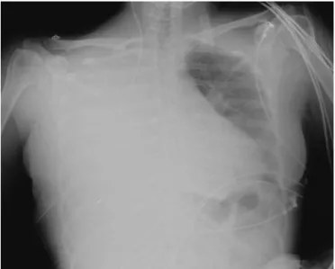
The chest radiograph showed opacity on the right hemithorax and the catheter tip was noted to be in the oppositedirectionofthecardiacsilhouette(Fig.1).
A chest tube was inserted with immediate drainage of 2500mL of serous fluid accompanied by symptomatic relieve.Thepatientwastransferredtotheward.Thechest tubewasremovedafter5daysandshewasdischarged,fully recovered.
Case
report
2
A67-year-oldwoman,ASAIII,presentedtotheemergency departmentbecauseofa2-weekhistoryofabdominalpain and diarrhea. A foreign body (chicken bone) was found jammed at the sigmoid. After unsuccessful attempt to removeitbycolonoscopy,shewasproposedforexploratory laparotomy.Herpastmedicalhistoryincludedmorbid obe-sity(weight110kg, height150cm),ischemicheartdisease
Figure1 PortablechestX-rayshowingrighthydrothorax9h afterinsertionofthecentralvenouscatheter.
with angina on moderate exertion, diabetes mellitus, chronicobstructivepulmonarydiseaseandhypothyroidism. Acentralvenouscatheterwasindicatedbecauseof dif-ficulty in obtaining a peripheral access and the possible need for vasopressor therapy.After induction of anesthe-sia,a7.0-Frenchtriplelumencentralvenouscatheterwas insertedintherightinternaljugularvein(Certofix® Trio
---B.Braun),usinganatomicallandmarksandSeldinger tech-nique. The insertion was achieved in one attempt by an experiencedanesthesiologistandaccordingtothehospital protocol.Theprocedurewasuneventfulexceptforaslight initialresistancetoretrievetheguidewire.Themiddleport catheterdidnotpresentpassiveblood returnanditcould onlybeachievedwithaspirationatmoderatenegative pres-sure.Amildincreasedresistanceduringinjectionwasalso observedinthisport.Theremainingportswerepermeable andwereeasilyreturningblood.Thecatheterwasfixedat 11cmlength.Aninfusionwithcrystalloidwasstarted.
The surgery proceeded and the patient underwent an opensigmoidectomy.
She was transferred to the intensive care unit in the immediate postoperative period to continue mechanical ventilatorysupport.Vasopressortherapywasnotneeded.
InthisunitachestX-raywasperformedandaloop for-mationofthecentralvenouscatheterwasnoted(Fig.2).
This catheter wasremoved without resistance. A new centralvenouscatheterwasplacedontherightsubclavian vein,usinganatomicallandmarksandSeldingertechnique, uneventfully.After36hthiscatheterwasremovedandthe patientwastransferredtotheward.Threedayslatershe needed a newcentral line because of difficult peripheral access.Therightinternaljugularveinwasused,onceagain usinganatomicallandmarksandSeldingertechnique, with-outcomplications. After10daysshewasdischarged,fully recovered.
Discussion
80 S.Pereiraetal.
Figure2 PortableX-rayshowingloopingofthecentralvenous catheter.
explainedbythecloseproximityofthesuperiorvenacava totheright pleura.3Thiscomplicationisusuallydescribed
incasesofprogressivevascularerosionofaninitial intravas-cular catheter,4---6 and may be associated with the poor
positioningof the tipor an unsafe catheterfixation with back-and-forthmovement.7Inourcasetheabsenceofblood
returnonaspirationofthedistalportsuggests,however,not erosionbutavascular lesionatinsertion,representing,as farasweknow,theonlycaseofacomplicationofthiskind. Vascular erosion with consequent hydrothorax after catheterization of the right internal jugular vein is extremelyrare,andhasbeen described in5% ofallcases ofcatheter-associatedhydrothorax.2Thismayberelatedto
thefactthatthe right internaljugularveinhas astraight coursetothesuperiorvenacava.8Afterreviewingthe
liter-aturewenotedthattheleft-sidedsubclaviancentralvenous placementisariskfactorforsuchcomplication.Dutleyetal. describedtherouteofcatheterizationcausinghydrothorax: leftsubclavianin46%,rightsubclavianin18%,leftinternal jugularin20%,rightinternaljugularin5%,externaljugular in6%andbrachialin5%.2
Clinicalmanifestationsofchestdiscomfortaresimilarto thosedescribedintheliteratureincasesofhydrothorax.A tensionhydrothoraxmayalsodevelop.7
In ourcase the restrictionin fluidtherapy intended at keepingalowcentralvenouspressureduringapartial hep-atectomyprobablycontributedtothedelayindiagnosis.
Theformation ofaloopincentralvenouscatheterisa rarecomplication,described in 2.9% of catheterizations.9
Loopingandknottinghasbeendescribed,mostlyduring pul-monaryarterycatheterizationorright-sidedsubclavianvein catheterization.10---12
Looping may occur due to the lockingof the catheter tipinthe ostiumof a tributaryvein, allowingconsequent progression of the catheter to form a loop.9 In our case
theloopingmayhaveresultedfromjammingthetipofthe catheteratthe ostiumof theright subclavianveinor the left brachiocephalic vein. The difficulty in retrieving the guidewire and the difficulty in aspirating blood from the
middleport suggestthatthecatheterwaspartlybent but keepingitspatency.
Severalstudieshavedemonstratedthatultrasound guid-anceduringcentralvenouscannulationcanincreasesuccess ratesanddecreasecomplications.13---15Thekeybenefitsfrom
theuseofultrasoundincludeincreasedoverallsuccessrate andreductioninneedlepuncturetime,reductionincarotid punctureandincarotidhematoma,reductioninhemothorax andpneumothorax.13
Hydrothoraxhasbeendescribedafterultrasoundguided internal jugular cannulation7 and we found no cases of
catheter or guide-wire looping diagnosed by ultrasound. Ultrasoundguidedcentralcannulationseemsunableto pre-venttheserarecomplications.
Conclusions
Mechanical complications associated with central venous catheterizationmayoccuratthetimeofinsertionor may developlater.Earlyradiologicalinvestigation,primarilywith achestX-ray,canaidtoidentifytheproblemandplanthe extractionofthecatheter.Inspecificsituationsother exam-inationsmightbeuseful,suchasangiographyorcomputed tomography.12
Inbothclinicalcasesthatwedescribedtheonlyfeature wasthedifficultyinbloodreturnataspirationinoneofthe ports,suggestingthatthisshouldnotbeundervalued.Evenif theprocedurewentuneventfullyintheeventofabsenceof freeflowonaspirationfromalllumens,thecathetershould notbeusedandneedstoberemoved.
Conflicts
of
interest
Theauthorsdeclarenoconflictsofinterest.
References
1.McGee DC, Gould MK. Preventing complications of central venouscatheterization.NEnglJMed.2003;348:1123---33. 2.DuntleyP,SieverJ,KorwesML,etal.Vascularerosionby
cen-tralvenouscatheters. Clinicalfeatures andoutcome. Chest. 1992;101:1633e1638.
3.Gibson F, BodenhamA. Misplaced central venous catheters: applied anatomy and practical management. Br J Anaesth. 2013;110:333---46.
4.RudgeCJ,BewickM,MccollI.Hydrothoraxaftercentralvenous catheterization.BrMedJ.1973;3:23---5.
5.Iberti TJ, Katz LB, Reiner MA, et al. Hydrothorax as a late complicationofcentralvenousindwellingcatheters.Surgery. 1983;94:842---6.
6.KunizawaA,FujiokaM,MinkS,etal.Centralvenous catheter-induceddelayedhydrothoraxviaprogressiveerosionofcentral venouswall.MinervaAnestesiol.2010;76:868---71.
7.MarounR,ChalhoubM,HarrisK.Rightinternaljugularvenous cannulationcomplicatedbytensionhydrothorax.HeartLung. 2013;42:372---4.
8.Bannon MP, Heller SF, Rivera M. Anatomic considerations for central venous cannulation. Risk Manag Healthc Policy. 2011;4:27---39.
9.Malatinsk´yJ,KadlicT,MájekM,etal.Misplacementandloop formationofcentralvenouscatheters.ActaAnaesthesiolScand. 1976;20:237---47.
Reportsofmisplacedcentralvenouscatheters 81
11.Bagul NB, Menon NJ, Pathak R, et al. Knot in the cavae: anunusual complication ofswan-ganz catheters. EurJVasc EndovascSurg.2005;29:651e653.
12.VetrugnoL,PiccoliG,CostaMG,etal.Thedosanddoknotsof centralvenouscatheterization.JClinAnesth.2012;24:148---50. 13.Karakitsos D, Labropoulos N, De Groot E, et al. Real-time ultrasound-guidedcatheterisationoftheinternaljugularvein:a prospectivecomparisonwiththelandmarktechniqueincritical carepatients.CritCare.2006;10:R162.
14.Leung J, Duffy M, Finckh A. Real-time ultrasonographically-guided internal jugular vein catheterization in the emer-gency department increases success rates and reduces complications: a randomized, prospective study. Ann Emerg Med.2006;48:540---7.