808
Brazilian Journal of otorhinolaryngology 77 (6) novemBer/DecemBer 2011 http://www.bjorl.org / e-mail: [email protected]
Ramsay Hunt syndrome following otoplasty
Marco Antônio Rios Lima
1, Jacinto de Negreiros Júnior
21 3rd-Year ENT Resident (R3).
2 MSc. Assistant Physician and Coordinator of the Medical Residency Program in Otorhinolaryngology of the Armed Forces Hospital in Brasília.
Armed Forces Hospital in Brasília.
Send correspondence to: Marco Antônio Rios Lima - S.M.T, conjunto 16, casa 5, Taguainga - DF. 72023-480.
Paper submited to the BJORL-SGP (Publishing Management System – Brazilian Journal of Otorhinolaryngology) on October 12, 2010; and accepted on November 07, 2010. cod. 7370
CASE REPORT
Braz J Otorhinolaryngol. 2011;77(6):808.
BJORL
Keywords: ear, external, herpes zoster, herpes zoster oticus.
.org
INTRODUCTION
The Ramsay-Hunt syndrome (RHS) is characterized by the association of herpes-zoster oticus with acute peripheral facial paralysis1. Its
physiopathology is associated with the reacti-vation of the varicella-zoster virus (VZV) in the geniculate ganglion of the facial nerve1,2.
Different surgical procedures have been described in the literature as triggers for virus reactivation1,2.
Below, we describe what we believe to be the first case reported of RHS happening after otoplasty, focusing on its diagnosis, treatment and evolution.
CASE PRESENTATION
A twelve year old boy, previously heal-thy, was submitted to bilateral otoplasty in order to correct protruding ears. There were no initial complications and the suture was removed 14 days after the procedure (PO).
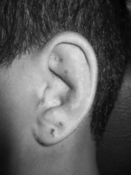
On the 25th day of post-op, he started having a little difficulty in shutting his left eye. The patient reported having noticed vesicles in his left ear pinna, which had been there for three days, together with ear pain and ipsila-teral hearing loss. Two days later, he reported difficulties in moving his face on the left side. He had had varicella infection at 4 years of age, without complications. Upon physical exam, he had House Brackmann level III left-side facial palsy and crusty lesions on his left ear pinna (Figure 1). Upon otoscopy, we noticed diffuse edema and hyperemia in his left external ear canal, making it impossible to see the tympanic membrane. His right ear pinna and ipsilateral otoscopy were normal. His mouth and anterior rhinoscopy did not show changes. He had a ne-gative Rinne test on the left side with the Weber test lateralized to the same side.
We started treating his RHS with De-xamethasone 4mg/day per os, during 7 days, with progressive weaning (28 days of steroid treatment according to our protocol), Acyclovir 2g/day (7 days) and eye lubrication drops on the left side. The external otitis was treated with ear drops (Ciprofloxacin 0.2% + hydrocortisone 1% - 10 days). We ordered a CBC and serology tests. One week later, he had complete symptom resolution in his left ear pinna as well as his ipsi-lateral external otitis. Tonal audiometry showed normal hearing thresholds in both ears. CBC sho-wed leukocytosis (16,100) without deviations.
Serology tests were positive for IgG and negative for IgM for Herpes I and II, mononucleosis and cytomegalovirus; negative serology for HIV. He progressively recovered facial movement and had complete palsy resolution upon the fourth week after the diagnosis. Herpes-zoster serology (enzyme immune assay) was carried out only one month after symptom onset, which revea-led negative IgM and positive IgG in high titers (2.556 – reference value is up to 0.2).
DISCUSSION
The RHS is rare in childhood, with an incidence of approximately 5 cases/100,000 inha-bitants/year. Nonetheless, it is the second most common cause of non-traumatic facial paralysis.3
Today it is believed that virus reactiva-tion is an important cause of facial paralysis in late post-op1. After the prime-infection, the VZV
remains latent in the sensorial ganglion of the facial (VII) and trigeminal (V) nevers2. Surgical
procedures in the vicinities of the facial nerve, such as in the middle and inner ears, trigger viral reactivation and facial paralysis because of the direct stimulation of the VII cranial nerve1. There
are also cases of late peripheral facial paralysis induced by the VZV after orofacial surgeries and dental procedures, and in these cases the
hypothesis is for an ascending viral infection through the lingual nerve2.
We may hypothesize that the otoplasty procedure triggered the viral reactivation through direct stimulation of the VII cranial nerve (ear pinna)4, or through the stimulation of the V nerve
(auricle-temporal branch)4, which would transmit
the stimulus through the chorda tympani nerve all the way to the facial nerve.
Diagnosis is fundamentally clinical5.
Complementary tests may corroborate or con-firm the diagnosis. In the present case, serologic diagnosis could not be established because of the difficulty in performing serology at the time of diagnosis. Nonetheless, high titers of anti-VZV IgG antibodies found one month after symptom onset are highly suggestive of virus reactivation. Serologic response is not always detected in RHS patients.2 There are reports of variable patterns
of immune response from the virus host6.
FINAL REMARKS
We described here a case of a patient who developed RHS in the late postoperative of bilateral otoplasty, having complete symptom remission after clinical treatment. The close follow up of patients submitted to otorhinola-ryngological and neck/facial surgeries is highly important in order to make an early diagnosis and to provide proper treatment aiming at opti-mizing prognosis.
REFERENCES
1. Gyo K, Honda N. Delayed facial palsy after middle-ear surgery due to reactivation of varicella-zoster virus. J Laryngol Otol. 1999;113(10):914-5. 2. Furuta Y, Ohtani F, Fukuda S, Inuyama Y,
Na-gashima K. Reactivation of varicella-zoster virus in delayed facial palsy after dental treatment and oro-facial surgery. J Med Virol. 2000;62(1):42-5. 3. Kim D, Bhimani M. Ramsay Hunt syndrome
presenting as simple otitis externa. CJEM. 2008;10(3):247-50.
4. Zorzetto NL. Anatomia da orelha. In: Costa SSD, et al. Otorrinolaringologia - Princípios e Prática. 2ª ed. Porto Alegre: Artmed; 2006. p.26. 5. Sandoval CC, Núñez FA, Lizama CM, Margarit SC,
Abarca VK, Escobar HR. Síndrome de Ramsay-Hunt en Pediatría: Reporte de cuatro casos y revisión de la literatura. Rev Chil Infectol. 2008;25(6):458-64. 6. Aizawa H, Ohtani F, Furuta Y, Sawa H, Fukuda
S. Variable patterns of varicella-zoster virus reac-tivation in Ramsay Hunt syndrome. J Med Virol. 2004;74(2):355-60.