* e-mail: [email protected]
Ca
2Ge
7O
16Nanowires Grown from CaO and GeO
2Li-Zhai Pei*, Yin-Qiang Pei, Yong Yang, Qian-Feng Zhang
Key Lab of Materials Science and Processing of Anhui Province, Institute of Molecular Engineering and Applied Chemistry,
School of Materials Science and Engineering, Anhui University of Technology, Ma’anshan, Anhui 243002, P. R. China
Received: June 27, 2011; Revised: September 7, 2011
Single crystalline Ca2Ge7O16 nanowires have been synthesized using CaO and GeO2 as the
raw materials. Various factors that affect the formation and size of the Ca2Ge7O16 nanowires have
been analyzed. The obtained products are characterized by X-ray diffraction, transmission electron microscopy and scanning electron microscopy. The results show that the calcium germanate nanowires
are composed of orthorhombic Ca2Ge7O16 phase with the length and diameter of several dozens of
micrometers and about 50 nm, respectively. Hydrothermal temperature plays an important role on the
formation and growth of the Ca2Ge7O16 nanowires. The formation process of the Ca2Ge7O16 nanowires
is initially interpreted according to the shape evolution of the products obtained from different growth conditions.
Keywords: Ca2Ge7O16 nanowires, CaO, GeO2, electron microscopy
1. Introduction
Ternary germanate nanowires have been attracted great attention as important one-dimensional (1D) nanomaterials for the application of electrochemical sensors, optical
devices, electron devices and catalysts1-4. Several kinds of
germanate nanowires, such as Zn2GeO4[5,6], PbGeO
3[7] and
strontium germanate8 have been reported previously. Among
these germanates, calcium germanate is a kind of excellent optical material exhibiting strong fluorescence emission at
620, 700 and 800 nm9. Perng et al.10 reported the synthesis
and photoluminescence of amorphous Ca5Ge2O9 nanowires
by dehydrating the hydrated Ca5Ge2O9 nanowires originated
by submersing Ge nanoparticles into calcium hydroxide aqueous solution. However, Ge nanoparticles with the size ranging from 10-50 nm need be firstly prepared by a vapor condensation technique taking the complexity and expensive apparatus for the synthesis of the calcium germanate nanowires. Very recently, different from the amorphous calcium germanate nanowires, crystalline calcium germanate nanowires have been synthesized by a simple hydrothermal
process using GeO2 and Ca(CH3COO)2·H2O as the raw
materials11. However, the obtained crystalline calcium
germanate nanowires consist of a mixed germanate phases
with tetragonal Ca2GeO4, orthorhombic Ca2Ge7O16 and
triclinic CaGe2O5 phases. It is difficult to gain single phase
using GeO2 and Ca(CH3COO)2·H2O as the raw materials.
In addition, the cost of Ca(CH3COO)2·H2O is high which
may confine the possible application of calcium germanate nanowires. Therefore, it is of important significance for the low-cost synthesizing calcium germanate nanowires with single phase.
CaO, as a kind of cheap Ca raw material, is slightly
soluble to form Ca(OH)2 in water becoming a proper Ca
source material for the synthesis of 1D Ca-based nanoscale
materials12,13. In the paper, single crystalline Ca
2Ge7O16
nanowires have been synthesized using GeO2 and CaO as the
raw materials. Using CaO instead of Ca(CH3COO)2 as the
Ca raw material also saves the cost of the calcium germanate nanowires. The effects of hydrothermal temperature and
time on the formation and size of the Ca2Ge7O16 nanowires
are demonstrated. And the growth process of the Ca2Ge7O16
nanowires is discussed.
2. Experimental
High pure GeO2 (purity: ≥99.99 wt. (%)) and CaO
(purity: ≥99.9 wt. (%)) were purchased from Sinopharm
Chemical Reagent Co., Ltd. of China. All source materials were used without further purification. In a typical
procedure, 0.16 g GeO2 and 0.22 g CaO were dissolved in
60 mL deionized water under vigorous stirring. Then, the mixture was placed in a 100 mL autoclave with a Teflon liner. The autoclave was maintained at 180 °C for 24 hours. Subsequently the autoclave was cooled naturally in air. The resulting white precipitates were filtered, washed with deionized water for several times and dried at 60 °C in air. Finally, the white powders were obtained.
The obtained products were characterized by X-ray diffraction (XRD), scanning electron microscopy (SEM), transmission electron microscopy (TEM) and high-resolution TEM (HRTEM). XRD pattern was carried out on a Bruker AXS D8 X-ray diffractometer equipped with a
The samples were scanned at a scanning rate of 0.05°/s in
the 2θ range of 20° ~ 80°. SEM observation was performed
using JEOL JSM-6490LV SEM with a 15-kV accelerating voltage. TEM and HRTEM samples were prepared by putting several drops of solution with calcium germanate nanowires onto a standard copper grid with a porous carbon film after the nanowire samples were dispersed into distilled water and treated for about 10 minutes using supersonic wave apparatus. TEM and HRTEM observations were performed using JEOL JEM-2100 TEM operating with 1.9Å point-to-point resolution operating at 200-kV accelerating voltage with a GATAN digital photography system.
3. Results and Discussion
Figure 1 shows the XRD pattern of the products obtained from the hydrothermal conditions of 180 °C for 24 hours. All of the diffraction peaks for the products
can be assigned to the orthorhombic phase of Ca2Ge7O16
(JCPDS card No. 34-0286). No characteristic peaks from
impurities are observed demonstrating that the high pure
Ca2Ge7O16 nanowires can be synthesized using CaO as the
Ca raw material.
The morphology and size of the Ca2Ge7O16 nanowires
are investigated by SEM, TEM and HRTEM. Figure 2 is
the SEM images of the Ca2Ge7O16 nanowires with different
magnifications synthesized at 180 °C for 12 hours. A large
amount of uniform Ca2Ge7O16 nanowires are achieved
according to Figure 2. No other morphologies are observed showing the highly pure nanowire-shaped structure of the products. The length of the nanowires is several dozens of micrometers, even longer than 100 micrometers. The nanowires with smooth surface are straight. The magnified
SEM image (Figure 2b) further shows that the Ca2Ge7O16
nanowires appear as uniform nanowire-shaped structure.
The Ca2Ge7O16 nanowires have a uniform diameter
distribution with average diameter of about 50 nm. The
morphology and size of the Ca2Ge7O16 nanowires are similar
to those synthesized using GeO2 and Ca(CH3COO)2·H2O as
the raw materials11.
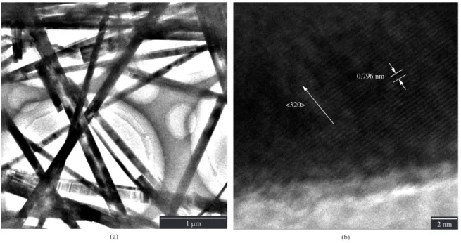
The detailed structure of the Ca2Ge7O16 nanowires is
further analyzed by TEM and HRTEM which are shown in Figure 3. Figure 3a shows the typical TEM image of the as-synthesized calcium germanate nanowires with the diameter of about 50 nm which is similar to those of the SEM observations. The nanowires are straight and have smooth surface. The diameter of the nanowires is uniform
throughout the length. It is noticed that the Ca2Ge7O16
nanowires have flat tips. The flat tips are similar to those of
the germanate nanowires prepared by other methods4,7,8,11.
Figure 3b displays the HRTEM image of single calcium germanate nanowire. The lattice fringes distinguished exhibit good single crystalline in nature demonstrating that
the Ca2Ge7O16 nanowires are composed of single crystalline
structure. The interplanar spacing of the crystalline is about 0.796 nm according to the HRTEM measurement and the subsequent calculation by the software of Digital Micrograph (Gatan Inc., Pleasanton, CA) applied in the HRTEM, which is the same as the interplanar spacing for
the {110} plane of orthorhombic Ca2Ge7O16. Combined
the XRD pattern with the HRTEM image of the nanowires,
Figure 1. X-ray diffraction pattern of the calcium germanate nanowires.
Figure 3. Transmission electron microscopy images of the calcium germanate nanowires. a) TEM image, and b) HRTEM image.
the strong diffraction peak of (320) indicates that the main growth orientation of the calcium germanate nanowires is <320> crystallographic direction. In addition, some nanowires are also observed to originate from <322> and <330> growth direction, respectively.
The growth conditions, such as hydrothermal temperature and time on the formation and growth of the calcium germanate nanowires are analyzed so as to research the possible formation process of the calcium germanate nanowires. Figure 4 is the SEM images of the calcium germanate nanowires obtained at 180 °C for different time. Calcium germanate nanowires with similar morphology can still be obtained when the reaction time is 0.5, 6, 12 and 48 hours, respectively. The length of the calcium germanate nanowires obtained from different time is similar with several dozens of micrometers. However, the diameter of the calcium germanate nanowires decreases obviously with the decrease of the reaction time at the same hydrothermal temperature. The average diameter of the calcium germanate nanowires is about 30 nm when the reaction time is 0.5 hours (Figure 4a). But the diameter of the calcium germanate nanowires also increases to about 300 nm with the reaction time increasing to 48 hours at 180 °C. The time dependence results show that the reaction time plays an important role on
Figure 5. SEM images of the products obtained from different hydrothermal temperature for 24 hours. a) 80 °C, b,c) 120 °C, and d) 160 °C.
the size of the calcium germanate nanowires. The diameter and length of the calcium germanate nanowires can be adjusted by controlling the reaction time.
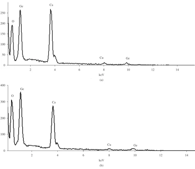
Figure 6. EDS spectra of the product obtained from 120 °C for 24 hours. a) EDS spectrum of the nanowires, and b) EDS spectrum of the nanoparticles in the nanowires.
The diameter of the calcium germanate nanowires decreases obviously with the decrease of the reaction time, which illustrates that the growth direction is along horizontal direction. However, the length of the nanowires increases with increasing the hydrothermal temperature, which means that the growth direction for these nanowires is along vertical direction. The growth velocity of the nanowires along the horizontal direction is far slower than that of the nanowires along the vertical direction.
The quantitative analysis of the chemical composition of the product obtained from the hydrothermal conditions of 120 °C for 24 hours (Figure 5b) using SEM-EDS experiment was performed by the software applied in the Link ISIS300 EDS. The EDS spectra of the nanowires and nanoparticles attached in the nanowires are shown in Figure 6. Elements Ca, Ge and O originate from the calcium germanate product and Cu arises from the copper sheet substrate. The atomic ratio of Ca:Ge:O of the nanowires is about 2:6.9:16 showing the composition of the calcium germanate nanowries is very
similar to Ca2Ge7O16. However, the atomic ratio of Ca:Ge:O
of the nanoparticles attached in the nanowires is about 2:6.8:22.6. Obviously the O content of the nanoparticles
attached in the nanowires is more than that of the nanowires.
It is considered that Ca2Ge6.8O22.6 in the nanoparticles further
reacts with H3GeO3 to form Ca2Ge7O16.
Figure 7 shows the XRD patterns of the products synthesized from 80 and 120 °C, respectively for 24 hours. Obviously, the intensity of the diffraction peaks increase with the increase of the reaction temperature. The XRD patterns of the products are still similar which is mainly
composed of orthorhombic Ca2Ge7O16. However, besides
the orthorhombic Ca2Ge7O16, some diffraction peaks of
the monoclinic CaGe2O5 (JCPDS card No. 21-0797) occur
in the XRD patterns of the product at the initial reaction stage (Figure 7a). With the increase of the hydrothermal temperature, the most of the diffraction peaks of the product disappears obviously. Only the diffraction peaks of
the orthorhombic Ca2Ge7O16 exist in the product with the
hydrothermal temperature further increasing to 180 °C. The results demonstrate that the two phases with orthorhombic
Ca2Ge7O16 and monoclinic CaGe2O5 structures form at the
initial formation stage of the calcium germanate nanowires, and temperature plays an essential role on the formation of
20 30 40 50 60 70 80
2θ (deg.)
Intensity (a.u.) (a) (b) (111) (021) (320) (221) (311) (330) (420) (002) (520) (431) (511) (322) (412) (332) (621) (710)
(522) (711) (721)
Monoclinic CaGe2O5
Figure 7. XRD patterns of the products obtained from different
hydrothermal temperature for 24 hours. a) 80 °C, and b) 120 °C.
Figure 8. Schematic illustration of the formation and shape evolution of the calcium germanate nanowires in the whole synthetic process.
It is clear Ca2Ge7O16 nanowires are formed by the
hydrothermal treatment of CaO and GeO2, but the reasons
for formation Ca2Ge7O16 nanowires with single phase is
still puzzling. It is noticed that the CaO and the phases formed at the initial reaction stage play essential role on
the formation and growth of Ca2Ge7O16 nanowires. Only
orthorhombic Ca2Ge7O16 and a small amount of monoclinic
CaGe2O5 structures form at the initial reaction stage of CaO
and GeO2. The monoclinic CaGe2O5 phase disappears with
the increase of the hydrothermal temperature. Therefore, based on the experimental results, the possible formation process of the calcium germanate nanowires are proposed, which is depicted in Figure 8. At the initial reaction
stage, H2GeO3 forms from the reaction of GeO2 and H2O.
Ca(OH)2 originates from CaO and H2O. Therefore, many
nanoscale spherical particles spontaneously appear in the supersaturated solution through the hydrothermal reaction
of H2GeO3 and Ca(OH)2 forming Ca2Ge7O16 and CaGe2O5.
Then the nanoparticles serve as the crystalline nuclei for the anisotropic growth of the calcium germanate nanocrystals. The linear growth is attributed to the preferential adsorption of nanoparticles to special crystal facets, which directs the growth of the nanoparticles into nanorods by controlling
the growth rates along different crystal axes14,15. With the
reaction going on, the smaller nanoparticles vanish at the site of the longer nanorods through an “Ostwald ripening” process due to their higher surface free energy compared
with that of the longer nanorods16,17 which is confirmed by
the SEM images of Figure 5b and 5c. With the increase of hydrothermal temperature and reaction time, the nanorods
grow continuously and CaGe2O5 phase disappears resulting
in the final formation of the Ca2Ge7O16 nanowires.
4. Conclusions
In summary, Ca2Ge7O16 nanowires have been synthesized
using CaO as the Ca source material by a simple hydrothermal process. The nanowires are composed of orthorhombic
Ca2Ge7O16 phase with average diameter of about 50 nm and
length of several dozens of micrometers, even longer than 100 µm. Hydrothermal temperature plays an essential role on the formation and growth of the calcium germanate nanowires.
Using cheap CaO instead of Ca(CH3COO)2 saves the cost of
the calcium germanate nanowires. The experimental results suggest that the calcium germanate nanowires are formed via an “Ostwald ripening” growth process.
Acknowledgements
This work was supported by the Natural Science Foundation of the Education Bureau of Anhui Province of China (KJ2011A042), Innovative Research Fundation of Postgraduate of Anhui University of Technology (2011005) and National Basic Research Program of China (863 Program, 2009AA03Z529).
References
1. Huang JH, Wang XC, Hou YD, Chen XF, Wu L and Fu XZ. Degradation of benzene over a zinc germinate photocatalst under ambient conditions. Environmental Science and Technology. 2008; 42:7387-7391. http://dx.doi.org/10.1021/ es800744e
2. Liu G, Zheng S and Yang G. In2Ge6O15(OH)2(H2dien): An open-framework indate germanate with one-dimensional 12-ring channels [J]. Angewandte Chemie International Edition. 2007;
46:2827-2830. PMid:17352442. http://dx.doi.org/10.1002/ anie.200604921
3. Bayya SS, Chin GD, Sanghera JS and Aggarwal ID. Germanate glass as a window for high energy laser systems. Optical Express. 2006; 14:11687-11693. PMid:19529589, http://dx.doi. org/10.1364/OE.14.011687
4. Dong YP, Pei LZ, Chu XF, Zhang WB and Zhang QF. Electrochemical behavior of cysteine at a CuGeO3 nanowires modified glassy carbon electrode. Electrochimica Acta. 2010; 55:5135-5141. http://dx.doi.org/10.1016/j.electacta.2010.04.020 5. Yan CY and Lee PS. Synthesis and structure characterization
Journal of Physical and Chemistry C. 2009; 113:14135-14139.
http://dx.doi.org/10.1021/jp9050879
6. Yan CY, Singh ND and Lee PS. Wide-bandgap Zn2GeO4 nanowire networks as efficient ultraviolet photodetectors with fast response and recovery time. Applied Physics Letter. 2010;
96:053108. http://dx.doi.org/10.1063/1.3297905
7. Wang N, Ding J, Li GC and Peng HR. Synthesis and properties of PbGeO3 nanostructures. Crystal Research and Technology.
2010; 45:316-320. http://dx.doi.org/10.1002/crat.200900516 8. Tsai MY and Perng TP. Synthesis and photoluminescence of
amorphous strontium germanate nanowires. In: Proceedings oh the 214th Electrochemical Society Meeting; 2008, Honolulu. Honolulu; 2008. p. 12-17.
9. Sharonov MY, Bykov AB, Myint T, Petricevic V and Alfano RR. Spectroscopic study of chromium-doped transparent calcium germinate glass-ceramics. Optical Communications. 2007;
275:123-128. http://dx.doi.org/10.1016/j.optcom.2007.02.058 10. Tsai MY, Yu CY and Perng TP. Synthesis and photoluminescence of amorphous Ca5Ge2O9 nanowires. Journal of Nanoscience and Nanotechnology. 2008; 8:6376-6380. PMid:19205209. http://dx.doi.org/10.1166/jnn.2008.383
11. Pei LZ, Yang Y, Fan CG, Yuan CZ, Duan TK and Zhang QF. Synthesis and characterizations of calcium germanate nanowires. CrystEngComm. 2011; 13. http://dx.doi.
org/10.1039/c1ce05070b
12. Pei LZ, Yang LJ, Yang Y, Fan CG, Yin WY, Chen J et al. A green and facile route to calcium silicate nanowires.
Materials Characterization. 2010; 61:1281-1285. http://dx.doi.
org/10.1016/j.matchar.2010.07.002
13. Xu T T, Zheng JG, Nicholls AW, Stankovich S, Piner RD and Ruoff RS. Single-crystal calcium hexaboride nanowires: Synthesis and characterization. Nano Letters. 2004,
4:2051-2055. http://dx.doi.org/10.1021/nl0486620
14. Zhou GT, Wang XC and Yu JC. Selected-control synthesis of NaV6O15 and Na2V6O16·3H2O single-crystalline nanowires.
Crystal Growth and Design. 2005; 5:969-974. http://dx.doi. org/10.1021/cg0496686
15. Lin LW. Synthesis and optical property of large-scale centimeters-long silicon carbide nanowires by catalyst-free CVD route under superatmospheric pressure. CrystEngComm. 2011, 13:1582-1591.
16. Zhu WC, Zhu SL and Xiang L. Successive effect of rolling up, oriented attachment and Ostwald ripening on the hydrothermal formation of szaibelyite MgBO2(OH) nanowhiskers. CrystEngComm. 2009; 11:1910-1919. http://dx.doi.org/10.1039/b905698j
17. Ma H, Yang XJ, Tao ZL, Liang J and Chen J. Controlled synthesis and characterization of porous FeVO4 nanorods and nanoparticles. CrystEngComm. 2011, 13:897-901.