*e-mail: [email protected]
1. Introduction
Photocatalytic degradation of toxic organic compounds has received a great attention for the past several years. Due to its strong oxidizing powder, cost-effective, and long-term stability against photo and chemical corrosion, TiO2 has been used widely in water puriication technology 1-4 However, the practical applications of TiO
2 are limited by its large band gap (3.2 eV), which can only active under the UV light irradiation5-7. Therefore, several strategies have been developed to shift the optical sensitivity of TiO2 from
UV to the visible-light region for the eficient use of solar
energy, such as element doping, metal deposition, surface sensitization, and coupling of composite semiconductors 8-11. Recently, co-doped titania with double non-metal 12,13,
metal-nonmetal elements 14-17 and double metal ions 18 have been attracted more attention, which could further improve the photocatalytic activity of TiO2. Among these, co-doping with transition metal and nonmetal elements such as Fe-N, V-N and W-C could effectively modify the electronic structures of TiO2 and shift its absorption edge to a low energy 16.
In addition, the structural properties of TiO2, such as crystalline phase, crystallite size, surface area and pore distribution, are important for its photocatalytic properties. In our previous work, we have reported that the use of hydrocarbon surfactants as the template and nonmetal doping precursor in the self-assembly sol–gel methods to tailor-design the structural properties of TiO2 from molecular
precursors 19-22, obtain the TiO
2 with high surface area, high porosity, small crystal size and narrow pore size distribution.
Speciically, these hydrocarbon surfactants can serve as a
nonmetal doping precursor to increase the photocatalytic activity of TiO2 under visible light irradiation 19,20.
In this study, novel manganese and carbon codoped TiO2 photocatalysts with nonionic surfactant Tween 80 as a pore template and carbon doping reagent were synthesized by the sol-gel method and characterized. The photocatalytic activity of the samples was evaluated by the photocatlytic degradation of Methyl orange (MO) under simulate daylight irradiation and the effect of the calcination temperature on photocatalytic activity and phase transformation was studied.
2. Materials and methods
2.1. Synthesis of Mn-C-TiO
2Mn-C-TiO2 were prepared by the self-assembly surfactant-based sol–gel method under mild conditions as follow. A nonionic surfactant Tween 80 (T80, polyoxyethylenesorbitan monooleate, Guoyao Chemical Co.) was employed as the
pore directing agent and carbon precursor in the modiied
sol-gel solution. 5 ml T80 was dissolved in 20 ml isopropyl alcohol (i-PrOH, 99.8%, Guoyao Chemical Co.) and then 3 ml titanium tetraisopropoxide (TTIP, 97%, Sigma-Aldrich) was added under vigorous stirring. Finally, 3 ml acetic acid (AcOH, Guoyao Chemical Co.) was added into the solution
Effect of calcination temperature on structural properties and
photocatalytic activity of Mn-C-codoped TiO
2Jianbo Caia, Wei Xina, Guanglong Liua*, Die Lina, Duanwei Zhua
aLaboratory of Eco-Environmental Engineering Research, College of Resources & Environment, Huazhong Agricultural University – HZAU, Wuhan 430070, China.
Received: July 1, 2015; Revised: November 19, 2015; Accepted: December 27, 2015
Mn-C-codoped TiO2 catalysts were synthesized by modiied sol-gel method based on the self-assembly technique using polyoxyethylenesorbitan monooleate (Tween 80) as template and carbon precursor and the effect of calcination temperature on their structural properties and photocatalytic activity were investigated. The XRD results showed undoped and Mn-C-codoped TiO2 calcined at 400 oC only include anatase phase and the rutile phase appears when the calcination temperature reached to 600 oC. UV-vis absorption spectroscopy demonstrates that the absorption spectra are strongly modiied by the calcination temperature. Moreover, the Mn-C-TiO2 calcined at 400 oC showed the lowest PL intensity due to a decrease in the recombination rate of photogenerated electrons and holes under light irradiation. The photocatlytic activity of Mn-C-codoped TiO2 were evaluated by the degradation of methyl orange (MO) under the simulate daylight irradiation and all the prepared Mn-C-codoped TiO2 samples exhibited high photocatalytic activities for photocatalytic decolorization of methyl orange aqueous solution. At 400 ◦C, the Mn-C-codoped TiO2 samples showed the highest photocatalytic activity due to synergetic effects of good crystallize ation, appropriate phase composition and slower
recombination rate of photogenerated charge carriers, which further conirms the calcination temperature
could affect the properties of Mn-C-codoped TiO2 signiicantly.
for the formation of water in the mixture. The sol-gel was aged at 65 oC for 24 hrs. To synthesize particles, the sol was dried at room temperature for 3 hrs and then calcined at 400, 500 and 600 oC for 3 hrs. For comparison, the control sample was prepared though the same method, without adding the corresponding dopants and calcined at 400 oC for 3hrs. The catalysts produced are denoted as TiO2, Mn-C-TiO2-400, Mn-C-TiO2-500 and Mn-C-TiO2-600.
2.2. Characterization of synthesized Mn-C-TiO
2The X-ray diffraction (XRD) analysis was performed on X’Pert PRO (D8 Advance) XRD diffractometer using Cu
Kα (λ= 1.5406 Å) radiation to study the crystal structure and
crystallinity of the Mn-C-TiO2 . The Brunauer–Emmett–Teller (BET) surface area, pore volume, porosity, Barret–Joyner–Halenda (BJH) pore size and distribution (based on nitrogen adsorption and desorption isotherms) were determined by a Tristar 300 (Micromeritics) porosimeter analyzer. All the tested powders were purged with nitrogen gas for 2 h at 150 oC using Flow prep 060 (Micromeritics). Fourier transform infrared (FT-IR) spectroscopy was carried out using Thermo
Scientiic Nicolet 6700 spectrometer to detect the presence
of carbon group on the samples. Measurement range was 4000-500 cm-1, with a 4 cm-1 resolution, 0.475 cm-1 /s scan speed and 32 scans. To investigate the light absorption and optical band gap of the synthesized TiO2 , the UV – vis absorption spectra were obtained with a UV – vis spectrophotometer (Shimadzu 2450 PC) mounted with an integrating sphere accessory (ISR1200) using BaSO4 as reference standard. An X-ray photoelectron spectroscope (XPS, PerkinElmer Model 5300) was employed to determining the binding energy of Mn and C. The conditions of the equipment include a takeoff angle of 45° and vacuum pressure of 10-8 to 10-9 Torr. The binding energies were referenced to the C1s peak at 284.6 eV. Photoluminescence spectra (PL) were
measured by Shimadzu RF-5301 PC spectroluorophotometer (EX = 240 nm, EM = 350-400 nm) at roonm temperature.
2.3. Degradation eficiency
The effect of calcination temperature on the degradation
eficiency of Mn-C-TiO2 was evaluated by the degradation of the degradation of methyl orange in water under the simulate daylight irradiation. Firstly, a particles suspension (0.5 g/L) solution was prepared and dispersed using an ultrasonicator (2510R-DH, Bransonic) for 24 h. Secondly, 50 µL methyl orange solution (50 g/L) was transferred to a 50 ml particles suspension placed in reactor to achieve
an initial concentration of 50 mg/L. Finally, 50μL HNO3 (0.05 mol L-1)was added into the solution. During irradiation
with two luorescent lamps (20W, Cole-Parmer) imitating
daylight, the reactor was mixed continuously to minimize mass transfer limitations. A 0.2 mL sample was withdrawn by 1 h. The photocatalyst was immediately removed from the samples after centrifugation. The progress of photocatalytic degradation was monitored through measuring the characteristic absorbance of the solution samples by a UV-760CRT
UV-Vis spectrophotometer (Shanghai Precision & Scientiic
Instrument Co., Ltd). The characteristic absorbance peaks of methyl orange shift from 460 to 517 nm under the acidic
condition. Therefore, λmax = 517 nm was choose to measure the content of methyl orange left in the sample.
3. Results and discussion
3.1. X-ray diffraction
XRD was used to investigate the crystal structure and crystal phase of the prepared Mn-C-TiO2 samples. Fig. 1 shows the effect of calcinations temperature on the phase structures of Mn-C-TiO2 and TiO2 samples. It can be seen
that the calcination temperature obviously inluences the
crystallization and phase composition of the TiO2. At 400 oC, only anatase TiO2 crystal phase could be identiied in Mn-C-TiO2 and TiO2 samples and no other dopant related crystal phases could be resolved. With the calcination temperature increasing, the peak intensities of anatase increase and the width of the (101) plane diffraction peak
of anatase (2 Theta=25.4o) become narrower. The rutile phase appeared when the calcination temperature reached to 600 oC. The average crystalline size calculated by applying the Scherrer formula on the anatase (101) diffraction peak was 12.28, 16.57 and 23.68 nm for the pure Mn-C-TiO2-400, Mn-C-TiO2-500 and Mn-C-TiO2-600 samples (shown in Table 1), respectively, which is increased as the calcination temperature increasing.
3.2. BET surface areas and pore structure
The N2 isothermal adsorption technique was employed to investigate the effect of calcination on the pore size variability. As shown in igure 2(a), The Barrett, Joyner and Halenda (BJH) pore size distribution of Mn-C-TiO2-400 was very narrow implying good homogeneity of the pores. With the calcination temperature increasing, the BJH pore size distribution of the Mn-C–TiO2 exhibited a systematic shift toward larger mesopores which can be associated with the severe collapse of the initial porous structure occurred for the calcinations temperature increasing. These results
are in agreement with recent work on the inluence of the
calcination temperature on the structural characteristic of TiO223,24.
Table 1 also shows the effects of calcination temperature on the physical properties of the TiO2 powders. It has been
Fig. 1. XRD spectra of synthesized TiO2 and Mn-C-TiO2 with
well know that the surface area plays an important role in the photocatlytic activity of TiO2, because the adsorption is the premise of photocatalytic reaction 25,26. In our case, Mn-C-TiO
2 powder calcined at 400 ◦C show a largest speciic surface area and their values reach 142.53m2/g. With increasing
calcination temperature, the speciic surface areas steadily
decreased due to the growth of TiO2 crystallite. At 600 oC,
the speciic surface area decreased to 15.68 m2/g. It is also easy to note that all powders show a monotonic increase in the average pore size with increasing calcination temperature. These results indicate that the calcination temperature
signiicantly could affect the physical properties of the TiO2 powders and this is similar to previous results for the effect of calcination temperature on the TiO2 powders 24 .
3.3. FT-IR analysis
To give additional evidence and further to conirm the
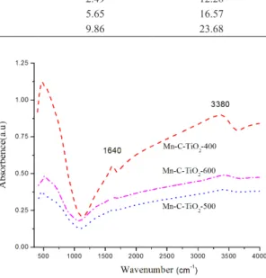
effect of calcinations temperature on the manganese and carbon doping, FT-IR characterizations were preformed. The infrared spectroscopy of Mn-C-TiO2 samples calcined at different temperatures was presented in Fig. 3. The bands at
1640 cm-1 and the wide bands at 3100-3700 cm-1 are resultant from O-H stretching of adsorbed water molecules and the surface hydroxyl groups on TiO2 have been recognized to play an important role in the photocatalytic process, as these groups can inhibit the recombination of photogeneration charges and interact with photogenerated holes to product reactive oxygen species 27. However, It can be seen that the intensity of these two peaks decreased with the calcination temperature increasing, which will reduce the photocatalytic
eficiency of Mn-C-codoped TiO2. Usually, the bands at 2330 cm-1 are assigned to the stretching vibrations of the
C=O bonds [19]. In our case, we can not ind this peak which
demonstrate that none carbon species exist on the surface of powder. In the region below 1000 cm-1, the peak was ascribed to absoption bands of Mn-O, which could be inferred that some manganese oxide may appear on the surface of TiO228.
3.4. UV-vis diffuse relectance spectra
It is well know that the photocatalytic activity of a semiconductor is related to its light absorption ability. Fig. 4 shows the UV-vis absorption spectra of the Mn-C-TiO2 samples. The typical onset of absorption near 380 nm can be assigned to the intrinsic band gap absorption of TiO229. We can
ind that there is a signiicant shift in the onset absorption
toward the higher wavelength for the Mn-C-TiO2 sample. The band gap energy was estimated by plotting F(R)1/2 as a function of the photon energy (hv) 19. The values of the band gap for Mn-C-TiO2-400 and reference TiO2 were accordingly determined by the linear extrapolation the high slope of the corresponding curve (Inset in Fig. 4). A signiicant reduction of the band gap energy for Mn-C-TiO2-400 (2.72 eV) was thus derived compared to 3.18 eV for the reference TiO2, which is very close to that of the anatase TiO2 phase (3.2 eV).
Fig. 2. N2 adsorption–desorption isotherms (a) and pore size distribution (b) of the as-prepared Mn-C-TiO2 powders calcined at different temperatures.
Fig. 3. FT-IR spectra of Mn-C-TiO2 powders calcined at different
temperatures.
Table 1. Structural characteristics of C-TiO2 with different oleic acid ratio and reference material
Sample Crystal phasea S
BET(m
2g-1)b Average proe size (nm)c Crystal size (nm)d
Reference A 15.52 5.34 25.41
Mn-C-TiO2-400 A 142.53 2.49 12.28
Mn-C-TiO2-500 A 48.37 5.65 16.57
The reason may due to the new electronic state in the middle of the TiO2 band gap, charge-transfer transition between the d electrons of the dopant and the CB of TiO2, the narrowed band gap resulted by C-doping, allowing visible light absorption 30. It has been reported that non-metal elements could reduce the band gap energy of TiO2 by mixing their p orbital of non-metal with O 2p orbital and the doping of various transitional metal ions into TiO2 could shift its optical absorption edge from UV to visible light range without a prominent change in TiO2 band gap 31. From our results, the red shift of Mn-C-TiO2 samples in the visible light region decreased with the calcination temperature increasing, which should be attributed to the carbon disappeared with the calcination temperature increasing.
3.5. XPS spectra
Fig. 5 shows the XPS spectra of Mn 2p and C1s of Mn-C-TiO2-400 sample, which has been reported in our previous paper 32. Fig. 5a shows the high-resolution Mn 2p XPS spectra of Mn-C-TiO2. The two Mn 2p peaks at the binding energy of 640.5 and 653.0 eV indicate the existence of MnO2 [28]. The C 1s XPS spectra of TiO2 and Mn-C-TiO2-400 were shown in Fig. 5b. Only one peak at 284.6 eV was found for pure TiO2, which arises from adventitious elemental carbon. But three broad but well separated peaks were observed in the C1s binding region at 281.3, 284.6 and 288.2 eV for the as-synthesized Mn-C-TiO2 samples. The peak at the lower binding energy has been accordingly assigned to the formation of Ti-C bonds in C-TiO2 in the previous reported 19. The highest C1s energy peak at 288.2 has been accordingly interpreted as the distinct feature of C-O bond formation in Mn-C-TiO2 that in principle arises from interstitial and/or substitutional (for Ti) C atoms, while the underlying carbonate species have been considered as the source of the extended optical absorption tail of Mn-C-TiO2 in the visible range 33. In the present case, the co-existence of the 281.3 and 288.2 eV C1s peaks corresponding to the binding energies of Ti-C and C-O bonds indicates both the presence of interstitial C atoms as well as carbon substitution for O and Ti atoms in the titania lattice.
3.6. Photoluminescence spectra
The PL emission spectra has been widely used to disclose
the eficiency of charge carrier trapping, immigration, and
transfer, and to understand the fate of electrons and holes in semiconductor since PL emission results from the recombination of free carriers 34-37. Fig. 6 illustrates that the PL spectra of Mn-C-TiO2 samples calcined at 400, 500 and 600 oC in the range of 350-400 nm under a 300 nm wavelength excitation source, respectively. An obvious emission intensity peaks
Fig. 4. UV-vis absorption properties of Mn-C-TiO2 with different calcination temperature (Insert is the banding energy of Mn-C-TiO2-400 and TiO2)
Fig. 5. XPS spectra of Mn-C-TiO2 nanopartile calcined at 400 oC.
(a) a spectrum of Mn 2p, (b) a spectrum of C 1s (insert is the C1s spectrum of reference sample).
Fig. 6. The photoluminescence spectra for Mn-C-TiO2 nanopartiles
could be observed for the Mn-C-TiO2 samples catalyst at 372 nm which was ascribed to the free exciton emission reported by Yu et al. 38. The three spectras showed nearly similar curve shape except for the peak intensities. As seen from the spectra, the PL intensity sharply increased as the calcination temperature increased. It was well known that the PL intensity was closely related to the surface defects of the materials. The sample calcined at 400 oC possessed the lowest PL intensity, revealing that the photo-generated holes and electrons had the lowest recombination rate in the samples.
3.7. Photocatalytic activity of samples in
photodegradation of Mn-C-TiO
2To evaluate the effect of calcination temperature on the photocatalytic activity of the Mn-C-TiO2 sample, the reactions of methyl orange degradation were preformed as photoreaction probes under the daylight irradiation. Fig. 7 shows the relationship of methyl orange degradation and calcinations temperatures. It could be found that the
degradation phenomenon occurred for pure TiO2 sample, which could be attributed to the adsorption and photocatalytic degradation induced by trace UV light in the reaction system. For the Mn-C-TiO2 sample, the sample calcined at 400 oC shows the highest photocalytic activity, which reached to the 85% after the irradiation for 4 hrs. With the calcination temperature increasing, the degradation rate of methyl orange decreased. It is well known that titania has three different crystalline phase: anatase, rutile and brookite and the rutile has the lowest photocatalytic activity. The decrease in the photocatalytic activity with the calcination temperature increasing is due to the following factors. First, according to XRD results, the phase transformation of anatase to rutile occurred when the calcination temperature reached to 600 oC. Second, the sintering and growth of TiO2 crystallites result
in the signiicant decrease of surface area of the Mn-C-TiO2 samples. Also, the carbon loss with calcination temperature increasing promoted the recombination of photo-generated holes and electrons. These causes may result in the decrease in photocatalytic activity.
4. Conclusions
In conclusion, we have synthesized the manganese and carbon codoped TiO2 as novel catalysts by surfactant-based sol–gel methods under mild conditions. For comparison, pure TiO2 were also prepared by the same method without adding the corresponding dopants. Detailed characterizations of the materials physicochemical properties were carried out. The Mn-C-TiO2 sample calcined at 400 oC shows the anatase phase and the rutile phase appears when the calcination temperature reach to 600 oC. The photocatalytic activity of Mn-C-TiO2 samples strongly depends on the calcination temperature. When the calcination temperature was from 400 oC to 600 oC, the photocatalytic activity of Mn-C-TiO
2 decreased due to the carbon disappeared and rutile phase TiO2 appeared. At 400 °C, the TiO2 powders shows the highest
photocatalytic activity due to high speciic surface area and
small particle size distribution. Moreover, the Mn-C-TiO2 sample calcined at 400 oC shows the lowest PL intensity due to the decrease in the recombination rate of photogenerated electrons and holes, which also can increase the photocalytic activity of manganese and carbon codoped TiO2 .
Acknowledges
This research was financially supported by the National Natural Science Foundation of China (41401547), Natural Science Foundation of Hubei Province of China (2014CFB283), China Postdoctoral Science Foundation (2013M540619, 2015T80855) and Open Project of State Key Laboratory of Freshwater Ecology and Biotechnology (2015FB04).
Fig. 7. (a) Absorbane spectra of methyl orange solution in presence of Mn-C-TiO2 calcined at 400
oC under the daylight irradiation. (b)
Photodegradation of methyl orange with Mn-C-TiO2 calcined at
different temperatures.
References
1. Savio AK, Fletcher J, Smith K, Iyer R, Bao JM, Hernández FC. Environmental effective photocatalyst CoO-Ti02 synthesized by thermal precipitation of C0 in amorphous. Applied Catalysis B. 2016;182:449-455.
2. Naldoni A, Allieta M, Santangelo S, Marelli M, Fabbri F, Cappelli
S, Bianchi CL, Psaro R, Santo VD. Effect of nature and location
of defects on bandgap narrowing in black TiO2 nanoparticles.
Journal of American Chemical Society.
3. Wu Q, Zhao J, Qin G, Wang C, Tong X, Xue S. Photocatalytic
reduction of Cr(VI) with TiO2 film under visible light.
Applied Catalysis B. 2013;142-143:142-148. DOI: 10.1016/j. apcatb.2013.04.056
4. Thomas J, Radhika S, Yoon M. Nd3+-doped TiO
2 nanoparticles
incorporated with heteropoly phosphotungstic acid: A novel solar photocatalyst for degradation of 4-chlorophenol in water Journal of Molecular Catalysis A. 2016;411:146-156. DOI: 10.1016/j.molcata.2015.10.021
5. Kuo YL, Su T, Kung F, Wu T. A study of parameter setting and characterization of visible-light driven nitrogen-modified commercial TiO2 photocatalysts. Journal of Hazardous Materials. 2011;190(1):938-944. doi:10.1016/j.jhazmat.2011.04.031 6. Song X, Hu Y, Zheng M, Wei C. Solvent-free in situ synthesis
of g-C3N4/{0 0 1}TiO2 composite with enhanced UV- and
visible-light photocatalytic activity for NO oxidation. Applied Catalysis B. 2016;182:587-597. doi:10.1016/j.apcatb.2015.10.007 7. Miao G, Ye F, Wu L, Ren X, Xiao J, Li Z, Wang H. Selective
adsorption of thiophenic compounds from fuel over TiO2/
SiO2 under UV-irradiation. Journal of Hazardous Materials. 2015;300:426-432. DOI: 10.1016/j.jhazmat.2015.07.027 8. Wang S, Liu C, Dai K, Cai P, Chen H, Yang C, Huang Q.
Fullerene C70–TiO2 hybrids with enhanced photocatalytic activity
under visible light irradiation. Journal of Material Chemistry
A. 2015;3:21090-21098. DOI: 10.1039/C5TA03229F
9. Luo X, Deng F, Min L, Luo S, Guo B, Zeng G, et al. Facile one-step synthesis of inorganic-framework molecularly imprinted
TiO2/WO3 nanocomposite and its molecular recognitive
photocatalytic degradation of target contaminant. Environmental Science & Technology. 2013;47(13):7404–7412. DOI: 10.1021/ es4013596
10. Papadimitriou VC, Stefanopoulos VG, Romanias MN, Papagiannakopoulos P, Sambani K, Tudose V, et al. Determination of photo-catalytic activity of un-doped and Mn-doped TiO2 anatase powders on acetaldehyde under UV and visible light. Thin Solid Films. 2011;520(4):1195-1201. doi:10.1016/j. tsf.2011.07.073
11. Gao T, Sun G, Cheng F, Dai K, Chen H, Deng K, et al. Enhanced visible-light-driven photoactivities of single-walled carbon
nanotubes coated with N doped TiO2 nanoparticles. RCS
Advances. 2015;5:28973-28979. DOI: 10.1039/C4RA16597G, Paper
12. Hassan ME, Chen J, Liu G, Zhu D, Cai J. Enhanced Photocatalytic Degradation of Methyl Orange Dye under the Daylight Irradiation over CN-TiO2 Modified with OMS-2. Materials.
2014;7:8024-8036. doi:10.3390/ma7128024
13. Lin L, Zheng RY, Xie JL, Zhu YX, Xie YC. Synthesis and characterization of phosphor and nitrogen co-doped titania. Applied Catalysis B. 2007;76(1-2):196-202. doi:10.1016/j. apcatb.2007.05.023
14. Shi J, Cui H, Zong X, Chen S, Chen J, Xu B, et al. Facile one-pot synthesis of Eu, N-codoped mesoporous titania microspheres with yolk-shell structure and high visible-light induced photocatalytic performance. Applied Catalysis A. 2012;435-436:86-92.
15. Ma Y, Zhang J, Tian B, Chen F, Wang L. Synthesis and characterization of thermally stable Sm,N co-doped TiO2 with highly visible light activity. Journal of Hazardous Materials. 2010;182:386-393.
16. Yang X, Cao C, Hohn K, Erickson L, Maghrang R, Hamal D, et al. Highly visible-light active C- and V-doped TiO2 for degradation of acetaldehyde. Journal of Catalysis. 2007;252:2296-302.
17. Xin W, Zhu D, Liu G, Hua Y, Zhou W. Synthesis and characterization of Mn–C–Codoped nanoparticles and photocatalytic degradation of methyl orange dye under sunlight irradiation. International
Journal of J Photoenergy. 2012(2012):767905. http://dx.doi. org/10.1155/2012/767905
18. Chen HY, Lo SL, Ou HH. Catalytic hydrogenation of nitrate on Cu-Pd supported on titanate nanotube and the experiment after aging, sulfide fouling and regeneration procedures. Applied Catalysis B. 2013;142-143:65-71. DOI: 10.1016/j. apcatb.2013.05.004
19. Liu G, Han C, Pelaez M, Zhu D, Liao S, Likodimos V, et al. Synthesis, characterization and photocatalytic evaluation of visible light activated C-doped TiO2 nanoparticles. Nanotechnology.
2012; 23(29):294003.
20. Liu G, Han C, Pelaez M, Zhu D, Liao S, Likodimos V, et al. Enhanced visible light photocatalytic activity of C-N-codoped TiO2 films for the degradation of microcystin-LR. Journal of Molecular Catalysis A. 2013;372:58-65.
21. Han C, Pelaez M, Likodimos V, Kontos AG, Falaras P, O’Shea K, Dionysiou DD. Innovative visible light-activated sulfur doped TiO2 films for water treatment. Applied Catalysis B. 2011;107(1-2):77-87. doi:10.1016/j.apcatb.2011.06.039
22. Pelaez M, de la Cruz AA, Stathatos E, Falaras P, Dionysiou DD. Visible light-activated N-F-codoped TiO2 nanoparticles for the photocatalytic degradation of microsystin LR in water. Catalysis Today. 2009;144:19-25.
23. Choi H, Antoniou MG, Pelaez M, de la Cruz AA, Shoemaker
JA, Dionysiou DD. Mesoporous nitrogen-doped TiO2 for
thephtocatalytic destruction of the cyanobacterial toxin microcystin-LR under visible light irradiation. Environmental Science Technology. 2005;41:7530-7535.
24. Pelaez M, Falaras P, Likodimos V, Kontos AG, de la Cruz AA, O’Shea K, et al . Synthesis, structural characterization and evaluation of sol-gel-bases NF-Ti02 films with visible light-photoactivation for the removal of microcystin-LR. Applied Catalysis B. 2010;99:378-387.
25. Avilés-García O, Espino-Valencia J, Romero R, Rico-Cerda JL, Natividad R. Oxidation of 4-Chlorophenol by mesoporous titania: effect of surface morphological characteristics. International Journal of Photoenergy. 2014(2014);210751. http://dx.doi. org/10.1155/2014/210751
26. Lu N, Yu HT, Su Y, Wu Y. Water absorption and photocatalytic activity of TiO2 in a scrubber system for odor control at varying
pH . Separation and Puriication Technology. 2012;90:196-203.
27. Huang Y, Ho WK, Lee SC, Zhang LZ, Fan XX, Zou ZG. Effect of carbon doping on the mesoporous structure of nanocrystalline titanium dioxide and its solar-light-Driven Photocatalytic
Degradation of Nox. Langmuir. 2008;24:3510-3516. DOI:
10.1021/la703333z
28. Liu G, Liao S, Zhu D, Cui J, Zhou W. Solid-phase photocatalytic degradation of polyethylene film with manganese oxide OMS-2. Solid State Science. 2011;13(1):88-94. doi:10.1016/j. solidstatesciences.2010.10.014
29. Wang X, Tang Y, Leiw M, Lim T. Solvothermal synthesis of Fe–C codoped TiO2 nanoparticles for visible-light photocatalytic removal of emerging organic contaminants in water. Applied Catalysis A. 2011;409-410:257-266. doi:10.1016/j.apcata.2011.10.011
30. Hamadanian M, Reisi-Vanani A, Behpour M, Esmaeily
AS. Synthesis and characterization of Fe,S-codoped TiO2
nanoparticles: Application in degradation of organic water pollutants. Desalination. 2011;281:319–324. doi:10.1016/j. desal.2011.08.028
32. Xin W, Zhu D, Liu G, Hua Y, Zhou W. Synthesis and characterization of Mn–C–Codoped Nanoparticles and photocatalytic degradation of methyl orange dye under sunlight irradiation. International Journal of Photoenergy. 2012(2012) :767905.
33. Matos J, Garcia A, Zhao L, Titirici MM. Solvothermal
carbon-doped TiO2 photocatalyst for the enhanced methylene blue
degradation under visible light. Applied Catalysis A. 2010;390(1-2):175-182. doi:10.1016/j.apcata.2010.10.009
34. Li Y, Zhang H, Hu X, Zhao X, Han M. Efficient Visible-Light-Induced Photocatalytic Activity of a 3D-Ordered Titania Hybrid Photocatalyst with a Core/Shell Structure of
Dye-Containing Polymer/Titania. The Journal of Physical
Chemistry. 2008;112:14973-14979. DOI: 10.1021/jp8055152
35. Li Y, Zhang H, Guo Z, Han J, Zhao X, Zhao Q, et al. Highly efficient visible-light-induced photocatalytic activity of nanostructured
AgI/TiO2 photocatalyst. Langmuir. 2008;24(15):8351-8357.
DOI: 10.1021/la801046u
36. Thomas J, Yoon M. Facile synthesis of pure TiO2 (B) nanofibers
doped with gold nanoparticles and solar photocatalytic activities. Applied Catalysis B. 2012;111-112:502-508. DOI: 10.1016/j. apcatb.2011.10.039
37. Xie W, Li Y, Sun W, Huang J, Xie H, Zhao X. Surface modification of ZnO with Ag improves its photocatalytic efficiency and photostability. Journal of Photochemistry and Photobiology A. 2010;216:149-155.