ISSN 0104-6632 Printed in Brazil
www.abeq.org.br/bjche
Vol. 32, No. 01, pp. 43 - 52, January - March, 2015 dx.doi.org/10.1590/0104-6632.20150321s00003124
Brazilian Journal
of Chemical
Engineering
RECOVERY OF CYCLODEXTRIN
GLUCANOTRANSFERASE (CGTase) USING
IMMOBILIZED METAL CHELATING
AFFINITY CHROMATOGRAPHY
M. Sivapragasam
1*and N. Abdullah
21
Industrial Biotechnology Laboratory, Institute of Biosciences, Universiti Putra Malaysia, Serdang 43400, Malaysia.
*
E-mail: [email protected]
2Department of Chemical & Environmental Engineering, Universiti Putra Malaysia, Serdang 43400, Malaysia.
Phone: + 60389566295, Fax: +60386567120 E-mail: [email protected]
(Submitted: November 18, 2013 ; Revised: May 4, 2014 ; Accepted: May 9, 2014)
Abstract - Immobilized metal affinity chromatography (IMAC) was chosen as a method of purification for the recovery of CGTase from E. coli homogenate. E. coli harbouring the Bacillus sp. G1 gene expressed extracellularly secreted CGTase into ampicillin supplied LB broth. Culture was pre-purified using SnakeSkin dialysis tubing (3.5 MWCO) with an enzyme activity of 147.80 U/mL. Three strategies (A, B and C) were employed for the purification of CGTase using column adsorption chromatography with Ni2+-Sepharose resin.
Strategy A employed an elution buffer of 50 mM EDTA, pH 7, Strategy B used 0.1 M imidazole, pH 7 and Strategy C employed 45 mM imidazole pH 7 as the elution buffer. Strategy C was found to be most suitable yielding a total CGTase recovery of 87.04% from an initial activity of 147.80 U/mL.
Keywords: Affinity chromatography; Binding capacity; CGTase; New chromatographic adsorbent; Nickel-Sepharose chelating.
INTRODUCTION
Cyclodextrin glucanotransferase (CGTase) is a class of enzymes consisting of three subtypes namely
α, and -CGTase. They are monomeric enzymes that are secreted extracellularly and catalyze trans-glycosylation reactions via their glucosyl residues, which are used as an acceptor in forming cyclodex-trins (CD). CD’s are widely used in the pharmaceuti-cal, medicine, food, textile, agriculture and the cos-metic industries. They have the unique property of solubilizing hydrophobic material and have the abil-ity to entrap volatile compounds by forming inclu-sion complexes. Purifying CGTase is often a compli-cated task due to its heterogeneity, complexity and instability. obtaining it requires downstream
proc-essing, which typically consist of a cascade of recov-ery steps. Current purification strategies include starch adsorption (Higuti et al., 2013, Atanasova et al., 2011, Kitayska et al., 2011, Vassileva et al., 2007, Yampayont
et al., 2006 and Martins and Hatti-Kaul, 2002), α
-cyclodextrin bound epoxy-activated Sepharose 6B affinity chromatography (Goh et al., 2012, Guru et al., 2012, Qi et al., 2007, Rahman et al., 2006 and Sian et al., 2005), ion exchange chromatography (Ibrahim et al., 2012, Savergave et al., 2008, Alves-Prado et al., 2007 and Doukyu et al., 2003), hydrophobic interac-tion chromatography (Shetty et al., 2011 and Charo-ensakdi et al., 2007) and aqueous two-phase separa-tion (Rosso et al., 2005).
high-affin-44 M. Sivapragasam and N. Abdullah
ity coordination binding between a group of amino acids (such as histidines, cysteine and tryptophan), with divalent metal ions (such as Zn2+, Cu2+, Ni2+, Co2+, Fe3+ and Ga3+ ) chelated to IMAC ligands pre-immobilised onto the resin (Ueda et al., 2003).
The IMAC interaction is based on the interaction of surface accessible side-chains of amino acid (mostly histidine) residues with the immobilized chelated metal ions on the resin (Westra et al., 2001). Various factors such as the nature of the chelating groups, metal ion, ligand density on the adsorbent, surface amino acid composition of the protein, mo-lecular size and the surrounding environment (e.g., pH, nature of buffer salts, ionic strength and tem-perature) affect protein adsorption in IMAC (Vunnum
et al., 1995, Porath, 1992, Arnold, 1991). Advantages
of IMAC include the ability of high protein loading, ligand stability, mild elution condition, low cost and simple regeneration and these have been intensively reviewed by several authors in the past (Yang et al., 2011, Prasanna et al., 2010, Gaberc-Porekar et al., 2001).
Based on the nature of the interactions between metal ions and proteins, the target proteins can be selectively eluted from IMAC resin by an elution buffer with either a suitable pH (via protein protona-tion) or an appropriate concentration of imidazole (competitive chelator). Some elution protocols also usedstrong chelating compounds, such as EDTA, urea, or guanidine hydrochloride to elute proteins with a strong affinity for IMAC (Sun et al., 2005). Most CGTase variants are known to contain ap-proximately 10 histidine residues in their primary structure. In previous studies, (Volkova et al., 2000) and (Cristancho et al., 2013) described the applica-tion of IMAC resin for the purificaapplica-tion of CGTase and found this approach to be simple, effective, high capacity, reproducible, stable and cost effective. In order to achieve higher selectivity and efficiency in IMAC separation, it is essential to understand the interaction between adsorbate and adsorbent during binding, washing and elution. Purification of CGTase was performed using Ni2+-Sepharose. Ni2+ seemed to be the most suitable in terms of selectivity for distribution of the histidine residues on the protein surface (Dalal et al., 2008) and it is important to choose first-row transition metal ions (Cu2+, Ni2+, Co2+ and Zn2+) (Clemmitt et al., 2000).
Concentration of salt and the pH of the buffer used for loading, washing and elution in the purifi-cation of CGTase have to be properly formulated. A well-defined binding/elution condition is crucial to enhance the purification performance and yield, while reducing the overall operation cost. In this study, the efficiency of immobilised metal affinity
chromatography was assessed in the recovery of CGTase from E. coli homogenate.
MATERIALS AND METHODS
Organism and Culture Conditions of CGTase
The alkalophilic bacteria Bacillus sp G1 was originally isolated from soil. CGTase was isolated and inserted into E. coli BL21 as per (Goh et al., 2008). The recombinant strain was then grown in Luria-Bertani broth supplemented with 50 µg/mL ampicillin (CALBIOCHEM, Massachusetts, USA). Upon harvesting and centrifugation, the supernatant was used as crude enzyme for the subsequent pre- purification steps.
Pre-Concentration of CGTase from E. coli
Feed-stock Via Snakeskin Dialysis Tubing
Twenty ml of E. coli feedstock supernatant con-taining CGTase was dialyzed against 100 mM sodium phosphate buffer, pH 7, in a SnakeSkin (Thermo Scientific, Illinois, USA) dialysis tube having an internal diameter of 3.5 cm. Experiments were per-formed at 4 °C in an ice bath overnight under gentle mixing. Pre-concentrated CGTase was used for sub-sequent purification steps.
Preparation of Ni2+ Loaded Sepharose Chelating Resin
Sepharose chelating resin (particle size of 45-165 µM) (GE Healthcare, Uppsala, Sweden) slurry was prepared in distilled water at a ratio of 75% settled resin volume to 25% distilled water. Adsorption was performed by mixing 30 mL of an aqueous solution containing 50 mM of nickel chloride (pH 5.5) with 15 ml of IMAC Sepharose resin (GE Healthcare, Uppsala, Sweden) and left to mix on the rotator for 24 hours.
Column Preparation
pumped through the bed prior to the loading of the CGTase feedstock.
Static Binding Capacity of CGTase Onto Ni2+ -Sepharose IMAC Resin Using an Adsorption Iso-therm Model
To each series of tubes, clarified CGTase solu-tions at concentrasolu-tions of 0-100% were prepared by dilution in 20 mM sodium phosphate buffer, pH 7. 200 μL of Ni2+ loaded IMAC resin were added to each tube at a 50:50 slurry ratio. Each tube was sealed and mixed end over end using a rotator for 2 hours at room temperature, 20 °C. The initial enzyme activity and protein assays were performed. After 2 hours, all the tubes were removed and centrifuged to allow settling. Clarified supernatants were collected and assayed for remaining total protein and enzyme activity.
Dynamic Binding Capacity of CGTase Onto Ni2+– Sepharose IMAC Resin
The Ni2+-Sepharose IMAC resin packed in a Tricorn 10/50 column was equilibrated with 5 CV (19.6 mL) of washing buffer, which was sufficient to achieve the target pH and conductivity. Protein was loaded onto the resin until breakthrough was ob-served based on absorbance at 280nm (A280). Un-bound protein was washed with 5 CV (19.6 mL) of washing buffer (20 mM sodium phosphate buffer, pH 7). All experiments were conducted at ambient temperature (25–27 °C) and a flow rate of 100 cm/h, corresponding to a residence time of 6 min.
Column Purification of CGTase Via Packed Bed Adsorption Approach Using Ni2+ –Sepharose IMAC Resin
All buffers were filtered prior to use through 0.2
μm Whatman filter paper with a vacuum pressure pump. A Tricorn 10/50 column (GE Healthcare, Uppsala, Sweden) was loaded with IMAC resin and attached to fast protein liquid chromatography (FPLC) (GE Healthcare, Uppsala, Sweden) equipment. A settled column of Ni2+-Sepharose loaded IMAC resin was equilibrated with 20 mM sodium phosphate buffer, pH 7, at a flow rate of 1.00 mL/min. 20 mL of clarified CGTase was injected once the baseline at A280 was observed. Non-bound protein was washed from the resin bed with 25 mL of 20 mM sodium phosphate buffer, pH 7. Elutions were performed via 3 methods. Elution buffers used were: 20 mM sodium phosphate buffer, 50 mM EDTA, pH 7 (gradient
elution); 20 mM sodium phosphate buffer, 1 M imi-dazole, pH 7 (gradient elution); and 20 mM sodium phosphate buffer, 450 mM imidazole, pH 7 (single step elution). Eluents from experiments were col-lected every one mL and assayed for enzyme activity and protein concentration.
Total Protein Determination
Protein concentration was quantified according to the Bradford assay (Bradford, 1976) with bovine serum albumin (BSA) as a standard (PIERCE, Illi-nois, USA). For the calibration of the standard curve, 2 mg/mL of BSA was prepared in aliqouts. To 20 µL of the protein sample, 1 mL of Bradford dye reagent was added and was incubated at room temperature for 5 min in a 1 mL cuvette. The absorbance reading was then taken at 595 nm. A standard curve with various BSA (Sigma Aldrich, Misouri, USA) concen-trations was generated and then employed to interpo-late the protein concentration of unknown samples.
CGTase Enzyme Assay
CGTase activity was determined using the phe-nolphthalein assay as described by Kaneko et al., (1987). The assay was performed by adding 0.1 mL of the sample to CGTase assay reagent containing 1 mL of 0.04 g soluble starch in 0.1 M phosphate buffer, pH 6.0. The mixture was then incubated at 60 °C for 10 min. The reaction was stopped by adding 3.5 mL of 30 mM NaOH (Sigma Aldrich, Misouri, USA) followed by 0.5 mL of 0.02% (w/v) phenol-phthalein in 5 mM Na2CO3 (Sigma Aldrich, Misouri, USA). This mixture was mixed using a rotator mixer for 15 min (STUART SB2, Fisher Scientific, Pitts-burgh, USA). The reduction in colour intensity was measured at 550 nm. Blanks lacking the CGTase were analysed simultaneously with each batch of samples. One unit of enzyme activity was defined as the amount of enzyme that formed 1µmol -CD (Sigma Aldrich, Misouri, USA) per minute under the defined conditions.
RESULTS AND DISCUSSION
Static Binding Capacity of CGTase from E. coli
Homogenate on the Ni2+ -Sepharose IMACResin Using an Adsorption Isotherm Analysis
46 M. Sivapragasam and N. Abdullah
resin to slurry ratio. Tubes were then subjected to ro-tational agitation for 2 hours. Figure 1 represents the graph of experimental data for the equilibrium iso-therm of CGTase on Ni2+ IMAC adsorbent and the least squares fit to the Langmuir equation. The ob-tained qm was 666.67 U/mL/ and the Kd was 5.3 x 10⁻¹ U/mL (Figure 1). The graph depicts a slow approach towards equilibrium with non-steep initial slopes. Values obtained from this study were seen to be higher in comparison to (Dalal et al., 2008) who ob-tained qm values of 56.2 U/mL/resin and a Kd values of 21.7 M when adsorption values were tested for green fluorescent protein (GFP) on Ni2+-STREAMLINE. Sharma et al. (2001) obtained higher Kd (2 x 10⁻⁵ M) but lower qm values (116-131 U/mL) when the ad-sorption of model proteins, such as lysozyme, oval-bumin and conaloval-bumin on Ni2+-IDA was studied.
Figure 1: Graph of experimental data of the equilib-rium isotherm of CGTase on Ni2+ -Sepharose IMAC adsorbent.
During an IMAC adsorption process, various in-teractions may take place, which includes, in this case, non-specific binding. The lower qm obtained could have been due to non-specific hydrophobic interactions that took place between the IMAC-Ni2+ -Sepharose and the CGTase (Bornhorst et al., 2000). Considering the physical properties of the enzyme CGTase, the isoletric point is 6.5 (carrying a net negative charge at pH > 6.5 and a net positive charge at pH < 6.5). The buffer involved (20 mM PBS) is at the pH 7. The ligand Ni2+ is also positively charged. This thus creates a slight negative charge on the sur-face of the enzyme. A slight ion exchange interaction might have happened, causing the monolayer on the surface of the adsorbent to constantly change, hence explaining a slight deviation from Langmuirian prin-ciples, as seen in Figure 1. Foo et al. (2010) reported that, for metal binding, a linearized equation such as the Langmuir isotherm, generated problems and faults. These arise from the complex transformation of data, which leads to the violation of the
funda-mentals underlying the Langmuir isotherm. However, among the many isotherm models, the Langmuir model stands as the most frequently used due to its simplicity (Yang et al., 2011), though said to be not suitable in the case of IMAC purification due to factors such as the formation of multiple coordina-tion bonds and low capacities of IMAC resins to bind to protein (Tsai et al., 2006, Vunnum et al., 1995). Although there are many arguments for the use of a Langmuir adsorption isotherm system in an IMAC operation, this model has been successfully used to describe the binding of protein onto resins (Dalal et al., 2008, Tsai et al., 2006, Hasar, 2003, Finette et al., 1997).
Dynamic Binding Capacity (DBC) of CGTase on Ni2+-Sepharose IMACResins Via Column Adsorp-tion Chromatography
The DBC of CGTase on Ni2+-Sepharose was de-termined at 10% breakthrough. Clarified CGTase was loaded onto a Tricorn 10/50 column pre-packed with 3.92 ml of Ni2+ -Sepharose resin. CGTase was loaded at 1 ml/min until a breakthrough (A280) was observed. DBC values were 6.43 mg/ml as seen in Figure 2. This value is coherent with results obtained by Bolanos-Garcia and Davies (2006) when purify-ing native proteins from E. coli using IMAC resin, who mentioned that DBC values of IMAC resins are within the range of 5-10 mg/mL. Results from this study were superior to those obtained by Clemmitt et al. (2000), who purified -galactosidase from E. coli
homogenate (via an expanded bed adsorption) with DBC values of 0.78 mg/mL. In another study of pu-rification of histidine-tagged nucleocapsid protein of Nipah virus via IMAC (Chong et al., 2009), a DBC value of 2.5 mg/ml was obtained using Nickel Sepharose FF, which was also lower compared to this study. Deviations such as these were explained
by Sharma et al. (2001) as being due to the suitabil-ity or deviation of the interaction of the protein with the resin, which depends heavily on the nature of the protein. One disadvantage associated with the IMAC is its vulnerability to potential binding interference by metal chelating species that sometimes are pre-sent in cell cultures. The consequences include a pronounced reduction in protein binding efficiency and leakage of immobilized ions from the packed column during a chromatographic run (Zhang et al., 2011).
Purification of CGTase from E. coli Homogenate
Using Ni2+-Sepharose IMAC Resin Loaded in a Packed Bed Adsorption Column Chromato-graphy
Purification of CGTase from E. coli homogenate using Ni2+-Sepharose IMAC resin was performed using three elution strategies which are:
Strategy A: Elution buffer: 20 mM sodium phos-phate buffer, 50 mM EDTA, pH 7 (gradient elution);
Strategy B: Elution buffer: 20 mM sodium phos-phate buffer, 0.1 M imidazole, pH 7 (gradient elu-tion);
Strategy C: Elution buffer: 20 mM sodium phos-phate buffer, 45 mM imidazole, pH 7 (single step elution).
Elution of Bound CGTase on Ni2+-Sepharose IMAC Resin Via Strategy A
Twenty ml of clarified E. coli homogenate con-taining the enzyme CGTase was loaded onto a packed column of Ni2+-Sepharoseresin. The column was then equilibrated and washed with 25 ml of 20 mM PBS, pH 7, with a fixed flow rate of 1 mL/min throughout. Once unbound protein was removed from the column, bound proteins were eluted via 20mM PBS, 50 mM EDTA, pH 7. This was performed via a 0-50 mM gra-dient elution with 20 mM PBS, pH 7, and 20 mM PBS, 50 mM EDTA, pH 7.
From the chromatogram (Figure 3), 20 mL of feedstock containing the CGTase enzyme were loaded onto the Tricorn 10/50 column containing the Ni2+ -SepharoseIMAC resin. About 20% enzyme loss was observed during the flowthrough step (Table 1). Protein concentrations (mg/mL) followed closely the enzyme activity (U/mL). A further 13% enzyme loss was observed during the washing step using the buffer of 20 mM PBS, pH 7. The elution buffer, 20 mM PBS, 50 mM EDTA, pH 7, was applied in a gradient manner from 0-50 mM EDTA. This was achieved using two buffers, 20 mM PBS, pH 7, and 20 mM PBS-50 mM EDTA, pH 7. The elution buffer were applied gradiently for 40 mL and resulted in an overall enzyme yield of 45%. The elution step gave a 1.01-fold purification relative to the initial feedstock.
Figure 3: Chromatogram of CGTase separation via Ni2+-Sepharose IMAC resin at 1 mL/min. Buffers used: equilibration buffer: 20 mM sodium phosphate, pH 7, washing buffer: 20 mM so-dium phosphate buffer, pH7, gradient elution 20 mM soso-dium phosphate, 50mM EDTA, pH 7.
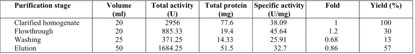
Table 1: Purification table for CGTase using Ni2+-Sepharose IMAC resin. (Strategy A).
Purification stage Volume
(ml)
Total Activity (U)
Total Protein (mg)
Specific activity (U/mg)
Fold Yield (%)
Clarified homogenate 20 2956 77.6 38.09 1. 100
Flowthrough 20 573 21.4 26.78 0.7 19
Washing 25 370 17.75 20.85 0.55 13
48 M. Sivapragasam and N. Abdullah
With every IMAC column, some leaching of metal ions occurs, depending on the type of chelating com-pound involved and the sort of elution. To assess this phenomenon, the elution samples collected during the elution peak were subjected to nickel ion leach-ing analysis usleach-ing an inductively coupled plasma-mass spectrometer (ICP-MS). For the effluent using 50 mM EDTA buffer, very high Ni2+, 158 mg/L, was co-eluted in the elution step. Metal leaching may generate charged groups which act as a cation ex-changer and bind to the positively charged groups on the surface of the proteins (Block et al., 2009) which accounts for low CGTase yields. Ni2+ compounds are also established human carcinogens (Kozlowski et al., 2000) and thus must be removed from the final product. Although the role of Ni2+ in carcinogenesis is not clear, some molecular models suggest interac-tion with histones in the cell nucleus, leading to DNA damage. Application of a strong chelating agent, such as EDTA, resulted in co-elution of the bound proteins. Elution with EDTA was also found to cause a co-elution of a small amount of enzyme before and after the main peak (Figure 3). This could also be due to the distribution of histidine residues on the surface of CGTase. Hemdan et al. (1989) mention that locations of histidines residues is criti-cal for the exploitation of IMAC chromatography. These histidine residues could be on the surface or interior, localized, accessible or non-accessible for coordination, distant or vicinal. Sometimes intra-molecular interaction such as hydrogen bonding may also occur, which results in a non-attachment of the histidine to the Ni2+-Sepharose resin. Thus, the EDTA was too strong as a chelating compound and eluted most of the Ni2+ ion from Ni2+ -Sepharose IMAC resin. A second competitive agent, imidazole, was there-fore chosen to replace EDTA in the elution buffer.
Elution of Bound CGTase on Ni2+-Sepharose IMAC Resin Via Strategy B
Twenty mL of clarified E. coli homogenate con-taining the enzyme CGTase was loaded onto a packed column containing Ni2+-Sepharoseresin. The column was then equilibrated and washed with 25 ml of 20 mM PBS, pH 7, with a flow rate of 1 mL/min. Once unbound protein was removed from the col-umn, bound proteins were eluted via 20 mM PBS, pH 7, 0.1 M imidazole. This was performed via a 0-100 mM gradient elution with 20 mM PBS, pH 7, and 20 mM PBS, 0.1 M imidazole, pH 7.
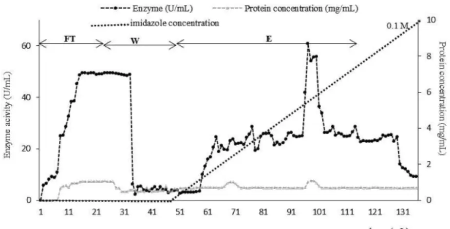
From the chromatogram in Figure 4, about 30% enzyme loss was observed in the flowthrough step (Table 2). In the washing step, a 13% enzyme loss was noted. This was the same as the results obtained when using the same washing buffer (20 mM PBS, pH 7) in the earlier purification step (Strategy A). The elution buffer used was 20 mM PBS, pH 7, 0.1 M imidazole. This was applied in a gradient manner using two buffers, i.e., 20 mM PBS, pH 7, and 20 mM PBS, 0.1 M imidazole, pH 7. Yields of 57% enzyme were obtained, which were higher than that obtained using Strategy A (0.05 M EDTA as chelating agent) with a 45% elution yield. However, a similar chro-matogram pattern (Figure 3 and Figure 4) with the formation of "shoulder" peaks (elution before and after the main peak) was observed, which resulted in a large amount of buffer loss. An apparent peak was observed at 45 mM imidazole concentration (Figure 4). It indicated that most of the elution oc-curred at 45 mM imidazole, which is more cost effective. Hence to reduce cost, buffer loss and the harmful effects of this chelating agent, subsequent elutions were done at 45 mM of imidazole in a sin-gle step manner.
Table 2: Purification table for CGTase using Ni2 -Sepharose IMAC resin (Strategy B).
Purification stage Volume
(ml) Total activity (U) Total protein (mg) Specific activity (U/mg)
Fold Yield (%)
Clarified homogenate 20 2956 77.6 38.09 1 100
Flowthrough 20 885.33 19.4 45.64 1.2 30
Washing 25 371.25 14.33 25.91 0.68 13
Elution 50 1684.25 51.5 32.7 0.86 57
Elution of Bound CGTase on Ni2+-Sepharose IMAC Resin Via Strategy C
Twenty mL of clarified E. coli homogenate con-taining the enzyme CGTase was loaded onto a packed column containing Ni2+-Sepharoseresin. The column was then equilibrated and washed with 25 ml of 20 mM PBS, pH 7, with a flow rate of 1 mL/min. Once unbound protein was removed from the col-umn, bound proteins were eluted via 20 mM PBS, 0.045 M imidazole, pH 7. This was performed via a single step elution.
The chromatogram in Figure (5) presented a good IMAC separation. A single step elution was done using 45 mM imidazole throughout. Purification of CGTase performed with 45 mM imidazole as its elution buffer showed a 5-fold purification during the elution step with yields of 49% (Table 3). The loss of enzyme was 13%, which indicated a good separation by Ni2+ -Sepharose IMAC resin. The re-maining enzyme in the Ni2+ -Sepharose IMAC col-umn was regenerated by washing the colcol-umn with 50 mM EDTA.
In the past, only two studies of the purification of CGTase using metal affinity chromatography were performed. In the first study by Berna et al. (1996), the enzyme CGTase was purified in a single step
metal affinity chromatography using two metals Cu (II), Zn (II) and a tandem combination of Cu (II) and Zn (II). 25 mM imidazole was used as the eluting agent coupled with 50 mM EDTA. When using the tandem combination of Cu (II) and Zn (II) there were no deleterious effects on enzyme activity. IMAC was demonstrated to be a viable technique that can out-perform biospecific affinity chromatography (such as
-CD). This is because metal affinity chromatogra-phy gives similar purity and activity recovery with minimal or no additional steps required, which is an additional advantage when it comes to industrial applications. In the second study by Volkova et al. (2000), CGTase was purified using Cu (II)-IDA-Agarose and desorbed with the addition of 25 mM of imidazole to the washing buffer in a single step elu-tion manner. The specific activity of the CGTase increased 15-fold in comparison to the initial value (273.6 U/mL). Results showed a 73% activity re-covery, which is lower that than obtained from this study (87%). The author suggested the use of IMAC because most of the CGTases are said to contain approximately 10 histidine residues in their primary structure. The affinity of CGTase to Cu (II)-IDA-Agarose is based on the coordination bond formation between the metal ion and the imidazole groups of the accessible histidine groups in the primary structure
E n z y m e a cti v ity ( U /m L ) P ro tei n c o n cen tr at io n ( m g /m L )
Enzyme (U/mL) Protein concentration (mg/mL)
F W E
E n z y m e a cti v ity ( U /m L ) P ro tei n c o n cen tr at io n ( m g /m L )
Enzyme (U/mL) Protein concentration (mg/mL)
F W E
50 M. Sivapragasam and N. Abdullah
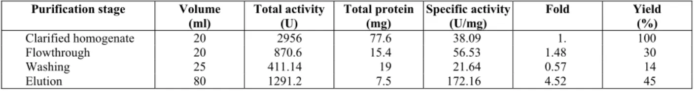
Table 3: Purification table for CGTase using Ni2+-Sepharose IMAC resin (Strategy C).
Purification stage Volume
(ml)
Total activity (U)
Total protein (mg)
Specific activity (U/mg)
Fold Yield (%)
Clarified homogenate 20 2956 77.6 38.09 1. 100
Flowthrough 20 870.6 15.4 56.53 1.48 30
Washing 25 411.14 19 21.64 0.57 14
Elution 80 1291.2 7.5 172.16 4.52 45
Total enzyme recovery: 87.04%
(Volkova et al., 2000). The enzyme is also said to be metal-independent since activity was retained with EDTA, but inhibition with Zn2+, Ni2+, Cu2+ and Fe3+ indicated the presence of histidine residues. In an-other study by Clemmitt et al. (2000), single step elutions were studied using various imidazole con-centrations for the purification of green fluorescent protein (GFP) via an IMAC expanded bed adsorp-tion. Imidazole with single step elution was found to yield a better separation and higher enzyme yields.
The ICP results showed only 0.4512 mg/L nickel ion was co-eluted with the CGTase. This amount of nickel is considered insignificant (< 0.5 ppm) and proved that 20 mM sodium phosphate with 45 mM of imidazole, pH 7, was a suitable elution buffer.
CONCLUSION
The equilibrium binding capacity of CGTase to-ward Ni2+ IMAC resin was high, 666.67 U/mL, and the dissociation constant is 5.3×10⁻¹ U/mL This indi-cated a strong binding capacity and affinity of the Ni2+ IMAC resin towards the enzyme. The dynamic binding capacity was 6.43 mg/mL of resin. The sepa-ration using 45 mM imidazole in the elution buffer gave 4.5-fold purification during the elution step and an 87% overall recovery of CGTase. Moreover, the nickel ion concentration in the eluted sample was only 0.4512 mg/L, which is approved for usage in the cosmetic and textile industry. All in all, IMAC separation has proven to be reliable, efficient and inexpensive in our preparatory scale test. It played an important role in reducing metal leaching into the final product, achieving higher purification and avoiding the denaturing of the enzyme during the process.
REFERENCES
Alves-Prado, H. F., Gomes, E., Silva, R., Purification and characterization of a cyclomaltodextrin glu-canotransferase from Paenibacillus campinasensis
strain H69-3. Appl. Biochem. Biotechnol., 136-140, 41-56 (2007).
Arnold, F. H., Metal-affinity separations: A new di-mension to protein processing. Biotechnol., 9, 151-156. (1991).
Atanasova, N., Kitayska, T., Bojadjieva, I., Yankov, D., Tonkova, A., A novel cyclodextrin glucano-transferase from alkaliphilic Bacillus
pseudal-caliphilus 20RF: Purification and properties.
Process Biochem., 46, 116-122 (2011).
Avci, A., Dönmez, S., Purification and characteriza-tion of a thermostable cyclodextrin glycosyltrans-ferase from Thermoanaerobacter sp. P4. Afr. J. Biotechnol., 11(45), 10407-10415 (2012). Berna, P., Moraes, F. F., Barbotin, J. N., Thomas, D.
and Vijayalakshmi, M. A., One step affinity puri-fication of a recombinant cyclodextrin glycosyl transferase by (Cu(II), Zn (II) tandem column) immobilized metal-ion affinity chromatography. Adv. Mol. Cell Biol., 15, 523-537 (1996).
Block, H., Maertens, B., Spriestesbach, A., Brinker, N., Kubicek, J., Fabis, R., Labahn, J., Schäfer, F., Immobilised metal affinity chromatography (IMAC): A review. Met. Enzymol., 463, 439-473 (2009).
Bolanos-Garcia, V. M., Davies, O. R., Structure analy-sis and classification of native proteins from E. coli commonly co-purified by immobilised metal affinity chromatography. Biochim. Biophys. Acta, 1760 (9), 1304-1313 (2006).
Bornhorst, J. A., Falke, J. J., Purification of proteins using polyhistidine affinity tags. Met. Enzymol., 326, 245-254 (2000).
Bradford, M. M., A rapid and sensitive method for the quantitation of microgram quantities of protein utilizing the principle of protein-dye binding. Anal. Biochem., 72, 248-254 (1976).
Chong, F. C., Tan, W. S., Awang Biak, D. R., Ling, T. C., Tey, B. T., Purification of histidine tagged nucleocapsid protein of Nipah virus using immo-bilised metal affinity chromatography. J. Chr. B., 877, 1561-1567 (2009).
an unclarified Escherichia coli homogenate within an immobilised metal affinity expanded bed. Biosep., 874, 27-43 (1999).
Cristancho, C. A. M., David, F., Franco-Lara, E., Seidel-Morgensten, A., Discontinuous and con-tinuous purification of single-chain antibody fragments using immobilised metal ion affinity chromatography. J. Biotechnol., 163, 233-242 (2013).
Dalal, S., Raghava, S., Gupta, M. N., Single-step purification of recombinant green fluorescent protein on expanded beds of immobilized metal affinity chromatography media. Biochem. Eng. J., 42, 3012-307 (2008).
Doukyu, N., Kuwahara, H., Aono, R., Isolation of
Paenibacillus illinoisensis that produces
cyclo-dextrin glucanotransferase resistant to organic solvents. Biosci. Biotechnol. Biochem., 2, 334-340 (2003).
Finette, G. M. S., Mao, Q. M., Hearn, M. T. W., Comparative studies on the isothermal character-istics of proteins adsorbed under batch equilib-rium conditions to ion-exchange, immobilized metal affinity and dye affinity matrices with dif-ferent ionic strength and temperature conditions. J. Chr. A., 763, 79-90 (1997).
Foo, K. Y., Hameed, B. H., Insights into the model-ling of adsorption isotherm systems. Chem. Eng. J., 156, 2-10 (2010).
Gaberc-Porekar, V., Menart, V., Perspectives of immobilized-metal affinity chromatography. J. Biochem. Biophy. Met., 49, 335-360 (2001). Goh, K. M., Mahadi, N. M., Hassan, O., Raja Abdul
Rahman, R. N. Z., Rosli, M. I., Molecular mod-eling of a predominant -CGTase G1 and analysis of ionic interaction in CGTase. Biotechnol., 3, 418-429 (2008).
Goh, H. P., Illias, R. M., Goh, K. M., Rational mutagenesis of cyclodextrin glucanotransferase at the calcium binding regions for enhancement of thermostability. Int. J. Mol. Sci., 13, 5307-5323 (2012).
Guru, M. M. S., Rajakumari, D. M., Jayashree, S., Fauzia, M., Kumar, D. J. M., Kalaichelvan, P. T., Production and purification of CGTase of alkalo-philic Bacillus isolated from Marneri pond in Ti-runelveli District, Tamil Nadu. J. Acad. Ind. Res., 2, 101-105 (2012).
Hasar, H., Adsorption of nickel (II) from aqueous solution onto activated carbon prepared from almond husk. J. Hazard. Mat. B. 97, 49-57 (2003). Hemdan, E. S., Zhao, Y. J., Sulkowski, E. and Porath,
J., Surface topography of histidine residues: A facile probe by immobilised metal affinity
chro-matography. Proc. Natl. Acad. Sci., USA, 86, 1811-1815 (1989).
Higuti, H. I., Grande, S. W., Sacco, R., Nascimento, A. J., Isolation of alkalophilic CGTase producing bacteria and characterization of cyclodextrin gly-cosyltransferase. Braz. Arch. Biol. Technol., 46, 183-186 (2003).
Ibrahim, A. S. S., Salamah, A. A. and Antranikian, G., A novel cyclodextrin glycosyltransferase from alkaliphilic Amphibacillus sp. NPST-10: Purifi-cation and properties. Int. J. Mol. Sci., 13(8), 10505-10522 (2012).
Kaneko, T., Kato, N., Nakamura, K., Horishoki, K., Spectrophotometric determination of cyclization activity of -cyclodextrin-forming cyclomalto-dextrin glucanotransferase. J. Jpn. Soc. Star. Sci., 34, 45-48 (1987).
Kozlowski, H., Bal, W., Kasprzak, K. S., Molecular models in nickel carcinogenesis. J. Inorg. Biochem., 79, 213-218 (2000).
Martins, R. F., Kaul, R. H., A new cyclodextrin glu-cosyltransferase froman alkaliphilc Bacillus
aga-radhaerens isolate: Purification and
characteri-zation. Enzyme Microb. Technol., 30, 116-124 (2002).
Porath, J., Immobilized metal ion affinity chroma-tography. Prot. Exp. Purif., 32, 263-281 (1992). Prasanna, R. R., Vijayalakshmi, M. A., Immobilized
metal-ion affinity systems for recovery and structure-function studies of proteins at molecu-lar, supramolecular and cellular levels. Pure Appl. Chem.,82, 39-55 (2010).
Qi, Q., Mokhtar, M. N., Zimmermann, W., Effect of ethanol on the synthesis of large-ring cyclodex-trin by cyclodexcyclodex-trin glucanotransferases. J. Inclu-sion Phenom. Macro. Chem., 57, 95-99 (2007). Rosso, A., Ferrarotti, S., Miranda, M. V.,
Krym-kiewicz, N., Nudel, B. C., Cascone, O., Rapid af-finity purification processes for cyclodextrin gly-cosyltransferase from Bacillus circulans. Bio-technol. Lett., 27, 1171-1175 (2005).
Savergave, L. S., Dhule, S. S., Jogdand, V. V., Nene, S. N., Garde, R., Production and single step puri-fication of cyclodextrin glycosyltransferase from alkalophilic Bacillus firmus by ion exchange chromatography. Biochem. Eng. J., 39, 510-515 (2008).
Sharma, S., Agarwal, G. P., Interactions of proteins with immobilised metal ions: A comparative analysis using various isotherm models. Anal. Biochem., 288, 126-140 (2001).
52 M. Sivapragasam and N. Abdullah
one step platform purification of cyclodextrin glucanotransferases. Prep. Biochem. Biotechnol., 41, 350-364 (2011).
Sian, H. K., Said, M., Hassan, O., Kamaruddin, K., Ismail, A. F., Rahman, R. A., Nik Mahmood, N. A., Illias, R. M., Purification and characterization of cyclodextrin glucanotransferase from alkalo-philic Bacillus sp. G1. Process Biochem., 40, 1101-1111 (2005).
Sun, X., Chiu, J. F., He, Q. Y., Application of immobilized metal affinity chromatography in proteomics. Expert Rev. Proteomics., 2, 649-657 (2005).
Tsai, S. Y., Lin, S. C., Suen, S. Y., Hsu, W. H., Effect of number of poly (His) tag on the adsorption of engineered proteins on immobilized metal affinity chromatography adsorbents. Process Biochem., 41, 89-95 (2006).
Ueda, E. K., Gout, P. W., Morganti, L., Current and prospective applications of metal ion-protein binding. J. Chr., A, 988, 1-23 (2003).
Vassileva, A., Atanasova, N., Ivanova, V., Dhulster, P., Tonkova, A., Characterization of cyclodextrin glucanotransferase from Bacillus circulans ATCC 21783 in terms of cyclodextrin production. Ann. Microbiol., 57, 609-615 (2007).
Volkova, D. A., Lopatin, S. A., Varlamov, V. P., One-step affinity purification of cyclodextrin
glu-canotransferase from Bacillus sp. 1070. Biocatal. 2000, Fund. Appl., 41, 67-69 (2000).
Vunnum, S., Gallant, S. R., Kim, Y. J., Cramer, S. M., Immobilized metal affinity hromatography: Mod-elling of nonlinear multicomponent equilibrium. Chem. Eng. Sci., 50, 1785- 1803 (1995).
Westra, D. F., Welling, G. W., Koedijk, D. G. A. M., Scheffer, A. J., Hauw, T. T., Welling-Westera, S., Immobilised metal-ion affinity chromatography purification of histidine-tagged recombinant pro-teins: A wash step with a low concentration of EDTA. J. Chr., B, 760, 129-136 (2001).
Yampayont, P., Iizuka, M., Ito, K., Limpaseni, T., Isolation of cyclodextrin producing
thermotoler-ant Paenibacillus sp. from waste of starch factory
and some properties of the cyclodextrin glyco-syltransferase. J. Incl. Phenom. Macro. Chem., 56, 203-207 (2006).
Yang, Y. H., Wu, T. T., Suen, S. Y., Lin, S. C., Equilibrium adsorption of poly(His)-tagged pro-teins on immobilized metal affinity chroma-tographic adsorbents. Biochem. Eng. J., 54, 1-9 (2011).