CliniCal sCienCe
Derivation and validation of the SLE Disease Activity
Score (SLE-DAS): a new SLE continuous measure with
high sensitivity for changes in disease activity
Diogo Jesus,
1ana Matos,
2,3Carla Henriques,
2,3,4Margherita Zen,
5Maddalena larosa,
5luca iaccarino,
5José antónio Pereira Da silva,
1,6andrea Doria,
5luís sousa inês
1,7To cite: Jesus D, Matos a, Henriques C, et al. Ann Rheum Dis 2019;78:365–371. Handling editor Josef s smolen
►additional material is published online only. To view please visit the journal online (http:// dx. doi. org/ 10. 1136/ annrheumdis- 2018- 214502). 1Rheumatology Department, Centro Hospitalar e Universitário de Coimbra, Coimbra, Portugal 2school of Technology and Management, Polytechnic institute of Viseu, Viseu, Portugal
3Centre for the study of education, Technologies and Health, Viseu, Portugal 4Centre for Mathematics, University of Coimbra, Coimbra, Portugal
5Rheumatology Unit, Department of Medicine, University of Padova, Padova, italy
6Coimbra institute for Clinical and Biomedical Research (iCBR), Faculty of Medicine, University of Coimbra, Coimbra, Portugal 7school of Health sciences, University of Beira interior, Covilhã, Portugal Correspondence to Dr luís sousa inês, serviço de Reumatologia, Centro Hospitalar Universitário de Coimbra, Coimbra 3000-075, Portugal; luisines@ gmail. com Received 29 september 2018 Revised 26 november 2018 accepted 27 november 2018 Published Online First 9 January 2019
© author(s) (or their employer(s)) 2019. no commercial re-use. see rights and permissions. Published by BMJ.
Key messages
What is already known about this subject?
► The SLE Disease Activity Index (SLEDAI) is the most widely used SLE disease activity measure and the core determinant in the SLE Responder Index (SRI) applied as primary endpoint in clinical trials.
► The performance of SLEDAI in detecting clinically meaningful changes in disease activity is limited.
What does this study add?
► This study presents the derivation and validation of the SLE Disease Activity Score (SLE-DAS), a new continuous global score to assess disease activity in SLE. The SLE-DAS provides a more accurate identification of clinically meaningful changes over time, with a much higher sensitivity as compared with SLEDAI-2K and similar specificity. The SLE-DAS presented higher performance in predicting damage accrual, as compared to SLEDAI-2K.
How might this impact on clinical practice or future developements?
► The use of the SLE-DAS in clinical practice and as an outcome in clinical trials will allow a much higher discriminative power to detect clinically meaningful changes in SLE disease activity. Importantly for its use, collection of SLE-DAS items requires a clinical workup time similar to SLEDAI, and much lower than the British Isles Lupus Assessment Group and SRI.
AbSTrACT
Objectives To derive and validate a new disease
activity measure for systemic lupus erythematosus (sle), the sle Disease activity score (sle-Das), with improved sensitivity to change as compared with sle Disease activity index (sleDai), while maintaining high specificity and easiness of use.
Methods We studied 520 patients with sle from two
tertiary care centres (derivation and validation cohorts). at each visit, disease activity was scored using the Physician Global assessment (PGa) and sleDai 2000 (sleDai-2K). To construct the sle-Das, we applied multivariate linear regression analysis in the derivation cohort, with PGa as dependent variable. The formula was validated in a different cohort through the study of: (1) correlations between sle-Das, PGa and sleDai-2K; (2) performance of sleDai-2K and sle-Das in identifying a clinically meaningful change in disease activity (ΔPGa≥0.3); and (3) accuracy of sleDai-2K and sle-Das time-adjusted means in predicting damage accrual.
results The final sle-Das instrument included 17
items. sle-Das was highly correlated with PGa (r=0.875, p<0.0005) and sleDai-2K (r=0.943, p<0.0005) in the validation cohort. The optimal discriminative Δsle-Das cut-off to detect a clinically meaningful change was 1.72. in the validation cohort, sle-Das showed a higher sensitivity than sleDai-2K (change ≥4) to detect a clinically meaningful improvement (89.5% vs 47.4%, p=0.008) or worsening (95.5% vs 59.1%, p=0.008), while maintaining similar specificities. sle-Das performed better in predicting damage accrual than sleDai-2K.
Conclusion sle-Das has a good construct validity and
has better performance than sleDai-2K in identifying clinically significant changes in disease activity and in predicting damage accrual.
InTrOduCTIOn
The accurate assessment of disease activity in patients with systemic lupus erythematosus (SLE) is a critical tool for clinical practice, observational studies and clinical trials.1 2 Over the last three
decades, multiple disease activity indices have been developed. Yet, all these instruments have important limitations, hindering their usefulness.3 Hence, the
standardised measurement of SLE disease activity remains a challenging task.4 5
The SLE Disease Activity Index (SLEDAI), which includes the SLEDAI 2000 (SLEDAI-2K) and Safety of Estrogens in Lupus Erythematosus National
Assessment (SELENA)-SLEDAI versions, is the most widely used SLE disease activity measure.6–8
Furthermore, SLEDAI is the core determinant in the SLE Responder Index 4 used as primary endpoint in recent clinical trials.9–13 However, the
perfor-mance of SLEDAI in detecting clinically meaningful changes in disease activity is limited, since each item of SLEDAI is scored dichotomically, with the same numerical weight regardless of the severity of change observed. Only remission of any lupus feature, but not partial improvement, is captured as a change in SLEDAI score; likewise, an increased severity of any previously active manifestation cannot be scored. In addition, potentially severe lupus manifestations, such as haemolytic anaemia,
on 14 June 2019 by guest. Protected by copyright.
http://ard.bmj.com/
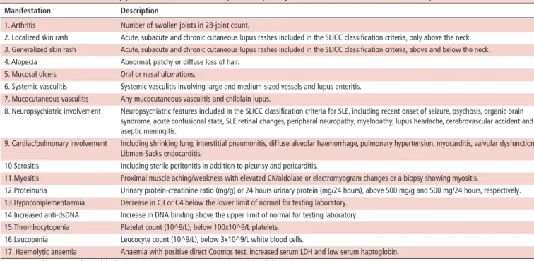
Table 1 Clinical and laboratory parameters attributable to systemic lupus erythematosus (SLE), assessed at each outpatient visit
Manifestation description
1. Arthritis Number of swollen joints in 28-joint count.
2. Localized skin rash Acute, subacute and chronic cutaneous lupus rashes included in the SLICC classification criteria, only above the neck. 3. Generalized skin rash Acute, subacute and chronic cutaneous lupus rashes included in the SLICC classification criteria, above and below the neck. 4. Alopecia Abnormal, patchy or diffuse loss of hair.
5. Mucosal ulcers Oral or nasal ulcerations.
6. Systemic vasculitis Systemic vasculitis involving large and medium-sized vessels and lupus enteritis. 7. Mucocutaneous vasculitis Any mucocutaneous vasculitis and chilblain lupus.
8. Neuropsychiatric involvement Neuropsychiatric features included in the SLICC classification criteria for SLE, including recent onset of seizure, psychosis, organic brain syndrome, acute confusional state, SLE retinal changes, peripheral neuropathy, myelopathy, lupus headache, cerebrovascular accident and aseptic meningitis.
9. Cardiac/pulmonary involvement Including shrinking lung, interstitial pneumonitis, diffuse alveolar haemorrhage, pulmonary hypertension, myocarditis, valvular dysfunction, Libman-Sacks endocarditis.
10.Serositis Including sterile peritonitis in addition to pleurisy and pericarditis.
11.Myositis Proximal muscle aching/weakness with elevated CK/aldolase or electromyogram changes or a biopsy showing myositis.
12.Proteinuria Urinary protein-creatinine ratio (mg/g) or 24 hours urinary protein (mg/24 hours), above 500 mg/g and 500 mg/24 hours, respectively. 13.Hypocomplementaemia Decrease in C3 or C4 below the lower limit of normal for testing laboratory.
14.Increased anti-dsDNA Increase in DNA binding above the upper limit of normal for testing laboratory. 15.Thrombocytopenia Platelet count (10^9/L), below 100x10^9/L platelets.
16.Leucopenia Leucocyte count (10^9/L), below 3x10^9/L white blood cells.
17. Haemolytic anaemia Anaemia with positive direct Coombs test, increased serum LDH and low serum haptoglobin. CK, creatine kinase; LDH, lactic dehydrogenase; SLICC, Systemic Lupus International Collaborating Clinics.
pneumonitis or gastrointestinal involvement, are not scored in SLEDAI.
These limitations have important implications on target-driven management of SLE in daily clinical practice, and in assessing the efficacy of new medications in clinical trials. In fact, despite many promising lupus treatments reaching early-phase human studies during last decades, none except belimumab has yet demon-strated efficacy in phase III trials using these outcome measures, in contrast with the clinician’s impression of efficacy.14–19 Thus,
there is an unmet need for tools able to accurately capture clini-cally meaningful changes in SLE disease activity.
Herein, we describe the development and validation of a new instrument for the measurement of SLE disease activity: the SLE Disease Activity Score (SLE-DAS).20 The aim of SLE-DAS
is to provide an accurate, continuous and user-friendly global measure of SLE disease activity with improved sensitivity to change and high specificity, in comparison to SLEDAI-2K.
PATIenTS And MeTHOdS
Study and settings
Longitudinal cohort study conducted at CHUC Lupus Clinic at Rheumatology Department, Centro Hospitalar e Univer-sitário de Coimbra (CHUC), Portugal (derivation cohort) and at Rheumatology Unit, University of Padova, Italy (validation cohort). This project adheres to the principles of the Declaration of Helsinki and was approved by the local ethics committees. Informed consent was obtained from all patients.
Patients
Consecutive patients with SLE fulfilling American College of Rheumatology (ACR’97) and/or Systemic Lupus International Collaborating Clinics (SLICC’12) classification criteria, enrolled in the CHUC Lupus Cohort from January 2014 to December 2017 and in the Padova Lupus Cohort, from January 2013 to June 2018, were studied.21 22 Data from the CHUC Lupus
Cohort were used for derivation and internal validation of the
SLE-DAS and data from the Padova Lupus Cohort were used for external validation.
Parameters assessed
Clinical and laboratory assessment considered all manifesta-tions included in SLEDAI-2K and in the current definimanifesta-tions of low disease activity (LDA) or remission, in a total of 17 vari-ables (table 1).8 12 13 Disease activity in the previous 30 days was
scored, by one senior rheumatologist with large experience in SLE management in each cohort, using Physician Global Assess-ment (PGA) (0–3) and SLEDAI-2K (0–105).8 Organ damage was
evaluated at each outpatient visit using the SLICC/ACR Damage Index for SLE (SDI).23
Moreover, in the Padova Lupus Cohort, the disease activity data at the last follow-up visit were compared with the previous one and the change was classified by the senior physician as reflecting: (1) clinically meaningful improvement; (2) no clini-cally meaningful change; and (3) cliniclini-cally meaningful worsening.
data analysis and statistics
On clinical grounds, we adopted the guiding principle that all variables listed in table 1 ought to be included in the final instru-ment, given that their relevance is well established in practice.
derivation of the disease activity score
For derivation of the SLE-DAS, data from the CHUC Lupus Cohort were used. Data from the visit with the highest disease activity during follow-up from each patient were selected for analysis.
Multivariate linear regression was applied, with PGA as depen-dent variable. All disease manifestations included in table 1 were considered as potential independent variables for the model. Because the precision of an estimated coefficient may be affected if the corresponding variable has little sample variation, and the regression model ability to predict future observations can be compromised increasing the number of variables, only variables
on 14 June 2019 by guest. Protected by copyright.
http://ard.bmj.com/
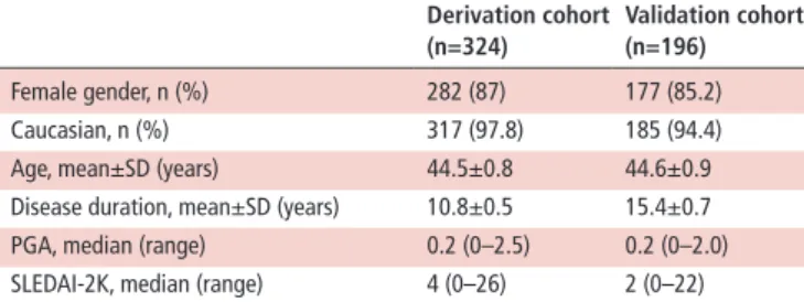
Table 2 Baseline characteristics of the patients included (n=520) derivation cohort (n=324) Validation cohort (n=196) Female gender, n (%) 282 (87) 177 (85.2) Caucasian, n (%) 317 (97.8) 185 (94.4) Age, mean±SD (years) 44.5±0.8 44.6±0.9 Disease duration, mean±SD (years) 10.8±0.5 15.4±0.7 PGA, median (range) 0.2 (0–2.5) 0.2 (0–2.0) SLEDAI-2K, median (range) 4 (0–26) 2 (0–22) PGA: Physician Global Assessment; SLEDAI-2K, Systemic Lupus Erythematosus Disease Activity Index 2000.
Figure 1 The final formula of Systemic Lupus Erythematosus Disease Activity Score (SLE-DAS). Alopecia, presence of alopecia (1=Yes, 0=No); Arthitis, presence of arthritis (1=Yes, 0=No); CardioPulm, presence of cardiac/pulmonary involvement (1=Yes, 0=No); GeneralRash, presence of generalized cutaneous rash (1=Yes, 0=No); Haemolytic, presence of haemolytic anaemia (1=Yes, 0=No); HypoC, presence of hypocomplementaemia (1=Yes, 0=No); IncreasedAnti-dsDNA, increased anti-dsDNA levels (1=Yes, 0=No); Leuc, leucopenia <3x10^9/L (1=Yes, 0=No); LeucCount, leucocyte count (10^9/L); LocalRash, presence of localized cutaneous rash (1=Yes, 0=No); MucocutVasculitis, presence of mucocutaneous vasculitis (1=Yes, 0=No); MucosalUlcers, presence of mucosal ulcers (1=Yes, 0=No); Myositis, presence of myositis (1=Yes, 0=No); Neuropsych, presence of neuropsychiatric involvement (1=Yes, 0=No); PlatCount, platelet count (10^9/L); PProt, proteinuria >500 mg/24 hours (1=Yes, 0=No); Prot, proteinuria (mg/24 hours); Serositis, presence of serositis (1=Yes, 0=No); SJC, swollen joint count (0–28); SystemicVasc, presence of systemic vasculitis (1=Yes, 0=No); Thromb, thrombocytopenia <100x10^9/L (1=Yes, 0=No).
with more than 10 observations were included in the multivar-iate linear regression model. The remaining manifestations were attributed a discretionary weight score by the senior investigator (LSI), taking into account the coefficients of the other variables in the regression model and also the relative impact of these vari-ables in the expert physician’s disease activity assessment.
Several different functions of disease manifestations were sequentially tested in looking for the model with the best possible adjustment to PGA. Urinary protein excretion, platelet and white cell counts were modelled considering their non-linear impact on disease activity assessment as reflected in PGA. Regarding arthritis, two variables were included in the model: a dummy variable indicating the presence or absence of arthritis, and the number of swollen joints, in order to improve the assessment of severity.
Internal validation
The construct validity was assessed by comparing the global score of the SLE-DAS with PGA and SLEDAI-2K, using Spear-man’s correlation coefficients , at the last follow-up visit. During follow-up, the ability of SLE-DAS to discriminate a clinically meaningful worsening or improvement in SLE disease activity (defined as a change in PGA≥0.3) was assessed using receiver operating characteristic (ROC) analysis and compared with the performance of SLEDAI-2K. The areas under the ROC curves (AUC) were used to evaluate and compare the discriminative ability of SLE-DAS and SLEDAI-2K. By analyses of ROC curves, ideal cut-off values of ΔSLE-DAS to detect clinically meaningful worsening/improvement were identified, and its sensitivity, spec-ificity, and positive and negative predictive values (PPV, NPV) were determined. McNemar’s test was used to assess whether there was a significant difference between the SLE-DAS and
SLEDAI-2K sensitivity and specificity for clinically meaningful changes.
external validation
The external validation was performed using data from patients enrolled in the Padova Lupus Cohort. Construct validity was assessed through Spearman’s correlation between SLE-DAS, SLEDAI-2K and PGA at last follow-up visit. Considering the last two assessments in the follow-up period, patients with clinically meaningful worsening or improvement were identified. ROC curves were used to evaluate the performance of SLE-DAS and SLEDAI-2K in detecting clinically meaningful changes. McNe-mar’s test was used to assess whether there was a significant difference between the SLE-DAS and SLEDAI-2K sensitivity and specificity for clinically meaningful changes.
Patients with more than two follow-up visits and regular follow-up (defined as less than 12 months between visits) were included in the analysis of the ability of adjusted mean of SLE-DAS (amSLE-DAS) and adjusted mean of SLEDAI-2K (amSLEDAI) to predict damage accrual. The amSLE-DAS and amSLEDAI were determined for each patient and each visit as the area under the curve (AUC) of the SLE-DAS and SLEDAI-2K values over time, divided by the total length of the time period until that visit.24
Cox regression with time-dependent covariates25 was applied to
assess the ability of the amSLE-DAS and amSLEDAI to predict damage accrual (ΔSDI≥1 during follow-up). The impact of each one of these variables in the hazard of damage was measured adjusting for other damage risk factors.26 Two models were
estimated, one with amSLE-DAS (model 1) and another with amSLEDAI (model 2). The amSLE-DAS and amSLEDAI models were compared using Akaike information criterion (AIC) and Bayesian information criterion (BIC). The likelihood ratio test was applied to evaluate the importance of amSLE-DAS in a model which already has the information given by amSLEDAI and vice versa. Non-rejection of the null hypothesis indicates that the variables not included in the reduced model do not contribute significantly to predict the outcome.
IBM SPSS Statistics, V.24, and R software, V.3.1.2 were used, and p<0.05 was considered statistically significant.27
reSulTS
Characteristics of the patients included
A total of 520 patients with SLE were included: 324 in the deri-vation cohort and 196 in the external validation cohort. Baseline characteristics of the patients included are presented in table 2. on 14 June 2019 by guest. Protected by copyright.
http://ard.bmj.com/
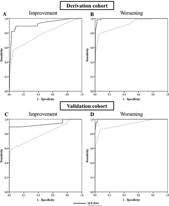
Figure 2 Receiver operating characteristic (ROC) curves comparing the performance of Systemic Lupus Erythematosus Disease Activity Score (SLE-DAS) and Systemic Lupus Erythematosus Disease Activity Index 2000 (SLEDAI-2K) to detect a clinically meaningful improvement (A, C) and worsening (B, D) in SLE disease activity. A and B correspond to the ROC curves of the derivation cohort and C and D correspond to the ROC curves of the external validation cohort.
determination of item weights and derivation of the Sle-dAS
Swollen joints, high urinary protein excretion, low platelet count, low white cell count, localized and generalized lupus rash, alopecia, mucosal membrane lesions, mucocutaneous vasculitis, low complement and increased levels of anti-dsDNA were observed in more than 10 patients and were included in the multivariate linear regression model. Urinary protein excretion, platelet count and white blood cell count were best modelled using the natural logarithmic function. These clinical and labo-ratory features were significantly associated with disease activity (as defined by PGA) in the multivariate model, and the R2 of the
model was 0.91.
The remaining variables, neuropsychiatric involvement, systemic vasculitis, cardiac/pulmonary involvement, myositis,
serositis and haemolytic anaemia were attributed discretionary weights, by expert decision as described above. These weights were incorporated into the final model.
The final SLE-DAS instrument is the sum of all the 17 weighted items (figure 1). The weighted scores of all these items and the detailed definition of each one can be found in the online supple-mentary table S1 and online supplesupple-mentary table S2, respectively.
Internal validation of Sle-dAS
In the derivation cohort, SLE-DAS score was highly correlated with PGA, as expected (rs=0.975, p<0.0005), and with SLEDAI-2K (rs=0.94, p<0.0005) at the last follow-up visit.
on 14 June 2019 by guest. Protected by copyright.
http://ard.bmj.com/
Table 3 Performance of SLE-DAS and SLEDAI-2K to detect clinically significant change in SLE disease activity
ΔSle-dAS≥1.72 ΔSledAI-2K≥4
Sens Spec PPV nPV Sens Spec PPV nPV
Derivation cohort
Clinically meaningful improvement 82.1 96.9 87.3 95.4 44.8 96.5 76.9 87.0 Clinically meaningful worsening 93.1 97.7 90.0 98.5 46.6 99.6 96.4 89.5 Validation cohort
Clinically meaningful improvement 89.5 100 100 98.9 47.4 99.4 90 94.5 Clinically meaningful worsening 95.5 98.2 87.5 99.4 59.1 99.4 92.9 94.9 SLE, systemic lupus erythematosus; SLEDAI-2K, SLE Disease Activity Index 2000; SLE-DAS, SLE Disease Activity Score.
NPV, negative predictive value (%); PPV: positive predictive value (%); SLE, systemic lupus erythematosus; SLEDAI-2K, SLE Disease Activity Index 2000; SLE-DAS, SLE Disease Activity Score; Sens, sensitivity (%); Spec, specificity (%).
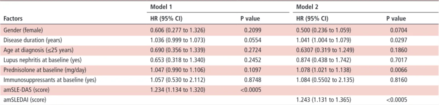
Table 4 Multivariate Cox regression analysis for damage accrual in the validation cohort Factors
Model 1 Model 2
Hr (95% CI) P value Hr (95% CI) P value
Gender (female) 0.606 (0.277 to 1.326) 0.2099 0.500 (0.236 to 1.059) 0.0704 Disease duration (years) 1.036 (0.999 to 1.073) 0.0554 1.041 (1.004 to 1.079) 0.0297 Age at diagnosis (≤25 years) 0.690 (0.356 to 1.339) 0.2724 0.6307 (0.319 to 1.249) 0.1860 Lupus nephritis at baseline (yes) 0.653 (0.318 to 1.340) 0.2452 0.874 (0.438 to 1.742) 0.7017 Prednisolone at baseline (mg/day) 1.047 (0.990 to 1.106) 0.1097 1.078 (1.021 to 1.138) 0.0066 Immunosuppressants at baseline (yes) 1.057 (0.530 to 2.112) 0.8748 1.084 (0.5502 to 2.135) 0.8160 amSLE-DAS (score) 1.234 (1.134 to 1.320) <0.0005
amSLEDAI (score) 1.243 (1.131 to 1.365) <0.0005
Model 1: model with amSLE-DAS as time-dependent covariate. Model 2: model with amSLEDAI as time-dependent covariate.
amSLEDAI, adjusted mean of SLEDAI-2K (time-dependent covariate); amSLE-DAS, adjusted mean of SLE-DAS (time-dependent covariate). Furthermore, SLEDAI-2K was also strongly correlated with PGA
(rs=0.973, p<0.0005).
The performance of SLEDAI-2K and SLE-DAS to discriminate a clinically meaningful improvement or worsening (both defined as ΔPGA≥0.3) was assessed by ROC analysis. SLE-DAS variation had a higher discriminative performance (AUC=0.927 (95% CI 0.885 to 0.969, p<0.0005) than SLEDAI-2K (AUC=0.787 (95% CI 0.718 to 0.857), p<0.0005) to detect a clinically meaningful improvement (figure 2). Variations in SLE-DAS also presented higher discriminative ability than SLEDAI-2K to detect a clinically meaningful worsening, with AUCs of 0.994 (95% CI 0.988 to 1.000, p<0.0005) and 0.914 (95% CI 0.87 to 0.959, p<0.0005), respectively (figure 2). A cut-off value of 1.72 points in the ΔSLE-DAS was identified as the optimal discriminant for either a PGA increase (worsening) or decrease (improvement).
A variation in SLE-DAS≥1.72 had a higher sensitivity to detect a clinically meaningful change in SLE disease activity compared with a SLEDAI-2K≥4 variation: for improvement (82.1% vs 44.8%, p<0.0005) and for worsening (93.1% vs 46.6%, p<0.0005), with similar specificities (96%–100%). Esti-mated sensitivities, specificities, PPV and NPV using cut-offs of decrease and increase in SLE-DAS≥1.72 and SLEDAI-2K≥4 points are shown in table 3.
external validation of Sle-dAS
In the external validation cohort, SLE-DAS score was strongly correlated with PGA (rs=0.875, p<0.0005) and SLEDAI-2K (rs=0.943, p<0.0005) at the last follow-up visit. SLEDAI-2K was also correlated with PGA (rs=0.839, p<0.0005).
For a clinically meaningful improvement, in ROC analysis, a change in SLE-DAS had a higher performance (AUC=0.938 (95% CI 0.852 to 1.000), p<0.0005) than SLEDAI-2K (AUC=0.807 (95% CI 0.681 to 0.933), p<0.0005) (figure 2). For a clinically
meaningful worsening, changes in SLE-DAS and SLEDAI-2K had an AUC of 0.998 (95% CI 0.994 to 1.000, p<0.0005) and 0.928 (95% CI 0.855 to 1.000, p<0.0005), respectively (figure 2).
A variation in SLE-DAS≥1.72 points showed a higher sensi-tivity to detect a clinically meaningful improvement and wors-ening in SLE disease activity than a change in SLEDAI-2K≥4 (89.5% vs 47.4% and 95.5% vs 59.1%, respectively, p=0.008 for both), maintaining similar specificities. Estimated sensi-tivities, specificities, PPV and NPV using cut-offs of increase or decrease in SLE-DAS≥1.72 and SLEDAI-2K≥4 points are reported in table 3.
A total of 192 patients had at least two visits with regular follow-up and were included in the analysis of the predictive value of amSLE-DAS and amSLEDAI for damage accrual. During a mean follow-up of 5 years, 44 (22.9%) patients had damage accrual. The amSLE-DAS and amSLEDAI were identified as independent predictors of damage accrual in the multivariate Cox regression analysis (table 4).
Three multivariate Cox regression models for damage accrual over follow-up were estimated: model 1 with amSLE-DAS and model 2 with amSLEDAI, both variables being considered as time-dependent covariates; and model 3 with amSLE-DAS and amSLEDAI as time-dependent covariates. Gender, disease duration, age at diagnosis, presence of lupus nephritis, pred-nisolone dose and immunosuppressants at baseline were also included in these models as potential confounders (table 4). Both amSLE-DAS and amSLEDAI are significant predictors of damage, but model 1 (with amSLE-DAS) presents better fit than model 2 (with amSLEDAI), as it has lower values of AIC (377.183 vs 385.015, respectively) and BIC (389.673 vs 397.504, respec-tively). Furthermore, the likelihood ratio test comparing model 1 (with amSLE-DAS) with model 3 (containing both amSLE-DAS and amSLEDAI) yields a p value of 0.694, which indicates
on 14 June 2019 by guest. Protected by copyright.
http://ard.bmj.com/
that there is no predictive advantage in including amSLEDAI when the model already considers amSLE-DAS information. On the contrary, the comparison of the model 3 with model 2 (containing only amSLEDAI) leads to the rejection of the null hypothesis (p=0.0047), which means that the prediction of damage improves significantly if we include amSLE-DAS in the model which already has amSLEDAI. Taken together, these anal-yses demonstrate that disease activity measured with SLE-DAS over time has a higher predictive value for damage accrual than SLEDAI-2K.
dISCuSSIOn
SLE-DAS is a continuous global score of disease activity in SLE, designed to more accurately measure disease activity of SLE and to better assess its changes over time. SLE-DAS presented a good construct validity, with very high correlations with both SLEDAI-2K, a validated instrument, and PGA, both in the deri-vation and in the validation cohorts. Criterion validity was demonstrated by the predictive value of SLE-DAS for damage accrual, which was better than that of SLEDAI-2K.
SLE-DAS performs much better than SLEDAI-2K in terms of sensitivity to detect a clinically meaningful change, while presenting a high specificity. Such a performance can have major implications in the interpretation of clinical trials applying the disease activity as the primary endpoint, and in daily clinical practice, where SLE-DAS could provide robust guidance for treatment in the individual patient.
The assessment of disease activity in SLE is a very challenging task due to the heterogeneity of the disease manifestations and the difficulty of incorporating in a single instrument all the poten-tially relevant variables and their relative weights in assessing severity.28 Using a purely dichotomous approach to score
vari-ables, without grading the intensity of their change, makes the currently available tools insensitive to relevant changes, both at individual and group levels.29 These limitations underlie the
diffi-culties that the scientific community has faced in establishing a defined clinical response as the outcome measure in randomised controlled trials or in defining remission and LDA as the targets in treat-to-target strategies.28 30 Actually, the current instruments
cannot separate remission from LDA as we do with DAS28 in rheumatoid arthritis.31 These observations led us to the concept
that a continuous measure of disease activity was indispensable to appropriately value change and to define cut-offs for remis-sion, LDA and moderate or high disease activity.
In the absence of a validated gold standard, we considered that measuring SLE activity should consider all variables that have deserved acceptance by the scientific community so far and should be modelled on experienced physicians, whose judge-ment is validated by high-quality health outcomes docujudge-mented in their patient cohorts.26 31 We started by acknowledging that
in clinical practice the judgement of disease activity in a patient with SLE is based on a combination of laboratory and clinical features as well as the physician overall impression of the patient status (scored in PGA).28 The derivation of the SLE-DAS was
modelled on the PGA and hence it was of utmost importance to ensure the accuracy of an expert clinician. Its construct validity was demonstrated in an external cohort with the PGA scored by a different expert clinician from another country and, most importantly, SLE-DAS was a better predictor of damage accrual than SLEDAI-2K, which argues for the objectivity of a better SLE-DAS performance.
In the SLE-DAS, the four parameters scored as continuous vari-ables (arthritis, proteinuria, leucopenia and thrombocytopenia)
are major drivers of its increased sensitivity to change. In addi-tion, we expect that our new scoring algorithm and item redef-inition, reduced from 24 (in SLEDAI) to 17 (in SLE-DAS), can also contribute to the improved performance of SLE-DAS. At least in some patients, it can be of major impact the inclusion of potentially severe features absent from SLEDAI (haemolytic anaemia, gastrointestinal and cardiopulmonary involvement), which were included in the current definitions of lupus LDA and remission.10 12 13 Assembling all these items into a
quantita-tive disease activity index provided us with a sensiquantita-tive and stan-dardised instrument to assess disease activity in SLE.
Limitations of our study include the use given to PGA, given its subjective nature. However, PGA is considered the gold stan-dard to assess SLE disease activity and has received renewed interest with the recent development of the Lupus Foundation of America-Rapid Evaluation of Activity, an index based on the assessment of a PGA for each organ system.32 Furthermore, in
the current study, PGA was scored always by the same senior rheumatologist over the follow-up, its validity being supported by a high correlation with SLEDAI-2K in both cohorts. Addition-ally, the SLE-DAS was highly correlated with SLEDAI-2K and PGA in the internal and external validation cohort, supporting that PGA was consistently scored. Another important limita-tion refers external validalimita-tion being performed, so far, in only one centre and mainly in Caucasian patients. Further studies are needed to confirm these results in multicentric, multiethnic cohorts. The prospective assessment of data at each clinical visit with consistently scored SLEDAI-2K, PGA and SDI, the internal and external validation including the damage accrual prediction are important strengths of our study.
In conclusion, SLE-DAS has good construct validity, a much higher discriminative power to detect clinically meaningful changes in SLE disease activity and a higher performance in predicting damage accrual, as compared with SLEDAI-2K. Future work from our group will focus in defining cut-off values for remission, LDA, moderate or high disease activity and devel-oping an online calculator.
Contributors DJ, JaPs, aD and lsi contributed to the conception and design of the project, analysis and interpretation of data, and drafting and critical revision of the manuscript. DJ and lsi also contributed to patient follow-up at CHUC lupus Cohort. aM and CH contributed to the design of the project, statistical analysis and interpretation of data, and critically revised the manuscript. MZ, Ml and li contributed to patient follow-up, and analysis and interpretation of data, and critically revised the manuscript. lsi and li scored the disease measures in CHUC lupus Cohort and Padova lupus Cohort, respectively.
Funding The authors have not declared a specific grant for this research from any funding agency in the public, commercial or not-for-profit sectors.
Competing interests none declared. Patient consent for publication Obtained.
ethics approval This study was approved by the ethics Committee of the ’Centro Hospitalar e Universitário de Coimbra’, Coimbra, Portugal and ’azienda Ospedaliera-Università degli studi di Padova’, Padova, italy. informed consent was obtained from all patients before any study procedures.
Provenance and peer review not commissioned; externally peer reviewed.
RefeRences
1 Mikdashi J, nived O. Measuring disease activity in adults with systemic lupus erythematosus: the challenges of administrative burden and responsiveness to patient concerns in clinical research. Arthritis Res Ther 2015;17:183.
2 Rodrigues M, inês l, Gordon C. systemic lupus erythematosus: pathogenisis and clinical features. Eular Textbook on Rheumatic Diseases London: BMJ 2018:577–604. 3 Romero-Diaz J, isenberg D, Ramsey-Goldman R. Measures of adult systemic lupus
erythematosus: updated version of British isles lupus assessment Group (BilaG 2004), european Consensus lupus activity Measurements (eClaM), systemic lupus activity Measure, Revised (slaM-R), systemic lupus activity Questionnaire for population studies (slaQ), systemic lupus erythematosus Disease activity index 2000
on 14 June 2019 by guest. Protected by copyright.
http://ard.bmj.com/
(sleDai-2K), and systemic lupus international Collaborating Clinics/american College of Rheumatology Damage index (sDi). Arthritis Care Res 2011;63–s37–46. 4 Griffiths B, Mosca M, Gordon C. assessment of patients with systemic lupus
erythematosus and the use of lupus disease activity indices. Best Pract Res Clin Rheumatol 2005;19:685–708.
5 Thanou a, Chakravarty e, James Ja, et al. Which outcome measures in sle clinical trials best reflect medical judgment? Lupus Sci Med 2014;1:e000005.
6 Bombardier C, Gladman DD, Urowitz MB, et al. Derivation of the sleDai. a disease activity index for lupus patients. The committee on prognosis studies in sle. Arthritis Rheum 1992;35:630–40.
7 Petri M, Kim MY, Kalunian KC, et al. Combined oral contraceptives in women with systemic lupus erythematosus. N Engl J Med 2005;353:2550–8.
8 Gladman DD, ibañez D, Urowitz MB. systemic lupus erythematosus disease activity index 2000. J Rheumatol 2002;29:288–91.
9 Furie Ra, Petri Ma, Wallace DJ, et al. novel evidence-based systemic lupus erythematosus responder index. Arthritis Rheum 2009;61:1143–51.
10 van Vollenhoven R, Voskuyl a, Bertsias G, et al. a framework for remission in sle: consensus findings from a large international task force on definitions of remission in sle (DORis). Ann Rheum Dis 2017;76:554–61.
11 inês l, Rodrigues M, Jesus D, et al. Risk of damage and mortality in sle patients fulfilling the aCR or only the sliCC classification criteria. a 10-year, inception cohort study. Lupus 2018;27:556–63.
12 Franklyn K, lau Cs, navarra sV, et al. Definition and initial validation of a lupus low Disease activity state (llDas). Ann Rheum Dis 2016;75:1615–21.
13 Zen M, iaccarino l, Gatto M, et al. Prolonged remission in caucasian patients with sle: prevalence and outcomes. Ann Rheum Dis 2015;74:2117–22.
14 Furie Ra, leon G, Thomas M, et al. a phase 2, randomised, placebo-controlled clinical trial of blisibimod, an inhibitor of B cell activating factor, in patients with moderate-to-severe systemic lupus erythematosus, the PeaRl-sC study. Ann Rheum Dis
2015;74:1667–75.
15 Merrill JT, van Vollenhoven RF, Buyon JP, et al. efficacy and safety of subcutaneous tabalumab, a monoclonal antibody to B-cell activating factor, in patients with systemic lupus erythematosus: results from illUMinaTe-2, a 52-week, phase iii, multicentre, randomised, double-blind, placebo-controlled study. Ann Rheum Dis
2016;75:332–40.
16 isenberg Da, Petri M, Kalunian K, et al. efficacy and safety of subcutaneous tabalumab in patients with systemic lupus erythematosus: results from illUMinaTe-1, a 52-week, phase iii, multicentre, randomised, double-blind, placebo-controlled study.
Ann Rheum Dis 2016;75:323–31.
17 Kalunian KC, Merrill JT, Maciuca R, et al. a Phase ii study of the efficacy and safety of rontalizumab (rhuMab interferon-α) in patients with systemic lupus erythematosus (ROse). Ann Rheum Dis 2016;75:196–202.
18 Khamashta M, Merrill JT, Werth VP, et al. sifalimumab, an anti-interferon-α monoclonal antibody, in moderate to severe systemic lupus erythematosus: a randomised, double-blind, placebo-controlled study. Ann Rheum Dis
2016;75:1909–16.
19 Furie R, Khamashta M, Merrill JT, et al. anifrolumab, an anti-interferon-α receptor monoclonal antibody, in moderate-to-severe systemic lupus erythematosus. Arthritis Rheumatol 2017;69:376–86.
20 Jesus D, Matos a, Henriques C. FRi0641 Detection of changes in sle disease activity is highly improved with sle-das as compared to sledai: derivation and preliminary validation of the sle disease activity score (sle-Das). Ann Rheum Dis 2018;77:842–3. 21 Hochberg MC. Updating the american college of rheumatology revised criteria for the
classification of systemic lupus erythematosus. Arthritis Rheum 1997;40:1725. 22 Petri M, Orbai aM, alarcón Gs, et al. Derivation and validation of the systemic lupus international collaborating clinics classification criteria for systemic lupus erythematosus. Arthritis Rheum 2012;64:2677–86.
23 Gladman D, Ginzler e, Goldsmith C, et al. The development and initial validation of the systemic lupus international collaborating clinics/american college of rheumatology damage index for systemic lupus erythematosus. Arthritis Rheum
1996;39:363–9.
24 ibañez D, Urowitz MB, Gladman DD. summarizing disease features over time: i. adjusted mean sleDai derivation and application to an index of disease activity in lupus. J Rheumatol 2003;30:1977–82.
25 Fisher lD, lin DY. Time-dependent covariates in the cox proportional-hazards regression model. Annu Rev Public Health 1999;20:145–57.
26 inês l, Duarte C, silva Rs, et al. identification of clinical predictors of flare in systemic lupus erythematosus patients: a 24-month prospective cohort study. Rheumatology
2014;53:85–9.
27 R Development Core Team. R: A language and environment for statistical computing. Vienna austria: R Foundation for statistical Computing, 2010.
28 Doria a, Gatto M, Zen M, et al. Optimizing outcome in sle: treating-to-target and definition of treatment goals. Autoimmun Rev 2014;13:770–7.
29 Zen M, Bassi n, nalotto l, et al. Disease activity patterns in a monocentric cohort of sle patients: a seven-year follow-up study. Clin Exp Rheumatol 2012;30:856–63. 30 Gatto M, saccon F, Zen M, et al. success and failure of biological treatment in
systemic lupus erythematosus: a critical analysis. J Autoimmun 2016;74:94–105. 31 Zen M, iaccarino l, Gatto M, et al. lupus low disease activity state is associated with
a decrease in damage progression in caucasian patients with sle, but overlaps with remission. Ann Rheum Dis 2018;77:104–10.
32 askanase a, li X, Pong a, et al. Preliminary test of the lFa rapid evaluation of activity in lupus (lFa-Real): an efficient outcome measure correlates with validated instruments. Lupus Sci Med 2015;2:e000075.
on 14 June 2019 by guest. Protected by copyright.
http://ard.bmj.com/