A comprehensive review of genetics and genetic
testing in azoospermia
Alaa J. Hamada,I Sandro C. Esteves,II Ashok AgarwalI
ICenter for Reproductive Medicine, Glickman Urological and Kidney Institute, Cleveland Clinic, Cleveland, Ohio, USA.IIANDROFERT – Andrology & Human
Reproduction Clinic, Campinas, Sa˜o Paulo, Brazil.
Azoospermia due to obstructive and non-obstructive mechanisms is a common manifestation of male infertility accounting for 10-15% of such cases. Known genetic factors are responsible for approximately 1/3 of cases of azoospermia. Nonetheless, at least 40% of cases are currently categorized as idiopathic and may be linked to unknown genetic abnormalities. It is recommended that various genetic screening tests are performed in azoospermic men, given that their results may play vital role in not only identifying the etiology but also in preventing the iatrogenic transmission of genetic defects to offspring via advanced assisted conception techniques. In the present review, we examine the current genetic information associated with azoospermia based on results from search engines, such as PUBMED, OVID, SCIENCE DIRECT and SCOPUS. We also present a critical appraisal of use of genetic testing in this subset of infertile patients.
KEYWORDS: Azoospermia; Male Infertility; Genetic Testing; Genetic Diseases; Klinefelter Syndrome; Y Chromosome.
Hamada AJ, Esteves SC, Agarwal A. A comprehensive review of genetics and genetic testing in azoospermia. Clinics. 2013;68(S1):39-60.
Received for publication onSeptember 5, 2012;Accepted for publication onSeptember 6, 2012 E-mail: [email protected]
Tel.: 1 216 444-9485
& INTRODUCTION
Infertility refers to failure of a couple to conceive following 12 months of unprotected regular intercourse, and this problem affects 10-15% of couples in the United States (1). Male factor infertility is partially or fully responsible for approximately 30-55% of cases of infertility (2,3). It was recently reported that 1 in 13 men of reproductive age requests medical assistance to have a child (4). Azoospermia, which is the complete absence of sperm in the ejaculate, accounts for 10-15% of male infertility cases and generally affects 1% of the male population (3,5,6). Azoospermia is divided into two major categories: obstructive azoospermia (OA), in which there is genital tract outflow obstruction, blocking passage of the sperm, and non-obstructive azoospermia (NOA), in which the testicle fails to produce mature sperm in the ejaculate. Although some reports indicate a higher incidence of NOA than OA (60 vs. 40%) (6) and (85.2 vs. 12.9%) (7), others have reported the opposite. (8). Genetic factors explain 21-29% of azoospermia (9), whereas 12-41% of azoospermic cases are idiopathic and most likely related to unknown genetic factors (7). Using a series of advanced diagnostic and
assisted reproduction therapeutic tools, clinical pregnancy and live birth rates were reported to range from 26-57% and 18-55% for NOA and OA, respectively (10-16).
Azoospermia of a genetic origin is primarily caused by a wide array of genetic disorders, such as chromosomal abnormalities, monogenic disorders, multifactorial genetic diseases, and epigenetic disorders. These conditions con-stitute the genetic basis of reproductive failure. Table 1 summarizes the genetic basis of azoospermia at the post-testicular (obstructive azoospermia), pre-post-testicular and testicular (non-obstructive azoospermia) levels. The aim of this article is to review the genetic causes of azoospermia and to critically appraise the available types of genetic testing and the utility of such tests for the diagnosis and management of azoospermic males.
& OBSTRUCTIVE AZOOSPERMIA OF GENETIC ORIGIN
Obstructive azoospermia comprises the genetic and acquired diseases that cause the obstruction of the reproductive tract pathway. The genetic causes of OA account for approximately 30% of cases (17). In a study that evaluated 179 men with OA, congenital bilateral absence of the vas deferens (CBAVD) was the most frequent condition linked to genetic abnormalities (17). Four genetic post-testicular diseases have been characterized and are described below. Young syndrome is another condition that is associated with OA and that may have a genetic background. Nevertheless, no definitive causative muta-tions have been discovered.
Copyrightß2013CLINICS– This is an Open Access article distributed under the terms of the Creative Commons Attribution Non-Commercial License (http:// creativecommons.org/licenses/by-nc/3.0/) which permits unrestricted non-commercial use, distribution, and reproduction in any medium, provided the original work is properly cited.
No potential conflict of interest was reported.
Cystic fibrosis
Cystic fibrosis (CF) is a life-threatening autosomal recessive disease that affects 1/2,500 Caucasian newborns and has a carrier frequency of 4% (18). CF is characterized by the presence of thick and viscid secretions in the lungs, pancreas, intestines, and liver. Such abnormal secretions result in the formation of plugs that obstruct the ductal lumens in these organs, causing dilatations, frequent infections, and fibrosis. The cause of the viscid secretions is a failure of epithelial cells to transport chloride, sodium ions and water to the lumen of the epithelial tubes. This failure is due to a mutation in the cystic fibrosis transmem-brane conductance regulator (CFTR) gene, which encodes a chloride channel protein.
The CFTR gene is located on chromosome 7 q31.2 (19) and has been implicated in the formation of excurrent seminal ducts (20). The CFTR gene is 190 kb in length and consists of 27 exons. Over 1,800 mutations in this gene have been reported (20). The CFTR protein is a glycosylated trans-membrane ATPase protein that functions as a plasma membrane channel for sugars, peptides, inorganic phos-phate, chloride, and metal cations. CFTR is expressed in the epithelial cells of exocrine tissues, such as the lungs, the pancreas, sweat glands, the head of the epididymis and
the vas deferens. This protein is composed of 1,480 amino acids and has a molecular weight of 168,173 Da. Several reports noted the role of CFTR in sperm maturation in the epididymis, as this protein is necessary for fluid absorption and the facilitation of sperm capacitation and fertilization ability (21-23).
The level of functionally active CFTR protein determines the clinical phenotypes of CFTR mutations. Normal indivi-duals should have an active CFTR protein level of 50-100%, whereas individuals with CF exhibit CFTR activity that is lower than 10% of normal levels (24). Only 2-3% of men with classic CF are fertile, and the majority are infertile (98%) due to congenital bilateral absence or atresia of the vas deferens, which results in azoospermia (25). Epididymal malformations are also common manifestation of CF and are characterized by the absence of the body and tail and blind-ended efferent ductules. Seminal vesicles in these men exhibit a spectrum of anomalies, ranging from atresia, hypoplasia and cystic dilatation. Obstructed ejaculatory ducts are also common. Patients with CF may exhibit varying combinations of these abnormalities.
In most patients with CF, testicular histology indicates normal spermatogenesis. A subset of patients, however, exhibit impaired spermatogenesis, and such impairment may be attributed to the role of the CFTR protein in gametogenesis (26,27). Abnormal sperm morphology has also been described. It is attributed to defective spermiogen-esis due to a lack of CFTR protein, which is also expressed in spermatozoa (28). The vast majority of patients with CF carry two major mutations in chromosome 7 (88%), whereas 11% exhibit compound heterozygous mutations, consisting of a severe mutation in one chromosome and a mild mutation in the other (18,29). The types of severe mutations that are frequently encountered in patients with CF encompass F508del (30%), G542X (3.4%) and G551D (2.4%) (30). The prevalence of these specific mutations vary with the ethnic background of the patient. For example,nF508 is the most common mutation in 70-90% of men with CF in North America and Northern Europe, compared with 50% in Southern Europe and less than 30% in Asians and Indians (31). CFTR mutations are also implicated in isolated seminal duct abnormalities. These abnormalities are referred to as primary genital or atypical forms of CF.
Congenital bilateral absence of the vas deferens
The congenital bilateral absence of the vas deferens (CBAVD) invariably results in azoospermia. CFTR is mutated in 60-90% of patients with CBAVD (32). CBAVD accounts for at least 6-25% of cases of obstructive azoos-permia and approximately 2% of infertility cases (33,34). Men with CBAVD generally exhibit either a single or two mild mutations in the CFTR gene (12%) or a combination of a severe and a mild mutation (88%) (18,35,36). Five patterns of CFTR mutations have been described: i) class I, which is characterized by a defect in protein synthesis, with a premature termination that results in a nonsense or truncated protein; ii) class II, which is caused by a defect in protein processing and localization of CFTR protein to the apical plasma membrane, e.g.,DF508; iii) class III, which is characterized by a defect in cAMP regulation of the channel opening, e.g., G551D (replacement of glycine with aspartic acid at position 551); iv) class IV, which is caused by a partial decline in chloride conductance; and v) class V, which is characterized by reduced levels of functional CFTR Table 1 -Genetic diseases and abnormalities that result
in azoospermia at the post-testicular (obstructive azoospermia), pre-testicular and testicular (non-obstructive azoospermia) levels.
Obstructive Azoospermia of Genetic Origin
Cystic Fibrosis
Congenital Bilateral Absence of the Vas Deferens
Congenital Unilateral Absence of the Vas Deferens (CUAVD) Congenital Bilateral Epididymal Obstruction and Normal Vasa Young Syndrome
Nonobstructive Azoospermia of Genetic Origin
Genetic Pre-testicular Causes of NOA Hypothalamic HH
Congenital HH
Adult-onset genetic hypothalamic HH
Pituitary Disorders Associated with Hypogonadism Generalized anterior pituitary hormone deficiency Selective gonadotropin deficiency
Genetic Testicular Disorders
Affecting Spermatogenesis and Androgen Production Klinefelter syndrome
XX male syndrome Mutation in X-linked USP 26 X-linked SOX3 mutation Bilateral anorchia Noonan syndrome
45 X/46XY mosaicism (mixed gonadal dysgenesis) Affecting Spermatogenesis
Y chromosome microdeletion Autosome translocations Monogenic disorders
Multifactorial disorders (e.g., cryptorchidism) Affecting Androgen Production or Action
Androgen receptor mutation
Steroidogenic acute regulatory protein StAR mutation 3BHSD type 2 deficiency
SRD5A2 mutation
Dysfunctional Cell Regulatory Pathways Epigenetic Defects
protein. Classes I, II and III have been associated with a complete lack of functional CFTR and severe CF manifesta-tions, including pancreatic insufficiency. Class IV and V mutations cause a mild CF phenotype due to residual CFTR protein activity. Men with compound heterozygosity (i.e., a mild and a severe mutation, such asDF508) may have either CBAVD or a mild form of CF.
The common CFTR mutations found in men with CBAVD are the following: i) DF508, in which the three nucleotides that encode the phenylalanine at position 508 are missing in the protein’s amino acid sequence.DF508 is a class II mutation observed in 21-40% of men with CBAVD; ii) polymorphisms within intron 8 (5T, 7T). Such polymorphisms reduce the production of the CFTR protein, resulting in a reduction in the splicing efficiency of the CFTR gene (accounts for 19-37% of CBAVD cases); iii) a missense R117H mutation in exon 4 (14% of CBAVD cases); and iv) a combination ofDF508/R117H, which represents the most common mutation in patients with CBAVD (40%) (36,37).
A recent meta-analysis by Yu et al. revealed that 78% of men with CBAVD carry a minimum of one CFTR mutation. In the aforementioned study, 46% and 28% of men carried two mutations and a single mutation, respectively (38). Moreover, compound heterozygousDF508/5T and DF508/ R117H mutations were present in 17% and 4% of CBAVD cases, respectively. The three most common mutations are 5T (25%), DF508 (17%), and R117H (3%) (38). Non-Caucasian men exhibit a higher incidence of single rather than double mutations (68 vs. 50%, p= 0.012), a higher frequency of 5T mutations (31 versus 20%,p= 0.009) and a lower frequency of A¨ F508 mutations (8 versus 22%,
p= 0.001) compared with Caucasian men (38).
Intracytoplasmic sperm injection (ICSI) is a useful method for the treatment of azoospermic men with the CFTR mutation. Partners who both carry the mutation should be advised to have a preimplantation genetic diagnosis (PGD) performed to avoid passing the abnormality to their offspring (39).
Congenital unilateral absence of the vas deferens (CUAVD)
This condition affects 0.5-1.0% of the male population and represents a heterogeneous disorder with respect to its etiology and clinical presentations. Whereas most men with CUAVD are fertile, a subgroup exhibits azoospermia or oligozoospermia. In the embryonic period, the vas deferens arises from the mesonephric duct. At the 7th week of development, this duct gives rise to the ureteric bud, which in turn induces the development of the kidney from the metanephros. An embryological insult to a single meso-nephric duct at or prior to the seventh week of development can result in unilateral vasal aplasia and ipsilateral renal agenesis. It has been estimated that 79% of individuals with CUAVD have an absent ipsilateral kidney (40).
CFTR mutations have been observed in men with CUAVD who exhibit no renal agenesis. Kolettis et al. reported either single CFTR mutations, such as Negative/ 621_G-T, or compound heterozygous mutations, such as
DF508/5T andDF508/7T/9T, in three out of four men with CUAVD and a distally obstructed contralateral vas defe-rens. The fourth patient, who was negative for a CFTR mutation, exhibited contralateral renal agenesis (41). In the aforementioned series, none of six men with CUAVD and
patent contralateral vas exhibited mutations in the CFTR gene, although three of these patients had ipsilateral renal agenesis (41). Other authors have reported that CFTR mutations may be observed even in men with CUAVD who have normal patent contralateral vas. Radpour et al. demonstrated that in the absence of renal agenesis, five of seven (70%) Iranian men with CUAVD and a patent contralateral vas had either single or compound hetero-zygous CFTR mutations (42). Studies from Canada (43), Spain (44), Germany (45), France (37), and Portugal (43) also reported variable rates of CFTR mutations among men with CUAVD (20%, 27%, 60-100%, and 75%, respectively). However, a critical point with respect to these data sets, besides the small sample sizes, is the lack of information regarding the patency of the contralateral vas deferens.
Congenital bilateral epididymal obstruction and normal vasa
CFTR mutations have been implicated in bilateral epididymal obstruction in azoospermic men in the presence of normal, bilaterally palpable vasa. Mak et al. identified CFTR mutations in 14/56 (25%) men with idiopathic epididymis obstruction (46). The most common identified mutations were IVS8-5T, DF508, R117H and L206W. Similarly, Jarvi et al. reported that as many as 47% of patients with bilateral epididymal obstruction carried CFTR mutations, such as IVS8-5T,DF508, and R117H (47).
Young syndrome
Young syndrome is a rare disease primarily characterized by a constellation of three components, that is, bilateral epididymal obstruction with azoospermia, bronchiectasis, and chronic sinusitis. The estimated prevalence is unknown, with newly discovered cases being described as case reports. Unfortunately, the origin of this disease is also unknown, although childhood exposure to mercury and genetic etiologies have been suggested (48,49). Its familial incidence in one case and its association with medullary sponge kidney in another suggest its inheritability (50,51); however, mutations have not been identified. Male inferti-lity is attributed to bilateral epididymal head dilatation and blockage by an expressible amorphous mass that is attributed to poor epididymal mucociliary clearance (49). The diagnosis of Young syndrome is made by the exclusion of the two other similar syndromes, namely, CF (screened for by testing for CFTR mutations) and immotile cilia syndrome, which is confirmed by prolonged nasal muco-ciliary clearance of the tested material (saccharine) (49,52). Functional rather than subtle ultrastructural epididymal and nasal ciliary defects are considered to be the basic mechanism of the disease, and epididymal aspirations revealed motile spermatozoa (52). Interestingly, epididymal obstruction often occurs in middle-aged men; therefore, previously successful parenthood may be anticipated in such syndromes (52,53).
& NON-OBSTRUCTIVE AZOOSPERMIA OF GENETIC ORIGIN
in the ejaculate. NOA accounts for approximately 60% of men with azoospermia and represents the most severe form of male factor infertility. Although NOA is caused by a multitude of factors, such as heat, radiation, drugs, varicocele, infections and cancer, genetic etiologies con-tribute significantly to the development of this disorder in 21-28% of cases (7,9,40,57-59). NOA is didactically divided into two major categories, i.e., pre-testicular and testicular. The genetic pre-testicular etiology encompasses hereditary hypothalamic-pituitary abnormalities, which lead to insuffi-cient gonadotropin action on testicular cells. This defect results in small testes that exhibit an immature histological pattern. In these cases, immature Sertoli cells or spermato-gonia type A (which primarily reside in the center of seminiferous tubules) and the absence of Leydig cells are often observed.
Genetic testicular causes of NOA include the following: i) chromosomal abnormalities, ii) Y chromosome microdele-tions, iii) failure of the primordial germ cells to reach the developing gonads, iv) lack of differentiation of the primordial germ cells to spermatogonia, and v) male germ line mutations that affect spermatogenesis. The lattermost cause is further divided into mutations that control transcription, signal transduction, apoptosis, cell response to stress factors, cytokines (cross-talk), immune sensitization of germ cells, meiotic divisions, and epigenetic factors. Genetic mutations of androgen receptors are also included in this category.
& GENETIC PRE-TESTICULAR CAUSES OF NOA
Pre-testicular causes of NOA result from either hypotha-lamic or pituitary disorders.
Hypothalamic hypogonadotropic hypogonadism
Genetic hypothalamic disorders essentially fall under the classification of hypothalamic hypogonadotropic hypogo-nadism (HH), which encompasses a broad spectrum of diseases characterized by various genotypes. A deficiency of gonadotropin-releasing hormone (GnRH) or its receptor is the fundamental endocrine abnormality that is detected in this disease. GnRH is a decapeptide that is synthesized by a loose network of neurons located in the medial basal hypothalamus (MBH) and in the arcuate nucleus of the hypothalamus. A subset of GnRH neurons is observed outside of the hypothalamus in the olfactory organ, reflecting the common embryological origin of these neurons (54). Developmentally, GnRH neurons originate from the olfactory placode/vomeronasal organ of the olfactory system and migrate along the vomeronasal nerves to the hypothalamus. Here, these cells extend processes to the median eminence and the pituitary gland (55). GnRH is synthesized as a precursor, 92-amino acid hormone and is then cleaved into a 69-amino acid prohormone. This prohormone is further cleaved at the nerve terminals to form the active decapeptide (55). GnRH receptors are plasma membrane-associated receptors that promote increases in intracellular calcium concentrations, which acts as a second messenger, upon binding to GnRH (55).
The essential function of GnRH is to stimulate the secretion of LH and FSH from the anterior pituitary gland at the time of puberty (56). The hypothalamic pulse generator triggers the pulsatile release of GnRH and is considered to be a regulatory mechanism of the action of
this hormone. Moreover, there is a brief postnatal surge of GnRH during the infantile period, which lasts for a few months and allows for the proper diagnosis of a suspected deficiency at an early age (57).
Genetic HH is primarily divided into two general categories based on the age of onset: congenital and adult-onset HH. Congenital HH is subdivided by the presence of an intact olfactory sense: anosmic HH (Kallmann syndrome) and congenital normosmic isolated HH (IHH). Prader-Willi syndrome is also a congenital disorder that presents with HH.
Congenital HH
This disease is primarily characterized by early-onset hypogonadism due to the dysfunctional release or action of GnRH. This defect results in delayed or absent pubertal development, with low sex steroid levels in the setting of low or normal gonadotropin levels. Normal hypothalamic pituitary gland anatomy on magnetic resonance imaging and the absence of other causes of HH, such as hemochro-matosis, are prerequisites for diagnosis (58). The congenital incidence of HH is 1-10/100,000 live births, with approxi-mately 2/3 being due to Kallmann syndrome and 1/3 to normosmic HH (59).
Kallmann syndrome (KS) is characterized by the presence of complete or partial anosmia in association with con-genital HH. The failure of the migration of GnRH neurons from the olfactory placode to their destination in the hypothalamus and the olfactory lobe is the basic embry-ological defect that characterizes this syndrome (59). However, the genetic bases of this condition have not been fully elucidated. Sporadic (2/3) and familial (1/3) varieties of this condition have been described (60). Hereditary studies reveal that familial KS is heterogenetic, with variable modes of inheritance (autosomal dominant, autosomal recessive, and X-linked) being observed. X-linked inheri-tance is the most common mode. It is not only the genotypic characteristics but also the phenotypic features of this syndrome that are variable. A diverse spectrum of physical manifestations is observed. Males are affected five times more frequently than females, and its incidence in males is approximately 1/8,000 (61). Unfortunately, the genetic origin of only 50% of familial cases and 10% of sporadic cases has been clarified (62). Six known genes account for only 25-35% of all cases of KS (60). These genes are as follows: KAL-1, FGFR-1, PROK-2, PROKR-2, CHD-7, and FGF-8. However, other genetic abnormalities have been described, such as chromosomal translocation 46 XY, t(10,12) (63) and copy number variations (CNVs) (64). Regions in which CNVs are observed account for less than 12% of the human genome and are defined as large segments of DNA on a particular chromosome that have been deleted or duplicated (65,66). Five distinctive chromo-somal regions have been implicated in Kallmann syndrome, and the majority of these CNVs involve the intronic regions of a particular gene, reflecting a possible disturbance in splicing mechanisms. These regions include the following: 1p21.1, 2q32.2, 8q21.13, 14q21.2, and Xp22.31 (64).
migration (59,67). The majority of the observed mutations in the KAL-1 gene are either nucleotide insertions or deletions that result in a frame shift mutation or a premature stop codon (59). However, in fewer than 20% of cases, these nucleotide insertions or deletions may cause amino acid substitution, disrupting the tertiary structure of anosmin 1 and attenuating its function (68). Rarely, contiguous gene syndrome, which includes the deletion of terminal regions of the short arm of the X chromosome (Xp22), may contribute to KS (69,70). Such deletions may cause, in addition to KS, short stature, chondrodysplasia punctata, mental retardation, and steroid sulfatase deficiency (69,70). The KAL-1 gene is responsible for the X-linked recessive mode of inheritance in familial KS and accounts for 10-20% of all cases of KS (71-73). Specifically, KAL-1 mutations account for 30-60% of familial cases and 10-15% of sporadic cases of KS (73-75). Nevertheless, this gene has not been observed in female KS patients or in isolated normosmic HH cases (76). Interestingly, several phenotypic character-istics have been associated with KAL-1 mutations, such as mid-facial clefting, unilateral renal agenesis in 30% of cases, specific neurological abnormalities such as synkinesis (mirror movement), cerebellar dysfunction, deafness, eye abnormalities and mental retardation (60).
The fibroblast growth factor receptor 1 (FGFR-1 or KAL-2, 8p11.2-p11.1) gene is the second most common genetic mutation that is associated with KS (77,78). This gene encodes a tyrosine kinase-linked membrane glycoprotein receptor that binds to extracellular acidic and basic fibroblast growth factors (79). A potential function of fibroblast growth factor is to facilitate GnRH neuron migration, differentiation and survival (80,81). The dysfunc-tion of the FGF receptor results in improper migradysfunc-tion and localization of GnRH neurons, potentially explaining the contribution of this mutation to normosmic HH. FGFR-1 mutations are observed in 10% of patients with KS (78,82). Mutations in this gene are observed in 11% of sporadic cases and 8% of familial cases (83). More importantly, the observed mode of inheritance in familial KS is autosomal dominant, with variable expressivity, incomplete pene-trance and an even male-to-female ratio (84). Variable expression of the FGFR-1 gene is reflected by the occurrence of anosmia alone, hypogonadism alone, or both in family members of the proband (i.e., the affected individual). Moreover, mutations in the FGFR-1 gene do not always result in KS (incomplete penetrance); this phenomenon may highlight the requirement of a loss of function mutation in the development of KS. More than 70% of mutations in FGFR-1 are missense point mutations that result in amino acid substitutions in the immunoglobulin-like or tyrosine kinase domains. Other mutations are either nonsense, frame shift or splice mutations (61). FGFR-1 mutation-induced KS is characterized by variably severe hypogonadism (from mild to complete) and certain morphogenic abnormalities, such as mid-facial clefting, synkinesis (20% of patients) and missing teeth (61).
Fibroblast growth factor 8 (FGF-8) is considered to be one of the ligands for FGFR-1 and is hypothesized to facilitate the migration and differentiation of GnRH neurons to the hypothalamus, as was described above. Mutations in the gene that encodes this protein can cause KS and normosmic HH (85,86). This gene has been mapped to chromosome 10q24 and is responsible for fewer than 2% of all cases of KS (60). An autosomal dominant mode of inheritance is also
demonstrated in familial cases with variable penetrance. Other genes that may be affected in KS include PROK2, PROKR2, and CHD7. Prokineticin 2 is an 81-amino acid protein that is encoded by the PROK2 gene, which has been mapped to chromosome 3p13. This protein has a putative role in the chemoattraction of GnRH neurons in their migration to and differentiation in the hypothalamus (59). This protein acts by binding to a specific G-protein-linked receptor that is encoded by PROKR2 and is located at 20p12.3 (59,61,62). Frame-shift mutations in PROK2 and missense mutations in PROKR2 account for 5-10% of cases of KS (59,61). Mutations in either of these genes exhibit homozygous (autosomal recessive), heterozygous (autoso-mal dominant) and compound heterozygous modes of inheritance (87). Furthermore, these mutations are asso-ciated with variable phenotypic manifestations, such as fibrous dysplasia, severe obesity, sleep problems and synkinesis. Similarly, mutations in these genes have been described in normosmic HH (88). Chromodomain helicase DNA binding protein 7 (CHD7) is a member of a family of proteins whose function is to organize chromatin remodel-ing (packagremodel-ing), a process that regulates gene expression (89,90). Tightly arranged chromatin is characterized by lower gene expression compared with loosely arranged chromatin. CHD7 is ubiquitously expressed in fetal tissues, the brain, the eyes, the inner ear, olfactory neural tissue and GnRH neurons. The gene that encodes this protein is located on chromosome 8q12.2 (62). Mutations in this gene have been linked to several diseases, such as KS, normosmic HH and CHARGE syndrome. CHARGE syndrome is associated with eye coloboma, heart defects, atresia of the nasal choana, retarded growth, genitourinary abnormalities, anosmia, and hypogonadism (KS) (89-91). The CHD7 protein is postulated to be an essential factor in the migration and differentiation of GnRH neurons. Seven mutations have been described for this gene in sporadic and familial KS and normosmic HH (59,92). CHD7 accounts for 6% of all cases of KS and 6% of sporadic KS cases. Moreover, familial KS due to CHD7 mutations exhibits an autosomal dominant mode of inheritance (92).
GnRHR, KISS1R, TAC3, TACR3, and DAX1 (59,60,76). However, chromosomal abnormalities (generally sporadic) have been observed in 3% of patients with normosmic HH. Such abnormalities include 46,XY/46,X,inv(Y)(p11.2q11.2) and mos46,XY,t(3;12)(p13;p13)/46,XY (76,93).
Mutations in the GnRH gene were recently determined to be a rare cause of normosmic HH in two human studies of familial HH (94,95). The gene maps to chromosome 8q21-p11.2 and encodes a large 92-amino acid precursor protein. An autosomal recessive mode of inheritance for this trait has been described, and homozygous frame-shift mutations have been observed in probands (94,95). However, muta-tions in the gene that encodes the G-protein-coupled receptor for GnRH (GnRHR1) are the most common genetic abnormality that is detected in patients with this disorder, accounting for 5-40% of normosmic HH cases (3.5-16% of sporadic cases and as many as 40% of familial cases) (93). This gene maps to chromosome 4q13.2-3, spans over 18.9 kb and encodes a 328-amino acid protein (96). Autosomal recessive homozygous or compound heterozygous muta-tions have been reported in familial cases (97-103). The majority of GnRHR1 mutations are missense mutations that cause single amino acid substitutions. Such mutations result in variable functional effects, ranging from mild impairment to complete inactivation of the receptor (104,105).
Recent studies have identified other ligand proteins and their receptors, such as Kisspeptin and neurokinin B, the functions of which are to regulate the differentiation of GnRH neurons and to initiate their function during puberty. The Kisspeptin (KSS1) gene has been mapped to chromo-some 1q32, and a missense mutation in this gene causes autosomal recessive inherited normosmic HH (62,106). Moreover, Kisspeptin receptor (KSSR1, chromosome 19p13.3) mutations also exhibit autosomal recessive patterns of inheritance, and both KISS1 and KISSR1 mutations contribute to fewer than 5% of normosmic HH cases (107). Mutations in neurokinin B (TAC3, chromosome 12q13-q21) and its receptor (TACR3, 4q25) have also been implicated in the pathogenesis of normosmic HH and exhibit autosomal recessive inheritance patterns (108-110). Convertase 1 is an endopeptidase that is encoded by the PCKS1 gene and is involved in i) the post-translational modification of pre-cursor GnRH and ii) the release of mature and active GnRH (59). Mutations in the gene that encodes this protein have been linked to anosmic HH, diabetes and obesity (111). Finally, an X-linked mode of inheritance of normosmic HH has been linked to DAX1 mutations, which cause congenital X-linked adrenal hypoplasia (112).
Prader-Willi syndrome (PWS) is a complex genetic disorder that is associated with various degrees of systemic involvement. This condition is caused by the lack of expression of paternally derived imprinted genes on chromosome 15q11-q13. This lack of expression is due either to the deletion of these genes, maternal uniparental disomy of chromosome 15 or the disruption of the paternally inherited chromosome 15 (9,90). Genomic imprinting refers to a phenomenon in which certain genes are expressed in a parent in an origin-specific manner. In this situation, an allele from a given parent is silenced to allow for the expression of non-imprinted genes from the other parent. Genomic imprinting is observed in fewer than 1% of genes. Obesity, hyperphagia, growth retardation, mild to moderate mental retardation, dysmorphic facial features, and sleep abnormalities are the characteristic
features of Prader-Willi syndrome. Hypothalamic HH is a consistent feature of all men with this syndrome (9). During infancy, 80-90% of affected children exhibit cryptorchidism, with a poorly developed scrotum and micropenis. Most adolescents will exhibit delayed or incomplete puberty; however, precocious puberty has been described in 4% of patients (9,24,90). Maternally derived supernumerary mar-ker chromosome (SMC) 15 is the most frequently observed supernumerary chromosome marker in humans (50%). This dicentric chromosome fragment arises from the two homo-logous chromosomes 15 and is now referred to as dic(15). This genetic defect was formerly referred to as inverted duplication of chromosome 15 or inv dup(15). The size of the fragment is variable; long fragments may contain the PWS critical region and may lead to the development of PWS, whereas short fragments do not contain this critical region, in which case PWS is not observed. However, azoospermia and mild facial dysmorphism, such as man-dibular anomalies, have been reported in men that carry the short fragment. Further genetic analysis of the genes on chromosome 15 is lacking but may aid in our understanding of the role of these genes in male infertility.
Adult-onset genetic hypothalamic HH
This disease category has recently been described and is restricted to men who successfully completed pubertal development (and who may already have children) and who subsequently exhibited disruption of the HPG axis. Testicular size in such patients is normal, but serum testosterone and gonadotropins levels are low. Moreover, an apulsatile pattern of LH secretion is observed in these patients. A single study of 10 men with adult-onset HH revealed a heterozygousPROKR2mutation in one patient. However, a good prognosis is expected in these men following treatment with respect to their future fertility potential and androgenization status (9).
Pituitary disorders that are associated with hypogonadism
Male hypogonadism that is attributed to genetic pituitary diseases is rare, and such conditions are divided into two major categories: a) generalized or combined anterior pituitary hormone deficiency, and b) selective gonadotropin deficiency.
Generalized anterior pituitary hormone deficiency
(114). Specifically, the implicated mutated transcription factor genes that cause hypogonadism are the following:
PROP1 (most common), LHX and SOX2. Familial and sporadic cases that exhibit autosomal recessive patterns of inheritance are the most common forms of CPHD (114). To accurately differentiate between hypothalamic and pituitary HH, a GnRH stimulation test is performed. In hypothalamic disorders, significant increases in gonadotropin levels are observed, whereas no response is observed in patients with pituitary hypogonadism.
Selective gonadotropin deficiency
FSH and LH are glycoproteins that are secreted by anterior pituitary gonadotropes to stimulate testicular spermatogenesis and testosterone production, respectively. Each glycoprotein molecule is composed ofaandbchains. Theachain represents a common chain for LH, FSH, human chorionic gonadotropin (hCG), and TSH, whereas the differentbchains of these hormones confer immunological and biological hormone specificity. Selective gonadotropin deficiency includes mutations in the genes that code for the synthesis of FSH and LH and their receptors. Specifically, mutations that affect hormone synthesis generally involve thebchain genes, given that mutations in theachain gene (CGA, 6q12-21) are generally embryonically lethal due to the lack of placental HCG synthesis (115).
FSH exerts its action through Sertoli cell receptors, stimulating their proliferation in the immature testis. FSH also stimulates and maintains spermatogenesis. FSH is composed of ana chain, which is composed of 92 amino acids and is non-covalently bound to thebchain (111 amino acids). Rarebsubunit (chromosome 11p13) mutations result in isolated FSH deficiency, delayed or normal puberty and small or normal-sized testes in association with severe oligozoospermia or non-obstructive azoospermia (116-118). Theb subunit gene is composed of three exons and two introns. To date, two missense and three stop codon mutations with autosomal recessive modes of inheritance have been detected (117,119-121). In contrast, FSHb knock-out mice are not infertile, indicating differential regulatory mechanisms in humans and mice (122,123). Furthermore, rare mutations in the gene that encodes the FSH receptor (FSHR), which is expressed in Sertoli cells, have variable effects on spermatogenesis. In a study of five men who were homozygous for FSHR mutations, none exhibited normal semen parameters. Specifically, three patients exhibited severe oligozoospermia and one exhibited moderate oligo-zoospermia; the fifth patient exhibited a low semen volume and teratozoospermia despite having a normal sperm count (124). The FSHR gene has been mapped to chromosome 2p21-16 and consists of 10 exons and 9 introns (125,126). This gene encodes the mature form of a G-protein-linked glycoprotein receptor, which is composed of 678 amino acids and is exclusively expressed in Sertoli cells (125). Single missense inactivating mutations that result in a valine to alanine substitution at position 189 (A189V) have been reported; these mutations exhibit an autosomal recessive mode of inheritance. Preliminary studies by Simoni et al. (127) and Ahda et al. (128) have revealed differences in the FSHR polymorphisms between fertile and infertile men. Further research is required to clarify the genetic back-ground of FSHR mutations.
LH initiates male pubertal development through its effect on LH receptors on Leydig cells, which stimulate the release
of testosterone. Although reported in only five men, recessively inherited missense mutations in the beta subunit (121 amino acids) of the LH gene (19q13.3) result in delayed or absent pubertal development and oligozoospermia or azoospermia (129-131). The hormonal profile of such patients indicates normal FSH levels, high LH immunor-eactivity and low testosterone (129,130). The detected LH does not exhibit biological activity. A variety of autosomal recessive mutations (missense, insertion, deletion and nonsense) have been observed in the gene that encodes the G-protein-linked LH receptor. Such mutations result in variable phenotypic traits and infertility. Interestingly, LH glycoprotein receptors (674 amino acids) respond not only to LH but also to hCG and are thus occasionally referred to as LhCGRs. These receptors are present not only in Leydig cells but also in sperm, seminal vesicles, the skin, the thyroid and other organs, where they have an unidentified physiological significance. The gene for the LH receptor has been mapped to chromosome 2p21 and consists of 11 exons and 10 introns (132). This genomic location is close to that of the FSHR gene, and mutations in the LH receptor gene result in Leydig cell hypoplasia (LCH) or agenesis (115,133). LCH exhibits a wide spectrum of manifestations, ranging from male pseudohermophroditism in cases of 46,XY (female external genitalia, undescended abdominal testes, absent breast development) to selective, milder under-virilization defects, such as micropenis, hypospadias, and cryptorchidism (115).
Genetic testicular disorders (GTDs)
Genetic testicular disorders that cause male infertility can be divided into three categories according to the specific altered function: 1) genetic testicular disorders that primar-ily affect spermatogenesis and androgen production; 2) genetic testicular disorders that primarily affect spermato-genesis; and 3) genetic testicular disorders linked to androgen synthesis or action.
GTDs that affect spermatogenesis and androgen production
Klinefelter syndrome. KLFS is the most common cause of hypogonadism and infertility in males (1 in 500).
Klinefelter syndrome is also the most common
attributed to small testicular growth despite the presence of Leydig cell hyperplasia.
Several studies have demonstrated that KLFS patients exhibit a high incidence of aneuploid gametes, rendering them at risk for producing offspring with chromosomal abnormalities (137). Interestingly, the successful fathering of 60 children has been achieved by testicular sperm extraction (TESE) and ICSI in men with KLFS. Karyotype studies that were performed in approximately 50 children revealed no chromosomal abnormalities (138,139). These genetic find-ings have been further clarified in a study by Sciurano et al., who detected spermatogenesis foci in testicular biopsies of 6/11 men with nonmosaic KLFS (140). Whereas the majority of seminiferous tubules are devoid of germ cells, 8-24% do contain germ cells. Sciurano et al. examined the chromoso-mal complements of 92 meiotic spermatocytes using fluorescent in situ hybridization (FISH). These spermato-cytes exhibited euploidy (normal chromosomal constitu-ents) and the ability to form haploid gametes (140). These novel findings may explain the high rate of normal children who are born following testicular sperm extraction and ICSI in men with KLFS. Nonetheless, even with this high rate of normally born children, there is still a risk of genetic defects in the offspring; therefore, it is advised that a preimplanta-tion genetic diagnosis (PGD) be offered prior to ART to ensure that the offspring are not aneuploid (39).
XX males. This genetic disorder is very rare, with an estimated prevalence of 1:10,000-1: 20,000 (141). The genetic event that causes this disorder involves the translocation of genetic material of the testis-determining region of SRY (or SOX A, a gene that lies on the Y chromosome) to the X chromosome during paternal meiosis. SRY encodes a transcription factor of approximately 204 amino acids (142). This translocation results in the successful differentiation of indeterminate gonads into testes; however, the lack of other genes involved in the initiation of spermatogenesis renders these males azoospermic. Furthermore, SRY-negative variants have also been
described, which are characterized by severe
undervirilization defects, such as undescended testes, hypospadias and bifid scrota (143). Phenotypically, these men are very similar (but with smaller statures) to patients with KLFS (144).
Mutations in X-linked USP26. The USP26 gene is a single-exon gene that maps to chromosome Xq26.2. This gene encodes the USP26 protease (913 amino acids), which is a deubiquitinating enzyme (145,146). The ubiquitination and deubiquitination of macromolecules are essential for the regulation of the cell cycle, maintenance of chromosomal structure and gene silencing (145). The removal of histones and the regulation of protein turnover during meiosis are important functions of this protein. More than twenty mutations in this gene have been reported. These
mutations result in the severe impairment of
spermatogenesis, and several result in hypogonadism (147). Studies have demonstrated a relationship between this gene and certain cases of non-obstructive azoospermia (148,149).
X-linked SOX3 mutations. The SOX genes are essential for development and control of embryonic ontogenesis in the human testes, neural tissues, cartilage, and neural crest cells. These genes are present only in vertebrates and give rise to SOX proteins that have a role in both the developing and adult gonads. These proteins share a common 79-amino
acid DNA binding domain that is characteristic of a large protein superfamily that is referred to as the high mobility group (HMG) due to the high migration rate of these proteins in polyacrylamide gels (150). SOX proteins specifically bind to the DNA minor groove and regulate gene expression by acting either as transcriptional activators or repressors (150). The name SOX is derived from the first-discovered Y-linked SOX gene, i.e., sex-determining region Y (SRY). The other genes in this family are therefore referred to as SRY-box (SOX) genes. The SOX genes are divided into several groups, from A to H, with group A consisting only of the Y-linked SRY (150). SOX3 is X-linked (Xq26.3) and belongs to the SOX B1 group. The SOX3 gene is specifically expressed in developing testicular and neural tissues and encodes the SOX3 transcriptional activator (151,152). Solmon et al. and Woods et al. correlated genetic mutations in the SOX3 gene with hypopituitarism and mental retardation (153,154). Recessively inherited polymorphic mutations in SOX3 have been observed in men with idiopathic oligozoospermia and in mice with severely impaired sperm production and hypogonadism (155,156).
Bilateral anorchia. Bilateral anorchia is a rare congenital disease with an estimated prevalence of 1 in 20,000 males. This condition is characterized by the absence of testicular tissue in 46,XY individuals (157). Because male infants who are born with this disorder exhibit normal genital differentiation, the absence of testicular tissue is most likely attributed to testicular regression that occurs in the second half of gestation. A micropenis is observed in half of these cases (157). The exact etiology of this disease is unknown; however, a subset of cases exhibit familial clustering, suggesting a genetic etiology. Philibert et al. recently noted the role of mutated steroidogenic factor-1 (NR5A1), which is a member of the nuclear receptor family, in the disease etiology. These genes regulate the transcription of other genes that control the development of adrenal and gonadal tissues (158). NR5A1 has been mapped to chromosome 9q33 and has been correlated with other human diseases, such as male infertility, hypospadias, ovarian insufficiency, and others (158).
45X/46,XY mosaicism (mixed gonadal dysgenesis).
Normal male external genitalia are observed in 90% of males with 45,X/46,XY mosaicism; abnormal, ambiguous and female genitalia are observed in the other 10%. Mixed gonadal dysgenesis (a streak gonad on one side and a testis on the other) is observed in 10-30% of patients with this type of mosaicism (162). Abnormal gonadal development results in azoospermia and low testosterone levels (163).
GTDs that affect spermatogenesis
Y chromosome microdeletion. The long and short arms of the Y chromosome contain many genes that regulate spermatogenesis and testes development, respectively. Microdeletions on the long arm of the Y chromosome (Yq) are well correlated with male infertility. Yq microdeletions are detected in approximately 13% of men with non-obstructive azoospermia and in 5% of men with severe oligozoospermia (sperm counts lower than 5 million/mL) (164,165). A microdeletion is defined as a chromosomal deletion that spans several genes but that is small in size and cannot be detected using conventional cytogenetic methods (e.g., karyotyping). Y chromosome microdeletions are clustered in intervals 5 and 6 of the long arm of the Y chromosome. This region at Yq11 is referred to as the ‘‘Azoospermia Factor’’ (AZF) region. The AZF region is further subdivided into three subregions that are termed AZFa, AZFb, and AZFc (Figure 1). The most common aberrations in the AZF region are multiple gene deletions in the AZFb and AZFc sub regions (166), which can produce a wide range of infertility phenotypes. The genes on the Y chromosome are generally divided into three types: ‘‘X transposed’’, X degenerated, and amplicons. Eight palindromes are recognized in the AZF region and six of these are related to male fertility. Six of the genes that are located in the AZF regions are expressed exclusively in the testes and are therefore referred to as ‘‘AZF candidate genes’’.
The AZFa region is the smallest portion of the AZF and spans approximately 400-600 kb of DNA. It is located in the proximal portion of interval 5. The AZFa region is characterized by a non-repetitive structure and a low deletion frequency. This subregion contains three genes: USP9Y, DBY (DDX3Y) and UTY. Two protein-coding genes are directly related to male infertility: USP9Y and DBY (recently termed DDX3Y). Complete and partial deletions of AZFa have been described. Complete deletions that remove both genes cause Sertoli cell-only syndrome (SCOS) and bilateral small-sized testes (167,168). It is estimated that 9-55% of SCOS cases are caused by deletions in the AZFa region (169-171). Partial deletions have also been reported, with particular involvement of USP9Y. The DBY gene is the major single-copy gene in the AZFa region and belongs to the DEAD BOX RNA helicase family. This family consists of a group of genes that encode proteins that specifically regulate RNA transcription, translation, and splicing in the G1 phase of the cell cycle. DBY expression is observed in the male germ line, whereas its expression in other tissues is uncertain (172). DBX, a DBY homologue, belongs to the same family as DBY and is expressed in male germ cells. Its expression pattern has been elucidated by immunohisto-chemistry techniques. The DBY gene and protein are specifically expressed in premeiotic germ cells, whereas the DBX gene and protein are expressed in postmeiotic germ cells (172). This differential distribution explains why
deletions of the DBY gene can result in SCOS and azoospermia. The USP9Y gene is a single-copy gene that encodes a ubiquitin-specific protease (a deubiquitinating enzyme). This enzyme binds to ubiquitin-protein conjugates and hydrolyzes the ubiquitin peptide chain. Such action confers stability to cellular proteins and protects them from cellular degradation by the proteasome complex (173). The expression of USP9Y is restricted to spermatids in humans and mice (174,175). USP9Y expression in male germ cell lines may lead to the stability of certain cellular proteins that are synthesized in primordial germ cells (PGCs) and that are important for germ cell survival. Point mutations in the USP9Y gene result in maturation arrest at the spermatid stage (176), oligozoospermia (177), oligoasthenozoospermia, and asthenozoospermia (178).
The AZFb subregion spans approximately 6 Mb and is located in the distal portion of interval 5 and the proximal portion of interval 6 (subinterval 5O-6B) (179). The meiotic arrest of spermatogenesis at the primary spermatocyte stage is usually observed when AZFb is deleted. AZFb contains 32 genes and overlaps with AZFc (179). As such, AZFb deletions often remove certain genes from the AZFc region (e.g., DAZ1 and DAZ2), as well as one copy each of BPY2, CDY1, and PRY. The primary protein-encoding genes in AZFb are RBMY and PRY. Six copies of RBMY are located in the distal portion of AZFb and are only expressed in germ cells (180). RBMY encodes four types of testis-specific RNA-binding proteins that are involved in mRNA processing, transport, and splicing (181). Due to its proximity to AZFc, certain deletions within AZFc can remove several copies of RBMY genes. Two copies of PRY are located in the AZFb region and presumably regulate apoptosis (167). Hypospermatogenesis is observed when all of the AZFb genes are deleted except for RBMY and PRY. Conversely, spermatogenesis is completely arrested when both the RBMY and PRY genes are deleted (182,183).
AZFc spans over 3.5 Mb and contains a large number of amplicons that are arranged as direct repeats, inverted repeats, or palindromes. Examples of these repeats include b, g, r, and P repeats. Seven distinct gene families, encompassing 23 genes, are observed in the AZFc region. These families include PRY (two copies), TTY (eight copies), BPY (three copies), DAZ (four copies), GOLGA2LY (two copies), CSPYG4LY (two copies), and CDY (two copies). Deletions in the AZFc region alone or deletions in this region that are combined with deletions in other AZF regions are the most common types and account for as many as 87% of Yq microdeletions. The incidence of these deletions is 1/4,000 males. Although AZFa and AZFb deletions result in azoospermia, deletions in the AZFc region can result in either azoospermia or oligozoospermia (184). AZFc deletions can explain approximately 12% of non-obstructive azoospermia and 6% of severe oligozoos-permia cases (185). Two scenarios have been described regarding the cause of complete AZFc deletions, namely, a new deletion in addition to a preexisting partial deletion or a complete deletion of a preexisting normal gene. AZFc deletions may jeopardize Y chromosome integrity, resulting in its loss and sex reversal. As such, AZFc deletions predispose one’s offspring to the 45,X0 karyotype (186) and to the mosaic phenotype 45,X/46,XY (187).
Partial or sub-deletions are denoted by gr/gr, b1/b3, and b2/b3. The gr/gr subdeletion removes nearly half of the AZFc region and results in different phenotypes in various populations. Such variability suggests that other confound-ing factors may be involved, such as ethnicity and environmental effects on gene expression. Although certain studies have recognized gr/gr deletions as a risk factor for impaired spermatogenesis (168,188-190), others failed to conclusively demonstrate this relationship (191-193). The deletion of b1/b3 removes the proximal portion of AZFc. Two of the six copies of RBMY1, the two functional copies of PRY (194), one of the three copies of BPY2 and two of the four DAZ genes are deleted. Nevertheless, large 1.8-Mb deletions have been observed in normozoospermic fertile individuals (195), whereas other researchers have identified this deletion in azoospermic men (196,197). The deletion of b2/b3 removes nearly half of the AZFc region (1.8-Mb DNA segment) and 12 genes, including two copies of DAZ and two copies of BPY. With respect to b1/b2, no conclusive impact on spermatogenesis has been determined (198,199).
The coding genes in AZFc include DAZ and CDY. DAZ plays important roles throughout germ cell development from embryonic life to adulthood; there are four copies of this gene on the Y chromosome (210,211). Postnatally, DAZ encodes proteins that have RNA recognition motifs (RRMs),
which are involved in the regulation of RNA translation (200) and control of meiosis. DAZ is essential for the maintenance of the PGC during embryogenesis (201). DAZ2 and DAZ3 each have a single RRM, whereas DAZ1 has two and DAZ4 has three RRMs. There are two autosomal homologues of DAZ: DAZL on chromosome 3q24.3 and BOULE on chromosome 2q33. DAZ gene expression was observed to be reduced in azoospermic patients (202), and partial deletions of DAZ genes appear to be related to oligozoospermia. Two copies of the chromodomain protein Y-linked (CDY) gene are located in the AZFc region. CDY is expressed exclusively in germ cells, where it encodes a protein that contains a chromodomain and a histone acetyltransferase catalytic domain, which is primarily observed in the nucleus of late spermatids. (167). This protein regulates histone hyperacetylation, which is essen-tial to proceed from a histone- to a protamine-based chromatin structure in spermatid nuclei.
number variations of the TSPY gene revealed that more copies were present in infertile patients (204). This finding warrants further investigation of TSPY to characterize its role in azoospermia.
Autosome translocations. Translocations can cause loss of genetic material at gene breakpoints, thereby corrupting the genetic message (205). Autosomal translocations were determined to be 4-10 times more likely in infertile males than in normal males (206,207). Robertsonian translocations, which occur when two acrocentric chromosomes fuse, are the most frequent structural chromosomal abnormalities in humans (32). Although the prevalence of Robertsonian translocations is only 0.8% in infertile males, this prevalence is 9 times higher than that in the general population (39,208). Such translocations can result in a variety of sperm production phenotypes, from normal spermatogenesis to azoospermia (39). Robertsonian translocations are more common in oligozoospermic and azoospermic men, with rates of 1.6 and 0.09%, respectively (209,210). Given the risk of passing on the translocations to offspring, preimplantational genetic screening may be advisable for couples who undergo ART (41).
Monogenic disorders. DAZL is a single-copy gene that is located on chromosome 3. DAZL belongs to the DAZ gene family and is the autosomal homologue of the DAZ gene that is located on the Y chromosome. DAZL is expressed in fetal primordial germ cells, fetal gonocytes and adult male and female germ cells in both the nucleus and cytoplasm. The DAZL protein may regulate protein synthesis and meiosis (211). Although mice with DAZL null mutations are sterile in both sexes and the male germ cells of these animals are arrested at the leptotene stage, no conspicuous mutations in human DAZL that result in sterility have been recognized. Nevertheless, Teng et al., when examining a group of azoospermic and oligozoospermic men in Taiwan, reported that a subset of these men were heterozygous for the single nucleotide polymorphism 386A_G (212). However, these results have not been confirmed in Caucasian men (228), and more studies are required in different populations to examine the role of DAZL mutations and polymorphisms in azoospermia.
The methylenetetrahydrofolate reductase (MTHFR) gene, which is located on the short arm of chromosome 1, encodes an enzyme that catalyzes the conversion of 5,10-methylene-tetrahydrofolate to 5-methyl5,10-methylene-tetrahydrofolate. This reaction is important for methionine and S-adenosylmethionine (SAM) synthesis from homocysteine, which is a toxic product, as well as for the synthesis of thymidine. SAM serves as a methyl donor for DNA methyltransferase, which controls DNA methylation, an important process in germ cell development. A recent meta-analytic study revealed that individuals who were homozygous for the single nucleotide polymorphism (SNP) 1298CC (i.e., the CC vs. AA genotype) or who carried a recessive allele (CC vs. AA/ AC) were at an increased risk of azoospermia (OR = 1.66 for CC vs. AA; OR = 1.67 for CC vs. AA/AC genotype) (213). This SNP inserts an alanine at position 429. In another meta-analysis, the presence of a MTHFR 677T mutation, in which a valine substitution occurs at amino acid 222 and thus encodes a thermolabile enzyme with reduced activity, is associated with a significantly increased risk of azoospermia (214).
Multifactorial disorders. Cryptorchidism causes an infertile phenotype and appears to be influenced by
genetic factors. Mutations in the INSL3 gene (insulin-like 3 on chromosome 19) and its receptor LGR8 (relaxin/insulin-like family peptide receptor 2 on chromosome 13) occur in approximately 5% of men with cryptorchidism and have been linked to the disease (215). Additionally, the first phase of normal testicular descent is controlled by INSL3 (216). The INSL3 gene may also participate in testicular dysgenesis syndrome (TDS) (217), which comprises a variety of disorders that may present alone or in combination. Such disorders include cryptorchidism, hypospadias, an elevated risk of testicular cancer, and infertility. It has been suggested that TDS results from a combination of genetic, environmental, and lifestyle factors (218).
GTDs that affect androgen action or production
Androgen receptor mutations. The androgen receptor (AR) gene is a single-copy gene that is located on Xq11-q12. This gene encodes a cytoplasmic protein that binds specifically to testosterone, and the resultant complex can activate the expression of certain DNA segments. AR is essential for meiosis, in which spermatocytes are converted into round spermatids, and for the appearance of secondary sex characteristics. The AR gene consists of eight exons. Exon 1 encodes the transactivation domain, which activates transcription; exons 2-3 encode the DNA binding domain (DBD); exons 5-8 encode the ligand-binding domain (LBD); and exon 4 encodes the hinge region that connects the DBD and the LBD. From 2004 to the present, the number of reported mutations in the AR gene increased from 605 to 1,029; these mutations have been linked to prostate cancer, male infertility and breast cancer (219). Point mutations, insertions or deletions, and altered CAG repeats can severely impair the amount, structure and function of the AR gene, causing androgen insensitivity syndrome (AIS). The phenotypic manifestation of AIS includes ambiguous genitalia, partial labialscrotal fusions, hypospadias, bifid scrota and gynaecomastia (220). Testosterone and LH levels are consistently elevated in patients with AIS. Although no mutations have been identified in more than 40% of patients, others reported point mutations and polymorphisms in the AR gene in azoospermic men who exhibit normal external genitalia. Alterations in two polymorphic trinucleotide repeats (CAG and GGC) in the 5’ region of exon 1 have been implicated in male infertility and azoospermia. Mirfakhraie et al. identified the transversion of 1510CRA
in exon 1 of the AR gene in a single patient with SCOS (221). Hose et al. discovered the novel mutation 212ARG in the
CAG repeat that resulted in a glutamine-arginine substitution in men with SCOS (222). Although GGC polymorphisms or repeat length alterations are inversely correlated with the transactivation ability of the receptor (223), no marked effects on male fertility potential have been observed.
gene spans 8 kb, consists of seven exons and six introns and maps to 8p11.2 (225). Because StAR is the first essential step in the synthesis of all of the steroid hormones in all steroid-synthesizing cells, the classic deficiency of StAR results in a lack of corticosteroids, mineralocorticoids and testosterone. Males with the classic form are born with feminized external genitalia. Given that the condition is life-threatening, the delayed administration of proper hormonal replacement may lead to fatal outcomes shortly after birth. However, a non-classic form has also been described and is characterized by partial protein activity. In such a form, males may be born with external genitalia; nonetheless, they may exhibit compromised fertility potential and azoospermia (226).
3BHSD type 2 deficiency. 3BHSD type 2 is one of the intracellular adrenal and gonadal enzymes that is necessary for the synthesis of all steroids. Mutations in the 3BHSD type 2 gene (located at 1p11-13) result in the salt-losing or non-salt-losing forms, which are characterized by female-like genitalia at birth in 46,XY males. These males may develop secondary sexual characteristics at puberty (227).
SRD5A2 mutation. SRD5A2 is a gene that maps to 2p23 and encodes 5-alpha reductase type 2. This enzyme is present in the external genitalia and prostate. Autosomal recessive homozygous mutations in this gene result in feminized external genitalia in 46,XY males. At the time of puberty, signs of masculinization develop due to the activity of 5-alpha reductase type 1 (SRD5A1) in the skin and liver. However, other features, such as prostatic hypoplasia, less body hair and a female frontal hair line, remain. These men are often infertile due to prostatic underdevelopment. Nevertheless, fertility has been reported in certain men due to partial enzymatic activity, which can be conferred by different types of mutations. Epidemiologically, this disease has been reported more frequently in an isolated area in the Dominican Republic (228).
Dysfunctional cell regulatory pathways
Genetic defects at the levels of germ or Sertoli cells in azoospermic men may result from impaired cellular regulatory pathways, culminating in the loss of the germ cells or the arrest of spermatogenesis (229-286). First, exaggerated apoptotic signals due to mutations in the genes that encode for enzymes with crucial roles in apoptosis, such as inducible nitric oxide synthase (iNOS), Fas, FasL, and active caspase 3, affect germ cell proliferation and may result in azoospermia with the SCOS phenotype (245-250). Second, impaired cross-talk or increased immune injury due to mutations in the genes that encode interleukins may lead to maturation arrest or SCOS (229-232). Third, the disrup-tion of Sertoli cell cytoskeletal integrity can result in an inefficient supportive role for spermatogenesis. An intact germ cell cytoskeleton is essential for the regulation of germ cell development; maturation arrest and SCOS are observed when the cell cytoskeleton is disrupted (233-243). Fourth, the cell cycle in mitosis or meiosis should pass through four important phases: G1, S, G2 and M. A defect in any of these phases can result in cell cycle arrest or apoptosis (244-249). Fifth, impaired signal transduction may undermine the response of testicular cells to activating factors, such as hormones and growth factors (248-257). Finally, selective defects in the genes encoding transcription factors in germ and/or supporting cells, such as DAX-1 and nuclear export factor (NF2), have been implicated in NOA (283,284).
Epigenetic defects. Disturbed germ cell nuclear histone acetylation and single-stranded DNA break repair have been implicated in SCOS and maturation arrest (258).
Genetic abnormalities at the primordial germ cell level
The specification, formation, and migration of primordial germ cells toward the developing gonads are all under complex genetic control (287-290). Moreover, several genes regulate the settlement process of primordial germ cells within the testes, the protection of these cells from apoptosis, the formation of gonocytes and the differentia-tion of germ cells into spermatogonia (291). Spermatogonia undergo proliferative phases that are regulated by many external and internal factors that are encoded by specific genes. Certain genes are activated in each germ cell stage, whereas others are repressed. Selective mutations in the genes that regulate human primordial cell lines are likely to affect male fertility status and may also explain certain cases of non-obstructive azoospermia.
& GENETIC TESTING IN AZOOSPERMIA
There are three groups of genetic tests used to detect genetic diseases in azoospermic men: a) cytogenetic tests that detect chromosomal aneuploidy and structural altera-tions, such as conventional karyotyping; b) polymerase chain reaction to detect Y chromosome microdeletions; and c) specific gene sequencing for mutational analysis of a specific gene.
Conventional karyotyping involves the collection of heparinized peripheral blood samples (approximately 5 ml) from the patient and the isolation of a plasma lymphocyte suspension. Lymphocytes are then transferred to culture media (RPMI) containing a mitotic stimulator (PHA) and incubated for 72 hours. After 70 hours, cell division is arrested at the metaphase stage using colchicine. The cells are then subjected to hypotonic treatment (KCl) and fixed with Karnovsky fixative overnight at 4
˚
C. Finally, the cells are spread on a clean, grease-free wet slide and subjected to GTG banding for karyotyping (292). The standard protocols are available in various practical guide-lines and should be optimized for different laboratory conditions (293). Cytogenetic analysis is the most frequently used diagnostic test in the evaluation of patients with azoospermia (294,295).use of a set of seven markers in men with idiopathic infertility who present with severe oligozoospermia or azoospermia: six STSs in the AZF locus, and one STS (sY14) in the SRY locus, the latter of which is the internal control. Y chromosome infertility is inherited in a Y-linked manner.
For specific gene sequencing and mutational analyses, the ‘‘dye terminator sequence’’ method is performed. This method is a high-throughput, automated, more efficient and more rapid method than the original Sanger method of sequencing. The principle, which is similar to that of Sanger’s method, depends on the premature termination of four separate sequencing reactions that contain all four of the standard deoxynucleotides (dATP, dGTP, dCTP, and dTTP) and the DNA polymerase. To each reaction is added only one of the four dideoxynucleotides (ddATP, ddGTP, ddCTP, or ddTTP), which are chain-terminating nucleotides that lack the 3’-OH group required for the formation of a phosphodiester bond between two nucleotides. Thus, DNA strand extension is terminated, resulting in DNA fragments of varying lengths. Next, these labeled DNA fragments are separated by gel electrophoresis on a denaturing polyacry-lamide-urea gel and read in a specific manner from the shortest to the longest (297).
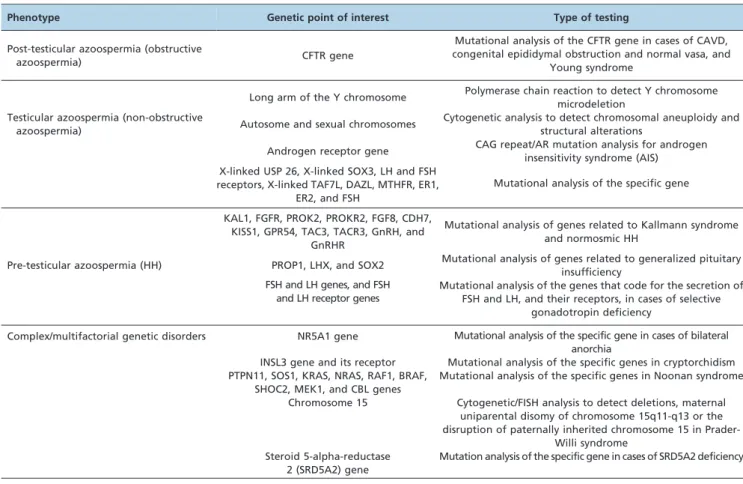
Table 2 summarizes the currently available genetic tests for azoospermic men who seek fertility counseling.
& EXPERT COMMENTARY
The aim of this article was to review the current knowledge of the genetic basis of azoospermia and to highlight the requirement for genetic testing in such conditions. Thousands of single or multiple genes are involved in establishing the male fertility potential, and many others are yet to be revealed. Currently, genetic testing ranges from the chromosomal level to specific gene mutations. Single gene disorders are important for under-standing the etiology of male infertility. Moreover, testing can aid in the improved management of couples who seek genetic counseling prior to conception.
The initial evaluation of infertile men generally com-mences with history taking and a thorough physical examination. These steps are followed by an initial seminal fluid analysis and, if required, endocrine profile testing and imaging analysis. Until approximately two decades ago, the understanding of the genetic basis of infertility was of limited value for it provided only a diagnosis. In the current era of ART, however, genetic testing have emerged as tools of paramount importance in helping clinicians not only to explore the specific genetic background of a disease but also to take the necessary precautions to prevent the transmis-sion of the disease to the offspring via assisted conception. Certain genetic defects are associated with increased morbidity, childhood cancer and genital ambiguity. As such,
Table 2 -Genetic testing that is currently available for the investigation of azoospermia
Phenotype Genetic point of interest Type of testing
Post-testicular azoospermia (obstructive
azoospermia) CFTR gene
Mutational analysis of the CFTR gene in cases of CAVD, congenital epididymal obstruction and normal vasa, and
Young syndrome
Long arm of the Y chromosome Polymerase chain reaction to detect Y chromosome microdeletion
Testicular azoospermia (non-obstructive
azoospermia) Autosome and sexual chromosomes
Cytogenetic analysis to detect chromosomal aneuploidy and structural alterations
Androgen receptor gene CAG repeat/AR mutation analysis for androgen insensitivity syndrome (AIS) X-linked USP 26, X-linked SOX3, LH and FSH
receptors, X-linked TAF7L, DAZL, MTHFR, ER1, ER2, and FSH
Mutational analysis of the specific gene
KAL1, FGFR, PROK2, PROKR2, FGF8, CDH7, KISS1, GPR54, TAC3, TACR3, GnRH, and
GnRHR
Mutational analysis of genes related to Kallmann syndrome and normosmic HH
Pre-testicular azoospermia (HH) PROP1, LHX, and SOX2 Mutational analysis of genes related to generalized pituitary insufficiency
FSH and LH genes, and FSH and LH receptor genes
Mutational analysis of the genes that code for the secretion of FSH and LH, and their receptors, in cases of selective
gonadotropin deficiency
Complex/multifactorial genetic disorders NR5A1 gene Mutational analysis of the specific gene in cases of bilateral anorchia
INSL3 gene and its receptor Mutational analysis of the specific genes in cryptorchidism PTPN11, SOS1, KRAS, NRAS, RAF1, BRAF,
SHOC2, MEK1, and CBL genes
Mutational analysis of the specific genes in Noonan syndrome
Chromosome 15 Cytogenetic/FISH analysis to detect deletions, maternal uniparental disomy of chromosome 15q11-q13 or the disruption of paternally inherited chromosome 15 in
Prader-Willi syndrome Steroid 5-alpha-reductase
2 (SRD5A2) gene
Mutation analysis of the specific gene in cases of SRD5A2 deficiency