Arq Neuropsiquiatr 2008;66(2-A):256-258
256
TransienT ischemic aTTacks in a child
wiTh posT-varicella arTeriopaThy and
mThFr homozigoTic muTaTion c677T
Pedro Beleza
1, João Fernandes
2, Ana Afonso
3, Helena Silva
3, Maria J. Jordão
1acidenTes isquÊmicos TransiTórios em criança com arTeriopaTia pós-varicela e muTação mThFr c677T homozigóTica
São Marcos Hospital, Braga, Portugal: 1MD, Department of Neurology; 2MD, Department of Pediatry; 3MD, Department of Neuroradiology.
Received 23 October 2007, received in inal form 2 January 2008. Accepted 20 February 2008.
Dr. Pedro Beleza – Serviço de Neurologia / Hospital São Marcos - Largo Carlos Amarante, Apartado 2242 - 4701-965 Braga - Portugal. E-mail: [email protected]
Acute arteriopathies represent 79% of stroke in chil-dren1. Chickenpox is the primary etiologic agent involved in 60% of cases and usually carries a good prognosis2. We report on a patient with multiple transients ischemic at-tacks (TIA) who presented a post-varicella artheriopa-thy (PVA) associated with MTHFR homozigotic mutation C677T. Moreover the diagnosis criteria and therapy of PVA is discussed.
case
A three year-old boy was admitted to the emergency room (ER) with sudden onset of transient motor deicit of both left limbs. The neurological and general examination was normal. His past medical history was remarkable for primary varicella infec-tion occurring ive months before. In the ER he underwent blood tests, brain CT and EEG which displayed normal results. While performing EEG, he presented another deicit episode lasting for ive minutes and similar to the one previously described. In the light of multiple TIA’s, anticoagulation with heparin was initiat-ed. However, he had three more TIA’s in the same territory, and at the ifth day after admission had sudden onset of a left pyra-midal syndrome with discrete hemiparesis.
The heart evaluation including electrocardiogram and echo-cardiogram was normal. Noteworthy, concearning the
analyti-cal work-up, we underscore the serologic evidence of a non-re-cent VVZ infection (IgG+, IgM–) and the presence of a MTHFR homozigotic mutation C677T with normal homocystein and folic acid. All the remaining study was normal or negative and includ-ed hemogram, biochemistry, immunologic study, metabolic, pro-trombotic and CSF (cytochemistry, serologic, microbiologic and virus PCR including VVZ). The MRI with Angio-MRI performed at the second day after admission showed a recent stroke in the territory of right lenticulostriate arteries and a decreased low on the M1-M2 transition of right MCA (Fig 1). A brain conven-tional angiography was performed to better study all the brain vascular system including the distal vessels. The intracranial
es-Fig 1. MRI at day 2 after admission reveals recent striatum infarction with T2-hyperintensity (A) and restricted diffusion (B, C).
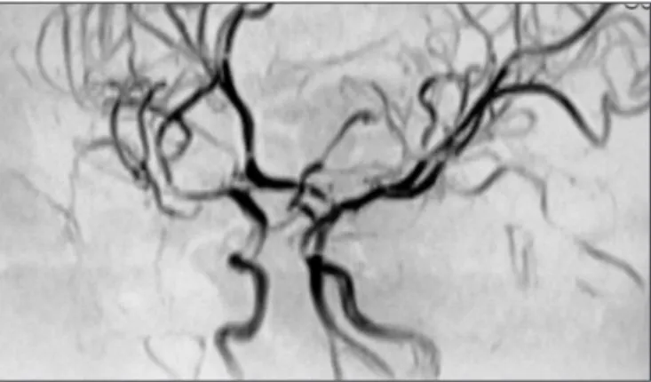
Fig 2. Digital subtraction angiography, right internal carotid injec-tion, frontal projection. Stenosis of distal M1 and proximal M2 seg-ments is conirmed.
Arq Neuropsiquiatr 2008;66(2-A)
257 Transient ischemic attacks Beleza et al.
tenosis of the right MCA was conirmed and other vascular ab-normalities were excluded (Fig 2). Despite showing therapeutic APTT levels he had another similar TIA at the 9th day. Thus, ace-tilsalicilic acid (ASA) 3 mg/Kg/day was associated. As he pre-sented the MTHFR homozigotic mutation C677T it was provided folic acid 2.5 mg/day. The diagnostic suspicion was a post-vari-cella arteriopathy, so he was treated with acyclovir 30 mg/kg/ day for eight days. Since, that time he had no more stroke re-currences. The follow-up MRI with AngioMRI performed at the 21th day yielded identical results to the previous one. He was dis-charged home treated with warfarin, ASA and folic acid. Three months later, a MRI with AngioMRI (Fig 3) revealed a complete reversion of stenosis and thus hypocoagulation was stopped.
discussion
The patient reported presents clinical and imagiologi-cal features compatible with PVA according to Lanthier et al.3: irst ever stroke ocurring within one year after primary varicella; stroke manifestations and cerebral infarct loca-tion consistent with unilateral vascular disease affecting the distal internal carotid arteery (ICA) or proximal seg-ments of anterior caratid artery (ACA) or medium carotid artery (MCA); cerebral vessel imaging showing vascular stenoses of these arterial segments. Moreover, corrobo-rating the diagnosis of PVA, the arteriopathy took a mono-phasic course with subsequent stenosis regression, which was proved to be the most common clinical evolution3. The mode of onset of stroke in our patient was with mul-tiple TIAs, which was demonstrated to predict an underly-ing artheriopathy4. This fact guided us to perform vascular imaging as soon as possible, which led to the diagnosis of PVA in the second day after admission. The follow-up of the arteriopathy was performed with AngioMRI since transcranial Doppler proved to have less sensibility in case of minimal arterial stenoses or in case of lesions located in arterial segments other than M15.
Primary varicella is known to be an important risk fac-tor for cerebral infarction in childhood inasmuch as in idiopathic arterial ischemic stroke (AIS), the incidence of varicella-related strokes increases 6 fold (to 50%) com-pared to population rates6. Although the absolute risk of varicella-associated AIS is estimated at only 1 in 15 0000 children7,8. Variations in immune susceptibility or in the strain of the VZV may account for different susceptibil-ity to varicella-related AIS. However, HLA-B51, a promis-ing immunogenetic marker for predisposition to vascular oclusion in response to an immunological study failed to show association with PVA9. The reasons for that such a small subgroup of children with varicella experience AIS are not yet known. Evidence of varicella-zoster virus with-in the vessel wall of diseased arteries is found at autopsy in children with PVA10 and in adults with cerebral arteritis associated with herpes zoster ophtalmicus11, supporting
that childhood PVA results from viral migration from the trigeminal ganglion and nerve to the major arteries. Re-garding therapy, a review on varicella-associated stroke showed no obvious beneit of antiviral drugs or cortico-steroid therapy because most children recovered nearly completely regardless therapy, although the authors ad-vocated antiviral therapy because of suspected recent vi-ral replication in such cases12. Other studies suggest that anticoagulant therapy in the initial phases of varicella-as-sociated AIS may be helpful in preventing local extension of the thrombus and embolization13.
Additionally, our patient presented MTHFR C677T homozygotic mutation with normal homocystein levels, which was demonstrated to determine susceptibility to ischemic stroke inasmuch as in a large pediatric series (n=148) the OR associated with the TT genotype was 2.614. Despite this associated inding we still considered the di-agnosis of PVA because it presents typical clinical and imagiological features which could not be explained only by the prothrombotic abnormality. The combination with chickenpox might have triggered the occurrence of stroke in our patient. Multiple risk factors were seen in 24% of stroke in children15. Their identiication may represent a change in therapeutic management and also predicts a poor outcome, namely stroke recurrence15. Thus is crucial to perform a complete investigation which should include vascular imaging, cardiac evaluation and prothrombotic testing16. Actually, although stroke in children is com-monly associated with the presence of both genetic and acquired risk factors17 this is one of the rare patients re-ported in the literature with stroke and PVA combined with a prothrombotic disorder18. The rarity of this inding, can be due to the underinvestigation of additional risk factors in children with varicella and stroke which would justify at least partially why only some children with vari-cella develop AIS, an issue that diserves documentation in further studies. In addition, another possible explanation is the clinical use of strict diagnostic criteria as deined for typical PVA by Lanthier et al.3 in a research context, thereby excluding cases with atypical PVA, such as those with multiple possible stroke aetiologies. Unfortunatelly, further studies did not arise the issue of what diagnos-tic criteria should be applied in clinical pracdiagnos-tice. We sug-gest that in clinical grounds PVA might be diagnosed ac-cording to the originally criteria proposed by Lanthier et al.3, although we would not consider it as a diagnosis of exclusion. Actually the co-existence of a metabolic or a prothrombotic disorder with an arteriopathy fulilling all the criteria previously cited of PVA3 should not prevent to perform a diagnose of PVA.
Arq Neuropsiquiatr 2008;66(2-A)
258
Transient ischemic attacks Beleza et al.
with stroke and PVA, which should always include pro-thrombotic testing. Actually the identiication of an asso-ciated prothrombotic abnormality can represent a change in the therapeutic management and also signify stroke recurrence. We also suggest that the clinical diagnosis of PVA should be based in the criteria proposed by Lanthier et al.3, although should not be considered a diagnosis of exclusion.
reFerences
1. Ganesan V, Prengler M, McShane MA, Wade AM, Kirkham FJ. Inves-tigation of risk factors in children with arterial ischemic stroke. Ann Neurol 2003;53:167-173.
2. Guillot M, El Hachem C, Amiour M, et al. [Varicella, acute postinfec-tious arteriopathy and cerebral arterial thrombosis in childhood: a unique clinical and etiologic framework to be fully acknowledged]. Arch Pediatr 2005;12(Suppl 1):S58-S60.
3. Lanthier S, Armstrong D, Domi T, deVeber G. Post-varicella arteriopa-thy of childhood: natural history of vascular stenosis. Neurology 2005; 64:660-663.
4. Braun KP, Rafay MF, Uiterwaal CS, Pontigon AM, DeVeber G. Mode of onset predicts etiological diagnosis of arterial ischemic stroke in chil-dren. Stroke 2007:38:298-302.
5. Rougeot C, Boissier C, Chabrier S. Post-varicella arteriopathy: beneits of using serial transcranial Doppler examinations. Eur J Paediatr Neu-rol 2006;10:152-153.
6. Sebire G, Meyer L, Chabrier S. Varicella as a risk factor for cerebral in-farction in childhood: a case-control study. Ann Neurol 1999;45:679-680.
7. deVeber G. The Canadian Pediatric Ischemic Stroke Study Group. Ca-nadian paediatric ischemic stroke registry: analysis of children with ar-terial ischemic stroke. Ann Neurol 2000;27(H-08).
8. Chickenpox in Canada, 1924-87. Cmaj 1988;138:133-134.
9. Kluger G, Hubmann M, Vogler L, Berz K. Lack of association between childhood stroke after varicella and human leukocyte antigen (HLA)-B51. Eur J Paediatr Neurol 2001;5:259-260.
10. Berger TM, Caduff JH, Gebbers JO. Fatal varicella-zoster virus antigen-positive giant cell arteritis of the central nervous system. Pediatr Infect Dis J 2000;19:653-656.
11. Gilden DH, Kleinschmidt-DeMasters BK, LaGuardia JJ, Mahalingam R . Cohrs RJ. Neurologic complications of the reactivation of varicella-zoster virus. N Engl J Med 2000;342:635-645.
12. Moriuchi H, Rodriguez W. Role of varicella-zoster virus in stroke syn-dromes. Pediatr Infect Dis J 2000;19:648-653.
13. Askalan R, Laughlin S, Mayank S, et al. Chickenpox and stroke in child-hood: a study of frequency and causation. Stroke 2001;32:1257-1262. 14. Nowak-Gottl U, Strater R, Heinecke A et al. Lipoprotein (a) and
genet-ic polymorphisms of clotting factor V, prothrombin and methylenetet-rahydrofolate reductase are risk factors of spontaneous ischemic stroke in childhood. Blood 1999;94:3678-3682.
15. Lanthier S, Carmant L, David M, Larbrisseau A, de Veber G. Stroke in children: the coexistence of multiple risk factors predicts poor outcome. Neurology 2000;54;371-378.
16. Pediatric Stroke Working Group. Stroke in childhood: November 2004. Available at: http://www.rcplondon.ac.uk/pubs/books/childstroke. 17. Barreirinho S, Ferro A, Santos M, et al. Inherited and acquired risk fac-tors and their combined effects in pediatric stroke. Pediatr Neurol 2003; 28:134-138.