Evaluation of Heterozygous Deletion of TP53 Gene
Evaluation of Heterozygous Deletion of TP53
Gene in Pleural Fluid Samples: A Case Series of 11 Patients
Plevral Sıvı Örneklerinde TP53 Geni Heterozigot
Delesyonunun Değerlendirilmesi: 11 Hastalık Bir Seri
DOI: 10.4328/JCAM.4551 Received: 13.04.2016 Accepted: 27.04.2016 Printed: 01.04.2016 J Clin Anal Med 2016;7(suppl 2): 165-7 Corresponding Author: Nigar Dirican, Department of Chest Diseases, Medical Faculty, Suleyman Demirel University, 32260, Isparta, Turkey.
T.: +90 2462119332 E-Mail: [email protected] Özet
Biz, malign hastalığı olmayan 2 hasta (pnömoni) ve malign hastalığı olan 9 has
-tada [Küçük hücreli akciğer karsinomu (n=3), küçük hücreli dışı akciğer karsino
-mu (n=4), non-hodgkin lenfoma (n=1) ve mide kanseri (n=1)] Tümör Protein 53 (TP53) genindeki heterozigot delesyonu tanımladık. Kromozomal aberant durum, sentromer ve 17p13.1 lokusuna özellikli problar kullanılarak loresan in situ hib
-ridizasyon yoluyla analiz edildi. Dokuz kanser hastasının 3’ünde, histolojik değer
-lendirme ve/veya kapalı plevral biyopsi ile malign plevral efüzyon tespit etmedik. TP53 geni heterozigot delesyonu malign hastalığı olanlarda bening plevral efüzyo
-nu olanlara kıyasla belirgin olarak yüksek olarak bulundu. So-nuç olarak, TP53 he
-terozigot delesyonu malignansi için bir belirteç olabileceğini öne sürmekteyiz, bu
-nunla birlikte bu öneriyi desteklemek için büyük hasta kohortları ile ileri çalışma -lar gerekmektedir.
Anahtar Kelimeler
Plevral Efüzyon; Malignite; TP53 Gen
Abstract
We described heterozygous deletion of tumor protein 53 (TP53) gene in 11 pa -tients including 2 tients with non-malignant diseases (pneumonia) and 9
pa-tients with malignant diseases [including small cell lung cancer (n = 3), non-small cell lung carcinoma (n = 4), non-Hodgkin’s lymphoma (n=1), and gastric carci
-noma (n=1)]. Chromosomal aberrant status was analyzed by luorescence in situ hybridization with centromere speciic and 17p13.1 locus speciic probes. In 3 of 9 cancer patients we did not ind malignant pleural efusion with histological examination and/or closed pleural biopsy. Heterozygous deletion of TP53 gene was found to be signiicantly higher in patients with malignant disease when compared to the patients with benign pleural luid. As a result, we suggest that
heterozygous deletion of TP53 may have indicator value for malignancy; however
further studies are warranted to conirm this suggestion in large patient cohorts.
Keywords
Pleural Efusion; Malignancy; TP53 Gene
Nigar Dirican1, Özkan Bagci2, Efkan Uz3, Önder Öztürk1, H.Ahmet Bircan1, Ahmet Dirican4 1Department of Chest Diseases, Faculty of Medicine, Suleyman Demirel University, Isparta, 2Department of Medical Genetics, Faculty of Medicine, Suleyman Demirel University, Isparta, 3Department of Medical Biochemistry, Faculty of Medicine, Suleyman Demirel University, Isparta, 4Department of Medical Oncology, Faculty of Medicine, Celal Bayar University, Manisa, Turkey
Introduction
Malignant pleural efusion (MPE) is a common complication in
patients with advanced cancers, occurring in 15% of
cancer-re-lated deaths. MPE is thought to be caused by the hyper perme
-ability of microvascular tissue or invasion of cancer cells into lymphatic vessels. Most pleural efusions (PEs) occur with con -comitant tumor. However, in a few patients, multiple
cytopatho-logic examinations of pleural luid are negative for tumor [1,2]. Diferential diagnoses of malign and benign pleural efusions
remain a challenge. The accuracy of cytological examination of
the diagnosis PEs is about 60% and pleural biopsy contributes 7–13% [3]. Video-assisted thoracic surgery is the gold standard
procedure in the diagnosis of PEs; however surgical procedures may have several complications. Therefore, avoiding invasive procedures with several serious complications lead clinicians to
try to ind non-invasive tests such as pleural luid biomarkers,
particularly through the genetic analysis of PEs.
In humans, TP53 gene is located on the short arm of
chromo-some 17 (17p13.1) [4]. TP53 is the most altered gene in can
-cer. TP53 mutation was found to be in more than 50% of hu
-man cancers. Mutations disabling the TP53 tumor suppressor
gene represent the most frequent events in human cancer and
typically occur through a double-hit mechanism involving a mis -sense mutation in one allele and a “loss of heterozygosity”
de-letion encompassing the other [5]. Dede-letion of TP53 gene has not been reported to date in the diagnosis of malignant pleural efusion. In the present study, we reported numerical chromo
-somal status in efusion cells derived from 11 patients with malignant and non-malignant diseases by using luorescence in situ hybridization (FISH) with centromere speciic and 17p13.1 locus speciic probes for chromosomes 17.
Material and Method
The present study was carried out at Süleyman Demirel Uni-versity Medical Faculty. All patients were male. Eleven pleural
efusion specimens were derived from 11 patients including 2
patients with non-malignant and 9 patients with malignant
dis-eases. In 3 of 9 patients with malignancy, the pleural efusion samples showed non-malignant feature. Closed pleural biopsy
or medical thoracoscopy performed in these 3 patients
indi-cated there was no malignancy in pleural luid. In all these ma
-lignant cases, metastatic diseases were deined by computed
tomography (CT) or magnetic resonance imaging (MRI). Cyto-logical evaluation was also performed in each sample.
Fluorescence in situ hybridization
Fluorescence in situ hybridization targeting TP53 locus was per
-formed for pleural luid samples. A direct luorochrome-labeled, dual-color DNA probe cocktail (Cytocell, UK) for P53 -17p13.1 and reference loci (D17Z1) was hybridized to slides of pleural luid samples. Hybridizations and washings were carried out ac -cording to the stringency conditions and the procedures
recom-mended by the manufacturers. Dual-color luorescent signals were detected and analyzed under epiluorescence microscopy equipped with speciic ilter sets. At least 200 interphase nuclei were analyzed and scored by independent investigators.
Case Series
The key features of these cases are summarized in Table 1. The
mean age was found as 75.3 years. Of the 9 malignant patients, 3 patients have small cell cancer, 4 patients have non-small cell
cancer, 1 patient has non-Hodgkin’s lymphoma, and 1 patient has gastric cancer. The diagnosis of 2 patients with benign dis -ease was pneumonia. There were lower levels of heterozygous
deletion in TP53 gene in patients with benign disease (cases
10-11) and patients with malign disease non-malignant
pleu-ral luid (cases 1-3) when compared to patients with malignant pleural luid (cases 4-9). TP53 gene deletion ratio in patients with benign pleural efusions was detected at less than 10% while the ratio in patients with malign efusion was found over 10% (table 1, igure 1).
Discussion
Cancer is one of the main causes of exudative PEs. Many of the
biochemical markers in pleural luid are currently being investi
-gated and/or utilized in the clinic. However, the diferential di
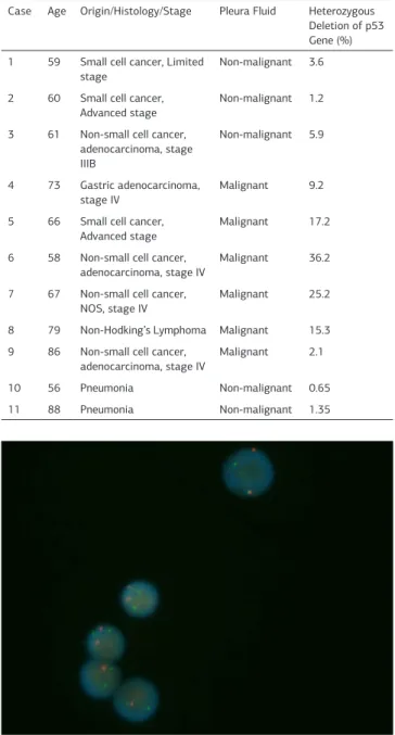
-agnosis is still diicult because the mechanism of PE formation Figure 1. Interphase luorescence in situ hybridization (FISH) results. FISH using P53 (TP53) gene deletion probe, reveals Green(G); chromosome D17Z1 locus,
Red(R); chromosome 17p13.1 (P53 gene). Blue; DAPI. Normal nuclei: 2G/2R, Nuclei with P53 deletion: 2G/1Rv
Table 1. The Heterozygous Deletion of P53 and Patients Characteristics
Case Age Origin/Histology/Stage Pleura Fluid Heterozygous Deletion of p53 Gene (%) 1 59 Small cell cancer, Limited
stage
Non-malignant 3.6
2 60 Small cell cancer, Advanced stage
Non-malignant 1.2
3 61 Non-small cell cancer, adenocarcinoma, stage IIIB
Non-malignant 5.9
4 73 Gastric adenocarcinoma,
stage IV
Malignant 9.2
5 66 Small cell cancer, Advanced stage
Malignant 17.2
6 58 Non-small cell cancer,
adenocarcinoma, stage IV
Malignant 36.2
7 67 Non-small cell cancer,
NOS, stage IV
Malignant 25.2
8 79 Non-Hodking’s Lymphoma Malignant 15.3
9 86 Non-small cell cancer,
adenocarcinoma, stage IV
Malignant 2.1
10 56 Pneumonia Non-malignant 0.65 11 88 Pneumonia Non-malignant 1.35
I Journal of Clinical and Analytical Medicine
166
is multifactorial and not completely understood. Furthermore,
detecting malignant and non-malignant efusion is still a chal
-lenging issue; biological behavior and diferentiation of each malignant cell are diferent. For this purpose, genetic studies of pleural efusion have been reported [6]. But the detection of TP53 gene heterozygous deletion in pleural luid has not been
reported to date. Therefore in the present paper, we
hypoth-esized that heterozygous deletion of TP53 gene could be a ben
-eicial candidate in the diferential diagnosis of MPEs.
We found higher levels of heterozygous deletion of TP53 in
PEs of malignant origin than in PEs of benign origin. However, malignant patients with benign pleural efusion had TP53 gene deletion similar to PEs of benign origin (table 1). Cora et al, found that there is an association between chromosomes an
-euploidies and pleural efusion cell status [6]. The FISH method was used in this study. In another genetic study of pleural luid, p16 gene homozygous deletion in pleural efusion was found to be strongly related to malignant status and higher metastatic potential [7]. The same method was used for genetic testing in
this study.
Fluorescence in situ hybridization is a cytogenetic technique that uses luorescent probes that bind to only those parts of
the chromosome with a high degree of sequence complementa-rity. Its role seems to increase in detecting the characterization of chromosomal rearrangements and marker chromosomes, the detection of micro deletions, and the prenatal diagnosis
of common aneuploidies [8-10]. By using an interphase-FISH technique, speciic genetic aberrations have been reported in efusion samples of patients with cancer [11,12]. The FISH technique, as seen in our series, may be a safe method in the diferential diagnosis of malignant and benign pleural efusion.
P53 is the most altered gene in cancer. More than 50% of
hu-man cancers are alicted with a TP53 mutation. The previous studies suggested that the use of p53-antibodies has potential diagnostic value for several cancers [13,14]. But TP53 mutation has not been evaluated in body luid samples for cancer diag
-nosis. For this reason our results may be important. However, the number of cases is quite small and it is not a homogeneous group. Despite these limitations, we believe that future genetic studies may be planned in the light of the present report.
Competing interests
The authors declare that they have no competing interests.
References
1. Rodriguez-Panadero F, Borderas Naranjo F, LópezMejias J. Pleural metastatic tumours and efusions. Frequency and pathogenic mechanisms in a post-mortem
series. Eur Respir J 1989;2(6):366–9.
2. Goldstraw P, Crowley J, Chansky K, Giroux DJ, Groome PA, Rami-Porta R et al.
The IASLC Lung Cancer Staging Project: Proposals fort the revision of the TNM stage groupings in the forthcoming (seventh) edition of the TNM classiication of
malignant tumors. J Thorac Oncol 2007;2(8):706-14.
3. Fiorelli A, Vicidomini G, Di Domenico M, Napolitano F, Messina G, Morgillo F et al. Vascular endothelial growth factor in pleural luid for diferential diagnosis of benign and malignant origin and its clinical applications. Interact CardioVasc
Thorac Surg 2011;12(3):420–4.
4. McBride OW, Merry D, Givol D. The gene for human p53 cellular tumor anti-gen is located on chromosome 17 short arm (17p13). Proc. Natl. Acad. Sci. U.S.A 1986;83(1):130-4.
5. Liu Y, Chen C, Xu Z, Scuoppo C, Rillahan CD, Gao J et al. Deletions linked to TP53 loss drive cancer through p53-independent mechanisms. Nature 2016;531(7595):471-5.
6. Cora T, Acar H, Ceran S, Bodur S. Analysis of chromosomes 9 and 11 aneuploidy
frequency in pleural efusion of patients with and without malignancy: interphase
FISH technique. Cancer Biol Ther 2005;4(2):248-51.
7. Gui S, Liu H, Zhang L, Zuo L, Zhou Q, Fei G et al. Clinical signiicance of the detection of the homozygous deletion of P16 gene in malignant pleural efusion.
Intern Med 2007;46(15):1161-6.
8. Tsukamoto M, Matsuyama H, Oba K, Yoshihiro S, Takahashi M, Naito K. Numeri
-cal aberrations of chromosome 9 in bladder cancer. A possible prognostic marker
for early tumor recurrence. Cancer Genet Cytogenet 2002;134(1):41-5. 9. Pinkel D, Landegent J, Collins C, Fuscoe J, Segraves R, Lucas J et al.
Fluores-cence in situ hybridization with human chromosome-speciic libraries: detection
of trisomy 21 and translocations of chromosome 4. Proc. Natl. Acad. Sci. USA 1988;85(23):9138–42.
10. Blennow E, Anneren G, Bui TH, Berggren E, Asadi E, Nordenskjold M. Char
-acterization of supernumerary ring marker chromosomes by luorescence in situ hybridization. Am. J. Hum. Genet 1993;53(2):433–42.
11. Jia D, Zhang Z, Liu S, Cheng S. Study on interphase cytogenetic abnormalities in malignant cells in pleural luids from lung cancer cases. Zhonghuo Yi Xue Ke Xue
Yuan Xue Bao 2000;17(4):244-7
12. Johnson TM, Kufel DG, Deward GW. Detection of hyperdiploid malignant cells in pleural efusions with chromosome-speciic probes and luorescence in situ hy
-bridization. Mayo Clin Proc 1996;71(7):643-8.
13. Yang ZC, Ling L, Xu ZW, Sui XD, Feng S, Zhang J. Are p53 Antibodies a Diagnos -tic Indicator for Patients with Oral Squamous Cell Carcinoma? Systema-tic Review and Meta-Analysis. Asian Pac J Cancer Prev 2016;17(1):109-15.
14. Soares JC, Soares AC, Pereira PA, Rodrigues Vda C, Shimizu FM, Melendez ME,
et al. Adsorption according to the Langmuir-Freundlich model is the detection mechanism of the antigen p53 for early diagnosis of cancer. Phys Chem Chem Phys 2016;18(12):8412-8.
How to cite this article:
Dirican N, Bagci Ö, Uz E, Öztürk Ö, Bircan HA, Dirican A. Evaluation of Heterozy-gous Deletion of TP53 Gene in Pleural Fluid Samples: A Case Series of 11 Patients. J Clin Anal Med 2016;7(suppl 2): 165-7.
Journal of Clinical and Analytical Medicine I 167