Case 4366
Sacrococcygeal Teratoma with malignant transformation
Gonçalves M*, Cunha TM**
* Hospital Central do Funchal, Radiology. 9004-514 Funchal, PORTUGAL.
** Instituto Português De Oncologia de Francisco Gentil de Lisboa (IPOFGCROL), Radiology. 1099-023 Lisboa, PORTUGAL.
Hospital Central do Funchal
Section: Genital (Female) Imaging Published: 2005, Dec. 30
Patient: 69 year(s), female
Clinical Summary
Female with a history of surgical excision of a pelvic tumor thirteen years before presented with constipation and obstruction to urinary flow.
Clinical History and Imaging Procedures
epitelial component was adenocarcinoma (Figs. 4 and 5). Following surgery the patient was treated with radiotherapy (150 Gy). At present (4 years after surgery) the patient has no evidence of recurrence.
Discussion
Teratomas, also called dermoid cyst, are congenital tumors comprising elements derived of more than one of the germ layers: endoderm, mesoderm and ectoderm1-6. They originate from pluripotential germ cells, normally located in the ovaries and testis or abnormally located in the midline, as result of sequestered rests during migration of embryonic germ cells from yolk sac to gonads1. Teratomas result from proliferation of tissue foreign to their anatomic site instead of metaplasia. Histopathologically teratomas can be classified in1: a) Mature (benign): well differentiated, predominantly cystic; b) Immature (benign or malignant): undifferentiated, predominantly solid; c) Malignant (germ cell origin): choriocarcinoma, germinoma, yolk sac tumor; d) With malignant transformation (non germ cells elements): carcinoma, sarcoma. Most common locations are: gonads, anterior mediastinum, retroperitoneum, sacrococcygeal, neck and intracranial3,5,6. Sacrococcygeal teratomas (SCT) are the most common teratomas in newborns (1/35 000?40 000 births), and 3?4 times more frequent in females, being rare in adults4,5,6,7. They are classified in four types according to the amount of lesion present internally or externally: type I exclusively external (90% of SCT in newborns/children are externally visible) and type IV exclusively internal (adults)4,7. 60% of SCT are mature and 50-70% are detected in the first days of life. The incidence of malignant SCT in infants is 10-50%. In adults are usually benign4,7. Tendency towards malignancy occurs with increasing age, intrapelvic component and the amount of solid tissue4. SCT may be asymptomatic, cause constipation, obstruction to urinary flow, low back pain, lower extremity paresis/paresthesias, or dystocia1,4. The differential diagnosis of adults' SCT include anterior meningocele, rectal or anal duplication cyst, anal gland cyst, pilonidal cyst, chordoma, neurofibroma, fibrosarcoma and giant cell tumor1,4. On physical examination a palpable mass may be detected, compressing pelvic organs. In malignant germ cell teratoma fetal oncogenes (alpha-fetoprotein, carcinoembryonic antigen and human chorionic gonadotropin) may increase in serum4. Plain films may demonstrate structural abnormalities in coccyx and sacrum or calcifications6. Ultrasound demonstrates a tumor, with cystic and solid components (Rokitansky nodule)3. Fat and calcification, may be difficult to identify5. CT and MRI identify fat and calcium. A fat-fluid level and calcification are highly suggestive. Teeth may exist. Enhancement of solid component is a criterion of malignancy5. Distinguishing mature from immature teratomas is very difficult3. Treatment consists of surgical excision with coccygectomy (coccyx may contain totipotential cells and therefore an increased risk of recurrence)
4,6. Recurrences (7,5-22% to 37% after total resection with or without coccygectomy, respectively) occur
complementary CT or MRI. Serum markers may be of interest in those patients with elevated markers preoperatively4.
Final Diagnosis
Sacrococcygeal teratoma with malignant transformation.
Figures
Figure 1 Fig. 1 - Contrast enhanced pelvic CT.
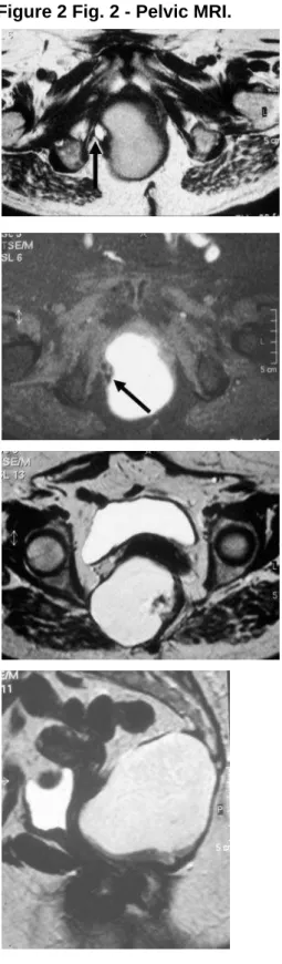
Figure 2 Fig. 2 - Pelvic MRI.
Fig. 2a - Axial T1-weighted image. A thin walled
high-signal mass, anterior to the sacrum, is displacing the
rectum and the vagina anteriorly. A discrete high-signal
intensity lamella, detected at the right periphery of the
cyst (arrow), corresponds to pure fat content, as it is
completely suppressed on a fat-suppressed sequence
(2b).
Fig. 2b - Axial fat-suppressed T1-weighted image. The
high-signal intensity focus on T1-weighted image is
completely suppressed (arrow).
Fig. 2c - Axial T2-weighted image. The lesion has
homogeneous high-signal intensity probably
corresponding to an high protein content cystic mass,
displacing the uterine cervix anteriorly.
Fig. 2e - Axial fat-suppressed T1-weighted scan after
gadolinium administration. The solid parietal vegetation
strongly enhances (arrow)
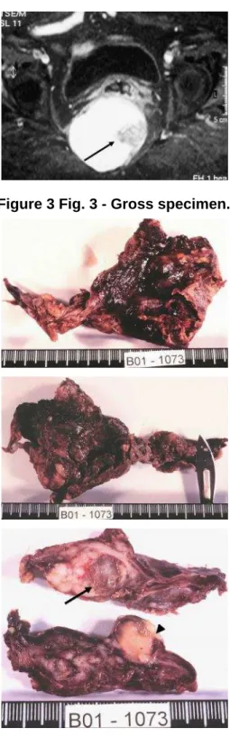
Figure 3 Fig. 3 - Gross specimen.
Fig. 3a - Gross specimen. A 11cm cystic tumor with
coccyx - cystic tumor inner surface.
Fig. 3b - Gross specimen. A 11cm cystic tumor with
coccyx - cystic tumor outer surface.
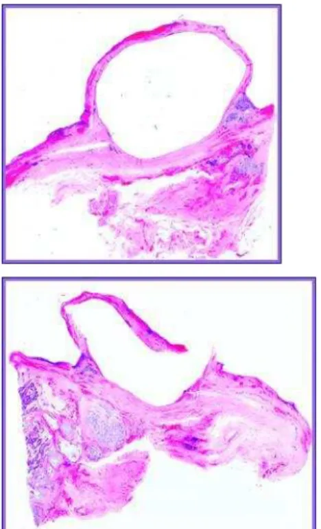
Figure 4 Fig. 4 - Histological section
Fig. 4a - Histological section showing a squamous cell
epithelium covering the small cystic cavity after
removing the content (H&E).
Fig. 4b - Histological section showing a squamous cell
epithelium covering the small cystic cavity after
removing the content (H&E).
Figure 5 Fig. 5 - Microscopic view.
Fig. 5 - The solid area identified on MRI corresponds to
an adenocarcinoma arising in a teratoma (H&E).
MeSH
Teratoma [C04.557.465.910]
A true neoplasm composed of a number of different types of tissue, none of which is native to the area in which it occurs. It is composed of tissues that are derived from three germinal layers, the endoderm, mesoderm, and ectoderm. They may be solid or cystic and are classified histologically as mature, immature, and malignant. (From Dorland, 27th ed & DeVita Jr et al., Cancer: Principles & Practice of Oncology, 3d ed, p1642)
[1] Ng EW, Porcu P, Loehrer PJ Sr. Sacrococcygeal teratoma in adults: case reports and a review of the literature. Cancer. 1999 Oct 1;86(7):1198-202. Review.
[2] Monteiro M, Cunha TM, Catarino A, Tome V. Case report: sacrococcygeal teratoma with malignant transformation in an adult female: CT and MRI findings. Br J Radiol. 2002 Jul;75(895):620-3.
[3] Ueno T, Tanaka YO, Nagata M, et al. Spectrum of germ cell tumors: from head to toe. Diel J, Ortiz O, Losada RA, Price DB, Hayt MW, Katz DS. The sacrum: pathologic spectrum, multimodality imaging, and subspecialty approach. Radiographics. 2001 Jan-Feb;21(1):83-104. Review.
[4] Audet IM, Goldhahn RT Jr, Dent TL. Adult sacrococcygeal teratomas. Am Surg. 2000 Jan;66(1):61-5.
[5] Gatcombe HG, Assikis V, Kooby D, Johnstone PA. Primary retroperitoneal teratomas: a review of the literature. J Surg Oncol. 2004 May 1;86(2):107-13. Review.
[6] Bull J Jr, Yeh KA, McDonnell D, Caudell P, Davis J. Mature presacral teratoma in an adult male: a case report. Am Surg. 1999 Jun;65(6):586-91.
[7] Diel J, Ortiz O, Losada RA, Price DB, Hayt MW, Katz DS. The sacrum: pathologic spectrum, multimodality imaging, and subspecialty approach. Radiographics. 2001 Jan-Feb;21(1):83-104. Review.
Citation
Gonçalves M*, Cunha TM**
* Hospital Central do Funchal, Radiology. 9004-514 Funchal, PORTUGAL.
** Instituto Português De Oncologia de Francisco Gentil de Lisboa (IPOFGCROL), Radiology. 1099-023 Lisboa, PORTUGAL. (2005, Dec. 30)