228 229
228 229
ABSTRACT: Viral coinfection in the oral cavity associated to HIV infection was evaluated in 180 children from birth to 13 years of age of both sexes. The oral examinations were performed at the Pediatric AIDS Outpatient Clinic, São Lucas Hospital and Clinic Hospital, both in Porto Alegre, Brazil and at the School of Dental Medicine, Univer-sity Hospital Center, State UniverUniver-sity of New York at Stony Brook, USA. The aim of this study was to identify the presence of viral infections in the oral cavity. PCR technique was used to determine opportunistic viral infections caused by CMV, EBV, and HSV in mucosal swabs. A high frequency of viral infection was detected in the oral cav-ity of HIV-infected children determined by the PCR technique. HIV-infected children with viruses had a favorable CD4+T lymphocyte count and unfavorable viral load.
DESCRIPTORS: AIDS-related opportunistic infections; Herpes simplex virus; Cytomegalovirus; Epstein-Barr virus; Viral load; CD4-positive T-lymphocytes.
RESUMO: A relação entre a infecção pelo HIV e a presença de diferentes tipos de vírus na cavidade bucal foi estu-dada em 180 crianças HIV-positivo, com idades entre zero e 13 anos de idade, de ambos os sexos. Os exames foram realizados nos Ambulatórios de Aids Pediátrica dos Hospitais São Lucas e de Clínicas, ambos em Porto Alegre, RS, Brasil e no Centro Hospitalar Universitário da Universidade Estadual de Nova Iorque, em Stony Brook (EUA). O objetivo desta pesquisa foi usar a técnica da PCR para detectar a presença dos vírus CMV, EBV e HSV na cavidade bucal desses pacientes, independentemente da presença ou não de manifestações estomatológicas relacionadas aos mesmos. Pode-se concluir que foi alta a freqüência de vírus detectados na cavidade bucal das crianças da amostra através da técnica da PCR e que a contagem média de linfócitos T-CD4+ das crianças com a presença dos
vírus encontrava-se próxima da normalidade, enquanto a Carga Viral do HIV encontrava-se elevada.
DESCRITORES: Infecções oportunistas relacionadas com o HIV; Vírus do herpes simplex; Citomegalovírus; Vírus Epstein-Barr; Carga viral; Linfócitos T CD4-positivos.
* PhD, Professor, Department of Pathology, Federal University of Santa Catarina.
** PhD, Head, Laboratory of Pneumology, Biomedical Research Institute; *******PhD, Head, Doctoral Program in Stomatology – Pontifical Catholic University of Rio Grande do Sul.
*** PhD, Head, Laboratory of Molecular Genetics; ****MD, Department of Pediatrics, Center of Health Science, University Hospi-tal Center; *****DDS, Department of Children’s Dentistry, School of DenHospi-tal Medicine; ******Registered DenHospi-tal Hygienist, MA, School of Dental Medicine – State University of New York at Stony Brook.
INTRODUCTION
Children and adults infected with the Human Immunodeficiency Virus (HIV) are prone to develop opportunistic viral infections in the oral mucosa
mainly by the Herpesviridae family members such as the Cytomegalovirus (CMV), the Epstein-Barr Virus (EBV), and the Herpes Simplex Virus (HSV),
Viral coinfection in the oral cavity of HIV-infected children:
relation among HIV viral load, CD4
+T lymphocyte count and
detection of EBV, CMV and HSV
Co-infecção viral na cavidade bucal de crianças infectadas pelo
HIV: relação entre carga viral, contagem de linfócitos T-CD4
+e
detecção de EBV, CMV e HSV
Liliane Janete Grando* Denise Cantarelli Machado** Silvia Spitzer***
228 229
228 229
Subjects
HIV-infected children between zero and 13 years of age were investigated. This study includ-ed 180 subjects: 143 Brazilians (Pinclud-ediatric AIDS Outpatient Clinic, São Lucas Hospital, Pontifical Catholic University of Rio Grande do Sul, and Clin-ic Hospital, Federal University of Rio Grande do Sul – both in Porto Alegre, Brazil); and 37 Ameri-cans (Pediatric Infectious Disease Clinic, Univer-sity Hospital Center, School of Dental Medicine, State University of New York at Stony Brook, NY, USA). All patients had CD4+T lymphocyte count (expressed in % value/cytometric technique) and HIV viral load (expressed in log10 value/NASBA method) determined.
Oral samples
Oral swabs were collected after obtaining a signed informed consent from the children’s par-ents or guardians. Oral swabs (saliva and epithe-lium cells) were collected with sterile cotton tipped applicators14 (Citmed, Citronelle, AL, USA) from the buccal mucosa bilaterally, regardless of the presence of oral lesions. Specimens were placed in test tubes (Eppendorf AG, Hamburg, Germany) containing 500 µl of sterile phosphate buffered saline (PBS – Merck KGaA, Darmstadt, Germany). DNA was extracted by the organic method (phenol: chloroform:IAA – Invitrogen Corporation, Carlsbad, CA, USA)22.
PCR assay
DNA extracted from the buccal swabs was used to amplify HSV (type 1 and type 2)18, CMV12, and EBV2 by two-step (semi-nested and nested) PCR amplifications. All specimens were tested for the presence of the β-globin gene to ensure the presence of DNA. All PCR reactions had positive (stored DNA obtained from a patient with HSV, CMV or EBV disease) and negative controls (stored DNA obtained from a patient without HSV, CMV or EBV disease) and a reaction mix. Some samples were lost during the laboratory process. The primer sequences used in the PCR reactions are shown in Table 1.
CMV was amplified with CMV-1/CMV-2 in the first and CMV-1/CMV-4 in the second round. EBV was amplified with EBV-1/EBV-2 in the first round and EBV-3/EBV-4 in the second. HSV was ampli-fied with primers HSV-1/HSV-2 in the first round and HSV-3/HSV-4 in the second round. The final products included a 220 bp fragment of the Major all of which are important etiologic agents of
mor-bidity14,19,23. Coinfections of HSV and CMV19; and CMV and EBV23 have been reported in oral ulcers of HIV-infected patients. Despite the unknown pathogenesis of these coinfections20, the detec-tion of more than one virus in the oral mucosa of HIV-infected patients may have important clini-cal implications and, therefore, requires further investigation.
Many authors have investigated bacterial, fungal and viral infections in oral lesions of HIV-infected patients utilizing the Polymerase Chain Reaction (PCR) technique6,13,22. This technique offers several advantages over other methods. It requires a low quantity of biological material16 and can detect the viral presence on “early” infec-tions24. PCR detection of HSV, EBV, and CMV is highly sensitive and specific, which can aid in the prevention of clinical manifestations of virus-as-sociated oral lesions through the selection of the appropriate therapy.
Many tests are used to evaluate the status of the immune system of HIV-infected patients, specially the CD4+T lymphocyte count and the HIV viral load5. The CD4+T lymphocyte count provides an estimation of the immune system status of the HIV-infected individual, and reflects the previous history of the disease20. The CD4+ T-lymphocyte count also indicates the necessity for prophylaxis for opportunist infections and helps to evaluate ini-tial antiretroviral therapy or treatment failure5,8,20. Children without evidence of immunodeficiency have CD4+T lymphocyte count around 25% and many authors have related the oral lesions in HIV-infected patients to low CD4+T lymphocyte count10,15,17.
In addition to the immune system abnormali-ties, HIV-infected Brazilian children can be affected by the lack of appropriate caregiver supervision4. The low education level of caregivers of the HIV-infected Brazilian children can be related to the poor compliance with the antiretroviral treatment, thus affecting the child’s health4,5.
The aim of this research was to detect the presence of some viruses (EBV, CMV and HSV) in the oral cavity of HIV+ children by the PCR tech-nique and study the relation among these virus types with the HIV viral load and CD4+T lympho-cyte count.
METHODS
230 231
230 231
Immediate Early Antigen (MIE) gene from CMV; a 209 bp product of the EBNA-1 gene from EBV and a 142 bp fragment of the D gene of HSV.
The PCR reaction mix contained 0.4 µM of the appropriate primer (Invitrogen Corporation, Carlsbad, CA, USA), 1 X PCR Buffer (Invitrogen Corporation, Carlsbad, CA, USA), 200 µM of each dNTP (Invitrogen Corporation, Carlsbad, CA, USA), 1.25 units of Taq DNA polymerase (Invitro-gen Corporation, Carlsbad, CA, USA) and 1.5 mM (HSV), 2.0 mM (CMV) or 2.5 mM (EBV) of MgCl2 (Invitrogen Corporation, Carlsbad, CA, USA) in
a final volume of 50 µl. Viral DNA, human DNA and reaction controls were included in each run. DNA amplification was performed in an automated thermal cycler (MJ Research, Waltham, MA, USA). Reactions were brought to 95°C for 10 min, fol-lowed by thirty cycles consisting of a denaturing step for 30 s at 94°C, annealing step for 30 s at 50°C (CMV), 60°C (EBV), 50°C (HSV first round) or 60°C (HSV second round), and an extension step for 30 s at 72°C. A final extension step at 72°C was carried out for 5 min. A total of 5 µl of the first round product was used in the second round of amplifications. Aliquots of 15 µl of the PCR product were analyzed on 2% agarose gel (Merck KGaA, Darmstadt, Germany) containing 0.5 g/ml of ethidium bromide (Merck KGaA, Darmstadt, Germany) and visualized under ultraviolet light (Bio-Rad Laboratories Inc., Hercules, CA, USA). The PCR sensitivities were the same in children from Brazil and from the USA: 0.5 pg for CMV; 50 ng for EBV; 5 × 10⁻⁵ pg for HSV.
RESULTS
It was possible to identify HSV in 116 children (79 Brazilians/37 Americans), CMV in 105 chil-dren (68 Brazilians/37 Americans) and EBV infec-tion in 177 children (140 Brazilians/37 Americans) by PCR.
Viral detection by PCR
HSV, CMV and EBV results obtained analyzing the Brazilian and American children are presented in Table 2. According to the Chi-squared test, there were no differences in CMV infection between Bra-zilian and American children (p = 0.110); there were more Brazilian children infected with EBV and HSV than American children (p = 0.001).
Figures 1, 2, and 3 show the reaction prod-ucts of the nested PCR for CMV, EBV and HSV after electrophoresis (Bio-Rad Laboratories Inc.,
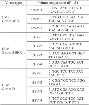
TABLE 1 - Primer sequences* used in the semi-nested and nested PCR technique.
Virus type Primer sequences (5’ - 3’)
CMV Gene: MIE
CMV-1 5’ CAT AAT CTC ATC AGG GGA GC 3’
CMV-2 5’ TTG GGC TAA CTA TGC AGA GC 3’
CMV-4 5’ AGC TGC ATG ATG TGA GCA AG 3’
EBV
Gene: EBNA-1
EBV-1 5’ ATC GTG GTC AAG GAG GTT CC 3’
EBV-2 5’ ACT CAA TGG TGT AAG ACG AC 3’
EBV-3 5’ AAG GAG GGT GGT TTG GAA AG 3’
EBV-4 5’ AGA CAA TGC ACT CCC TTA GC 3’
HSV Gene: D
HSV-1 5’ TGC TCC TAC AAC AAG TC 3’
HSV-2 5’ CGG TGC TCC AGG ATA AA 3’
HSV-3 5’ ATC CGA ACG CAG CCC CGC TG 3’
HSV-4 5’ TCT CCG TCC AGT CGT TTA TCT TC 3’
*The primer sequences were obtained from Krajden et al.12
(1996) (CMV); Cinque et al.2 (1993) (EBV) and Powell et al.18
(1990) (HSV). CMV: Cytomegalovirus; EBV: Epstein-Barr Virus; HSV: Herpes Simplex Virus.
TABLE 2 - Frequency (f) and percentage (%) of Brazilian and American children as detected by PCR.
Virus type Children with different virus types
Brazil f/n (%) USA f/n (%) Total f/n (%)
CMV 13/68 (19.12%) 3/37 (8.11%) 16/105 (15.24%)
EBV 79/140 (56.43%) 10/37 (27.07%) 89/177 (50.28%)
HSV 50/79 (63.29%) 6/37 (16.22%) 56/116 (48.28%)
Total 142/143 (99.30%) 19/37 (51.35%) 161/180 (89.44%)
230 231
230 231
Hercules, CA, USA) in 2% TAE-agarose gel (Merck KGaA, Darmstadt, Germany) containing 0.5 g/ml of ethidium bromide and visualized under ultravio-let light. Table 3 shows the results of the Sensitiv-ity Test for CMV, EBV and HSV.
Figure 4 shows an example of PCR sensitiv-ity (CMV) test. Although there is a difference in the sample size between Brazilian and American patients, when comparing the data obtained from the two countries, no statistical differences were observed between the number of Brazilian and American children with viral infections in the oral cavity (Chi-squared test, p = 0.659).
Relationship between viral infection in the oral cavity; CD4+T lymphocyte count and HIV viral load
The average ± standard deviation of CD4+T lymphocyte count (%) and HIV viral load (log10) were determined to evaluate a possible relation-ship between viral infection in the oral cavity and the general health status of the patient. Table 4 shows that the mean CD4+T lymphocyte count for all patients infected with viruses (HSV, CMV and/or EBV) were higher than 25%, thus suggest-ing no evidence of immunossuppression20. There was no statistical difference between mean CD4+T lymphocyte count of patients with different virus types in their oral cavity as shown by the Variance Analysis (ANOVA) test.
The mean HIV viral load of all patients infected with viruses (HSV, CMV and/or EBV) was higher than 1,000 copies of HIV/ml (Table 5). There were no statistical differences among the mean viral load of patients with different viruses in the oral cavity according to the Variance Analysis (ANOVA).
FIGURE 2 - Amplicons of nested PCR for EBV (Epstein-Barr Virus). Column 1: molecular weight marker VIII. Column 2: positive control. Columns 3 and 6: positive samples. Columns 4, 5 and 7: negative sample. Col-umn 8: negative control.
1
2
3
4
5
6
7
8
EBV (209 bp) 190 bp
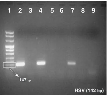
FIGURE 3 - Amplicons of nested PCR for HSV (Herpes Simplex Virus). Column 1: molecular weight marker VIII. Column 2: positive control. Columns 4 and 7: positive samples. Columns 3, 5, 6 and 8: negative sam-ples. Column 9: PCR reaction mix without DNA.
1 2 3 4 5 6 7 8 9
HSV (142 bp) 147 bp
FIGURE 1 - Amplicons of nested PCR for CMV (Cytomeg-alovirus). Column 1: molecular weight marker VIII. Column 2: positive control. Columns 3 and 5: positive samples. Column 4: negative sample. Column 6: emp-ty column. Column 7: negative control. Column 8: PCR reaction mix without DNA.
1
2
3
4
5
6
7
8
190 bp
232 233
232 233
TABLE 3 - Results of the Sensitivity test for CMV, EBV and HSV.
Virus type
Sensitivity Test
Brazil USA
CMV 0,5 pg 0,5 pg
EBV 50 ng 50 ng
HSV 5 × 10⁻⁵ pg 5 × 10⁻⁵ pg
CMV: Cytomegalovirus; EBV: Epstein-Barr Virus; HSV: Herpes Simplex Virus.
FIGURE 4 - PCR sensitivity for CMV (Cytomegalovirus). Column 1: molecular weight marker VIII. Column 2: 10 ng. Column 3: 5 ng. Column 4: 50 pg. Column 5: 0.5 pg. Column 6: 0.05 pg. Column 7: PCR reaction mix without DNA.
1
2
3
4
5
6
7
CMV (220 bp) 190 bp
Table 6 shows the frequency of HSV, CMV and EBV in the oral cavity of our study cohort. It was observed that the number of Brazilian chil-dren (29.37%) without viruses was similar to the number of American children (45.95%), according to the Chi-squared test (p = 0.372). Sixty-three Brazilian children (44.06%) and 17 American chil-dren (45.95%) had at least one virus type present. Seven American (18.92%) and 35 Brazilian chil-dren (24.48%) had two different virus types at the same time. Only 3 Brazilian children (2.10%) had HSV, CMV and EBV oral coinfection.
DISCUSSION
Since CMV viruses are endemic in children populations from developing countries1,24, a higher number of CMV infections was expected in Brazil. The development of the antiviral therapy could be responsible for the control of CMV infection in HIV-infected individuals9. Since 1984, CMV-related ul-cers in HIV-infected children had been reported1,6,9. The pathology of CMV in oral ulcerations remains unknown19.
EBV is the etiologic agent of an illness that can affect children and young adults, called Infectious Mononucleosis3,24. Approximately 90% of the global population has acquired EBV in a non-symptomat-ic way24 and the virus remains latent in the lymph nodes and pharynx cells. In immunodeficient pa-tients, Infectious Mononucleosis could be severe and the EBV recurrence can cause lymphoma21, Kaposi’s Sarcoma24 and hairy leukoplakia7,8.
HSV infection is considered the most frequent viral infection in HIV-infected patients. The early HSV infection and its high frequency in HIV-infect-ed children correlates with the rapid evolution of the disease, suggesting a worsening prognosis for the patient. In immunodeficient patients, as well as in HIV-infected patients, HSV infection can be
severe, clinically atypical, more painful and with a long-term duration11.
Twenty-five percent of CD4+T lymphocyte count means no evidence of immunosuppression. One thousand copies of HIV/ml (log10 = 3.0) is con-sidered a significant viral load20. The absence of clinical manifestations in the HIV-infected children of this study was possibly due to the presence of their high CD4+T lymphocyte count. The children who had viruses detected in their oral cavities had more than 1,585 copies of HIV/ml (log10 = 3.2), which is considered a high viral load20.
Before the AIDS era, Herpesviridae coinfec-tion in human tissue was rare. Presently, this is not the case. Coinfection with HSV and CMV has been described in many anatomic sites, such as the nervous system, skin, esophageal area, and lips. CMV infection often presents severe, painful and long-term duration oral ulcerations19. Viral and fungal coinfection has been also observed, in addition to viral coinfection with CMV and EBV in oral ulcers of HIV-infected patients23.
CONCLUSION
232 233
232 233
load. Multiple viral coinfections were also observed in the oral cavity of our cohort. Early viral infec-tion was detected in the oral cavity of the patients, despite the absence of clinical manifestations.
Considering the complexity of the viral infec-tion therapy in HIV-infected patients, it is very im-portant to identify the opportunistic agents present in the oral cavity as soon as possible. The clinician must consider the caregiver concerns, medication
REFERENCES
1. Britt WJ, Alford CA. Cytomegalovirus. In: Fields BN, Knipe DM, Howley PM, editors. Virology. 3rd ed. Philadelphia:
Lip-pincott-Raven; 1996. p. 2493-512.
2. Cinque P, Brytting M, Vago L, Castagna A, Parravicini C, Zanchetta N, et al. Epstein-Barr virus DNA in cerebroespinal fluid from patients with AIDS-related primary lymphoma of the central nervous system. Lancet 1993;342(8868):398-401. 3. Evans TG. Epstein-Barr Virus Disease. In: Dolin R, Masur
H, Saag MS. AIDS Therapy. New York: Churchill Living-stone; 1999. p. 500-6.
4. Grando LJ, Yurgel LS, Machado DC, Nachman S, Ferguson F, Berentsen B, et al. Associação entre manifestações esto-matológicas e características socioeconômicas e culturais de crianças brasileiras e norte-americanas infectadas pelo HIV. Pan Am J Public Health 2003;14(2):112-8.
history, clinical and laboratory findings in order to provide appropriate care for these children.
ACKNOWLEDGMENTS
Financial support: Pontifical Catholic Univer-sity of Rio Grande do Sul, Porto Alegre, RS, Brazil and State University of New York at Stony Brook, NY, USA.
5. Grando LJ, Yurgel LS, Machado DC, Silva CL, Menezes M, Picolli C. Manifestações estomatológicas, contagem de linfócitos CD4+T e carga viral de crianças brasileiras e norte-americanas infectadas pelo HIV. Pesqui Odontol Bras 2002;16(1):18-25.
6. Greenberg MS, Glick M, Nghiem L, Stewart JCB, Hodinka R, Dubin G. Relationship of cytomegalovirus to salivary gland dysfunction in HIV-infected patients. Oral Surg Oral Med Oral Pathol Oral Radiol Endod 1997;83(3):334-9. 7. Greenspan D, Greenspan JS, Conant M, Petersen V,
Silver-man S Jr, de Souza Y. Oral “hairy” leucoplakia in male ho-mosexuals: evidence of association with both papillomavirus and a herpes-group virus. Lancet 1984;2(8407):831-4. 8. Greenspan JS, Barr CE, Sciubba JJ, Winkler JR. Oral
manifestations of HIV infection. Definitions, diagnostic
TABLE 6 - Frequency (f) and percentage (%) of Brazilian and American children with virus coinfection.
Viral coinfection present in the oral cavity of the same HIV-infected children*
Country Brazil (n = 143)
f/n (%) USA (n = 37)f/n (%) Total (n = 180)f/n (%) 0 virus 42/143 (29.37%) 17/37 (45.95%) 59/180 (32.78%)
1 or more virus types
1 virus type 63/143 (44.06%) 17/37 (45.95%) 80/180 (44.44%) 2 virus types 35/143 (24.48%) 7/37 (18.92%) 42/180 (23.33%) 3 virus types 3/143 (2.10%) - 3/180 (1.67%) *Chi-squared test (p = 0.372).
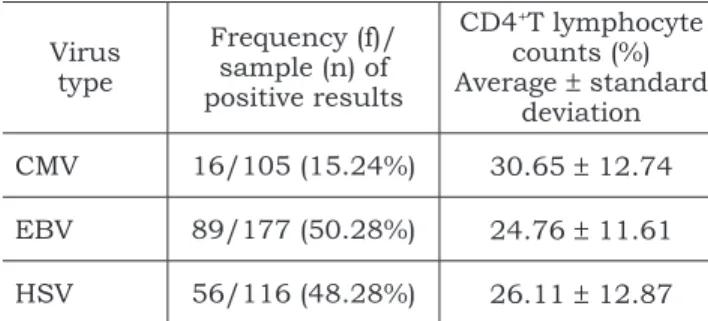
TABLE 4 - Relationship between virus type and CD4+T
lymphocyte counts(in % unit) of Brazilian and Ameri-can children (average ± standard deviation).
Virus type
Frequency (f)/ sample (n) of positive results
CD4+T lymphocyte
counts (%) Average ± standard
deviation CMV 16/105 (15.24%) 30.65 ± 12.74
EBV 89/177 (50.28%) 24.76 ± 11.61
HSV 56/116 (48.28%) 26.11 ± 12.87 CMV: Cytomegalovirus; EBV: Epstein-Barr Virus; HSV: Herpes Simplex Virus.
TABLE 5 - Relationship between virus type and viral load(in log10 unit) of Brazilian and American children
(average ± standard deviation).
Virus type
Frequency (f)/ sample (n) of positive results
Viral Load (log10)
average ± standard deviation CMV 16/105 (15.24%) 4.21 ± 2.30
EBV 89/177 (50.28%) 4.24 ± 0.90
234 PB criteria, and principles of therapy. Oral Surg Oral Med Oral
Pathol Oral Radiol Endod 1992;73(2):142-4.
9. Heinic GS. Oral cytomegalovirus infection in association with HIV infection: a review. In: Greenspan JS, Greenspan D. Oral manifestations of HIV infection. Chicago: Quintes-sence; 1995. p. 225-8.
10. Howell RB, Jandinski JJ, Palumbo P, Shey Z, Houpt MI. Oral soft tissue manifestations and CD4 lympho-cyte counts in HIV-infected children. Pediatr Dent 1996; 18(2):117-20.
11. Kline MW. Oral manifestations of pediatric human immunodeficiency virus infection: a review of the literature. Pediatrics 1997(3);97:380-8.
12. Krajden M, Shankaran P, Bourke C, Lau W. Detection of cytomegalovirus in blood donors by PCR using the digene SHARP signal system assay: effects of sample preparation and detection methodology. J Clin Microbiol 1996;34(1):29-33. 13. Mabruk MJE, Antonio M, Flint SR, Coleman DC,
Toner M, Kay E, et al. A simple and rapid technique for the detection of Epstein-Barr virus DNA in HIV-associated oral hairy leukoplakia biopsies. J Oral Pathol Med 2000;29(3):118-22.
14. MacPhail LA, Hilton JF, Heinic GS, Greenspan D. Di-rect immunofluorescence vs culture for detecting HSV in oral ulcers: a comparison. J Am Dent Assoc 1995;126(1):74-8. 15. Margiotta V, Campisi G, Mancuso S, Accurso V,
Ab-badessa V. HIV infection: oral lesions, CD4+ cell count and viral load in an Italian study population. J Oral Pathol Med 1999;28(4):173-7.
16. Mullis K, Faloona F, Scharf S, Saiki R, Horn G, Erlich H. Specific Enzymatic Amplification of DNA in vitro: the
Polymerase Chain Reaction. Cold Spring Harb Symp Quant Biol 1986;51 Pt 1:263-73.
17. Nicolatou O, Theodoridou M, Mostrou G, Velegraki A, Legakis NJ. Oral lesions in children with perinatally acquired human immunodeficiency virus infection. J Oral Pathol Med 1999;28(2):49-53.
18. Powell KF, Anderson NE, Frith RW, Croxson MC. Non-invasive diagnosis of herpes simplex encephalitis. Lancet 1990;335(8685):357-8.
19. Regezi JA, Eversole LR, Barker BF, Rick GM, Silver-man S Jr. Herpes simplex and cytomegalovirus coinfected oral ulcers in HIV-positive patients. Oral Surg Oral Med Oral Pathol Oral Radiol Endod 1996;81(1):55-62.
20. Revised pediatric HIV classification system. Pediatr Dent 1996;18(2):104-5.
21. Rickinson AB, Kieff E. Epstein-Barr Virus. In: Fields BN, Knipe DM, Howley PM, editors. Virology. 3rd ed.
Phila-delphia: Lippincott-Raven; 1996. p. 2397-436.
22. Sambrook J, Fritsch EF, Maniatis T. Appendix E: Commonly used techniques in molecular cloning. Purifica-tion of Nucleic Acids. In: Sambrook J, Fritsch EF, Maniatis T. Molecular Cloning: a laboratory manual. 2nd ed. New
York: Cold Spring Harbor; 1989. p. E 3-4.
23. Syrjänen S, Leimola-Virtanen R, Schmidt-Westhausen A, Reichart PA. Oral ulcers in AIDS patients frequently as-sociated with cytomegalovirus (CMV) and Epstein-Barr virus (EBV) infections. J Oral Pathol Med 1999;28(5):204-9. 24. Webster-Cyriaque J, Edwards RH, Quinlivan E,
Pat-ton L, Wohl D, Raab-Traub N. Epstein-Barr Virus and Human Herpesvirus 8 prevalence in Human Immunodefi-ciency Virus-associated oral mucosal lesions. J Infect Dis 1997;175(6):1324-32.