Sabrina Carvalho GomeS(a)
Rachel RomaGna(b)
Vanessa RoSSi(b)
Paula Chiattone CoRVello(b) Patrícia Daniela melchiors anGSt(a)
(a) Department of Periodontology, Dental
School, Universidade Federal do Rio Grande do Sul - UFRGS, Porto Alegre, RS, Brazil.
(b) Dental School, Universidade Luterana do
Brasil - ULBRA, Canoas, RS, Brazil.
Supragingival treatment as an aid to
reduce subgingival needs: a 450-day
investigation
abstract: This study investigated the clinical effects of using a
suprag-ingival bioilm control regimen (SUPRA) as a step prior to scaling and root planing (SRP). A split-mouth clinical trial was performed in which 25 subjects with periodontitis (47.2 ± 6.5 years) underwent treatment (days 0–60) and monitoring (days 90–450) phases. At Day 0 (baseline) treatments were randomly assigned per quadrant: SUPRA, SRP and S30SRP (SUPRA 30 days before SRP). The full-mouth visible plaque index (VPI), gingival bleeding index (GBI), periodontal probing depth (PPD), bleeding on probing (BOP), and clinical attachment loss (CAL) were examined on days 0, 30, 60, 90, 120, 270, and 450. Baseline data were similar among all groups. From days 0 to 60, the groups showed similar signiicant decreases in VPI and GBI. Reductions in PPD for the SRP (3.39 ± 0.17 to 2.42 ± 0.16 mm) and S30SRP (3.31 ± 0.11 to 2.40 ± 0.07 mm) groups were greater (p< 0.05) than those for the SUPRA group. This pattern was also observed for BOP. Attachment gain was simi
-lar and greater for the SRP (3.34 ± 0.28 to 2.58 ± 0.26 mm) and S30SRP (3.25 ± 0.21 to 2.54 ± 0.19 mm) groups compared to the SUPRA group. Results were maintained from day 90 forward. Overall, the S30SRP treatment reduced the subgingival treatment needs in 48.16%. Perfor -mance of a SUPRA step before SRP decreased subgingival treatment needs and maintained the periodontal stability over time.
Keywords: Clinical Trial; Dental Scaling; Root Planing.
introduction
Subgingival bioilm control is a sine qua non condition for successful
periodontal treatment.1,2 However, supragingival bioilm control has been strongly associated with the long-term maintenance of subgin
-gival treatment outcomes.3,4 Some authors have noted the importance of the supragingival condition in modulating the subgingival area.5,6 However, the exact mechanisms underlying this relationship are not completely understood.
Therapies focused solely on supragingival control7,8 have been demon
-strated to signiicantly reduce subgingival inlammatory markers, such as bleeding on probing (BOP) and periodontal probing depth (PPD). Use of a supragingival bioilm control regimen (SUPRA) reduced the PPD by an average of 2.4 mm in sites with 6.6 mm of PPD initially.7 This reduc-tion is somewhat comparable or even greater than those achieved by sub
-Declaration of interests: The authors certify that they have no commercial or associative interest that represents a conflict of interest in connection with the manuscript.
Corresponding author:
Sabrina Carvalho Gomes
E-mail: [email protected]
http://dx.doi.org/10.1590/S1806-83242014.50000004 Epub Jan 24, 2014
Submitted: Dec 03, 2012
gingival instrumentation. For example, Darby et al.9 reduced PPD by 1.5 ± 1.4 mm in sites with 5.9 ± 1.3 mm of PPD initially, after scaling and root planing (SRP). Similarly, Silva et al.10 report a reduction of 1.36 mm in deep pockets treated with a combination of SRP plus metronidazole and of 1.77 mm with SRP plus amoxicillin. In addition to improving inlam
-matory markers, SUPRA7 produced signiicant gains in clinical attachment, which is an important tool for monitoring long-term outcomes.11
Despite the importance of these observations, the complementary effects of SUPRA performed before and separately from the SRP intervention remain unexplored. It was hypothesized that SUPRA may
be helpful to reduce the subgingival intervention
needs. This study evaluated the effect of a supragin
-gival control regimen as a step performed as a prior intervention to SRP.
methodology
Study design and sample selection
This randomized, blinded, split-mouth clinical
trial included 25 patients selected consecutively at
Universidade Luterana do Brasil - ULBRA (May 2008 to October 2009). The sample size was calculated con
-sidering a mean difference of 1.0 ± 1.0 mm in PPD reduction,7 power of 80%, and α of 5%. The inclusion criteria were as follows:
• absence of systemic conditions that could inter
-fere with periodontal indicators (e.g., diabetes, cardiac conditions requiring antibiotic prophy
-laxis, etc.);
• no chemical control of supragingival bioilm; • no antibiotic or anti-inlammatory therapy in
the 3 months preceding inclusion in the study; • no pregnancy;
• ≥ 4 teeth in each quadrant, not including 3rd molars, teeth with furcation involvement, or endoperiodontal lesions;
• ≥ 2 sites (not in the same tooth and from dif
-ferent quadrants) with a diagnosis of bioilm-induced gingivitis and chronic periodontitis, with ≤ 2 mm difference in PPD and CAL be
-tween sites; and
• absence of conditions that could interfere with long-term treatment response.
ethical considerations
This study was approved by the Ethics Com
-mittee of Universidade Luterana do Brasil (ULBRA; 2007-324H). Participants signed an informed con
-sent form prior to inclusion in the study. Patients were monitored according to progression of clinical attachment loss (CAL).12
experimental interventions
treatment allocation
The following protocols were randomly assigned after the exams on day 0 (baseline). An author who was not involved with the clinical procedures (SCG) drew 2 coins to make the assignments:
• SUPRA (one quadrant): supragingival control as
the sole intervention;
• SRP (two quadrants): SUPRA plus subgingival control at day 0;
• S30SRP (one quadrant): SUPRA control for 30 days, followed by SRP intervention.
Data collection
Two examiners who were blinded to group allo
-cation (PC and VR) collected data on days 0, 60, 90, 120, 270, and 450, in the full mouth at 6 sites per tooth. The weighed kappa for interexaminer agreement was 0.75 for PPD and 0.68 for CAL (± 1 mm). The follow
-ing parameters were assessed: visible plaque (VPI) and gingival bleeding indexes (GBI),13 PPD, CAL, and BOP (Williams probe; Neumar, São Paulo, Bra
-zil). The PPD and CAL values were rounded up to the nearest millimeter.
treatment (days 0–60) and monitoring (days 90–450) phases
From days 0 to 60, appointments were conducted by one specialist in periodontics (RR); from days 90 to 450, they were conducted by another specialist (VR). All appointments were completed within a 45- to 60-minute session. During treatment, SUPRA consisted of calculus removal and oral hygiene instructions. SRP consisted of subgingival hand instrumentation under local anesthesia (Gracey curettes, Hirschfeld iles; Neumar, São Paulo, Brazil).
seen at an average interval of 2.2 ± 1.64 months. Throughout the study, patients underwent oral hygiene instructions on an individual basis and were given a soft multitufted toothbrush (Colgate Palmolive,
São Paulo, Brazil), interdental brush, and/or dental loss, according to their individual needs. No adverse effects were observed during the study.
Statistical analysis
Three analytic strategies were considered: 1. overall means, considering 6 sites in each tooth; 2. analysis of PPD, BOP, and CAL means, accord
-ing to PPD categories (1–3 mm, 4–6 mm, and 7+ mm) at day 0; and
3. analysis considering the worst site of each tooth.
Intra- and inter-group comparisons were con
-ducted. An analysis considering the smoking habit was also performed. In case of losses in follow-up, intention-to-treat analysis was used.
Linear models were used to calculate means and standard errors (SEs). Huber-White’s sandwich vari
-ance estimator was used to adjust SEs for intracluster correlation. Wald’s tests were used to estimate p
-val-ues, which were adjusted for multiple comparisons. All statistical analyses (signiicance set at 5%) were performed by using Stata SE 10.1 software (Stata Corporation, College Station, USA).
Results
No patients were lost due to CAL progression. Of the 25 patients, three were lost to follow-up, due to changed address, refusal to attend follow-up vis
-its, and pregnancy. Two patients did not attend the 450-day follow-up visit. The carry-forward technique was used in all of these cases, with data collected at the 60- and 270-day visit, respectively, being repeated. The inal sample comprised 25 patients (mean age, 47.24 ± 6.47 years): 18 females (72%) and 14 smokers (56%). In a comparative analysis between smokers and never-smokers, no signiicant differences were observed for periodontal indicators during the study.
Supragingival assessment revealed signiicant and similar reductions in the percentage of VPI between days 0 and 60 for all groups. These results were main
-tained until day 270. At day 450, a difference in VPI
was observed between the SRP and S30SRP groups. However, this difference did not have an impact on GBI, which showed no differences between the groups (data not shown).
Table 1 shows the absence of differences in subgin
-gival indicators among all groups at day 0. All treat
-ments yielded reductions in PPD during the treat
-ment phase (days 0–60; p < 0.05), with the greatest reductions being observed for the SRP and S30SRP groups. Results were maintained throughout the study. The BOP signiicantly decreased in all groups, with more pronounced results for the SRP and S30SRP. The CAL data also showed signiicant overall reduc
-tions from 0 to 60 days, which were maintained over the study, with greater reductions observed for the SRP and S30SRP.
When the PPD categories were analyzed (Table 2), the greatest reductions were observed for the SRP and S30SRP groups, with no differences between them. However, the mean PPD values in the SUPRA group were also reduced signiicantly. This analy
-sis by category also showed improvements in BOP for all groups, with the best results in the SRP and S30SRP for the 4–6 mm and 7+ mm sites. Reduc
-tions were maintained throughout the experimen
-tal period. At day 450, signiicant differences were observed between the SUPRA and S30SRP for both the 4–6 mm and 7+ mm PPD categories, although the SUPRA and SRP were statistically similar (Table 2). In the 4–6 mm category, gains in clinical attachment were signiicantly greater in the SRP and S30SRP com
-pared to the SUPRA. The same pattern was observed for the 7+ mm PPD category (Table 2). When only the worst site of each tooth was included in the analysis, no statistical difference between the 3 groups was observed over the study period (Table 3).
Discussion
The present study describes the results from treatment and monitoring phases of 3 periodon
-ter was also able to promote reductions and to main
-tain the results over time. Finally, performance of the SUPRA regimen prior to subgingival control (S30SRP) reduced the subgingival needs by about 48% as measured by the reduction of number of sites with BOP.
The main advantage of the present experimental design is that each patient served as his or her own control, which enabled us to eliminate interindi
-vidual variations in periodontal healing.14 Hujoel15 suggested that, in a split-mouth study, it is impor
-tant to ensure that sites being submitted to different therapies have the same clinical condition at base
-line. In the present study, attention was given to this issue, and subjects were included if they had simi
-lar periodontal conditions in quadrants (i.e., ≤ 2 mm of difference in PPD and CAL values between the sites). The baseline data underscore the similarity between the groups.
A signiicant reduction in VPI was observed for all groups, which remained low throughout the study. GBI was also reduced in a similar and signiicant pattern and remained without intergroup differ
-ences over the experiment. These indings suggest that the data did not suffer from the carry-across effect; the presence of sites not submitted to SRP did not interfere with the supragingival bioilm forma
-tion, which may, in turn, inluence the subgingival environment.5-7
Full-mouth analysis showed a reduction in all subgingival indicators (p < 0.05) for all groups. Although the greatest reductions were detected in groups treated with SRP, supragingival control alone yielded signiicant reductions, comparable to those observed in other studies that employed subgin
-gival intervention. Santos et al.16 observed a mean estimated reduction in PPD of 3.3 to 2.7 mm after 3 months after SRP plus antibiotics. Our mean PPD table 1. Mean ± standard error for periodontal indicators according to the experimental groups during the study.
Variable Day SUPRA SRP S30SRP
PPD 0 3.55 ± 0.19Aa 3.39 ± 0.17Aa 3.31 ± 0.11Aa
30 3.19 ± 0.19Ab 2.71 ± 0.17Bb 2.87 ± 0.12Bb 60 2.86 ± 0.18Ac 2.42 ± 0.16Bc 2.40 ± 0.07Bc 90 3.00 ± 0.19A 2.52 ± 0.15B 2.36 ± 0.07B 120 2.95 ± 0.18A 2.45 ± 0.14B 2.29 ± 0.07B 270 2.78 ± 0.17A 2.30 ± 0.11B 2.17 ± 0.07B 450 2.88 ± 0.19A 2.36 ± 0.10B 2.32 ± 0.08B
BOP 0 72.38 ± 3.52Aa 72.55 ± 4.58Aa 72.93 ± 2.66Aa
30 41.43 ± 3.70Ab 25.00 ± 2.26Bb 37.61 ± 3.25Ab 60 31.31 ± 4.75Ac 16.86 ± 2.90Bc 21.15 ± 2.08Bc 90 30.25 ± 4.03A 30.93 ± 4.32A 22.00 ± 2.60A 120 27.73 ± 3.72A 25.57 ± 3.46A 21.59 ± 3.29A
270 20.33 ± 2.50A 17.59 ± 2.36A 12.31 ± 1.60B 450 30.91 ± 4.16A 20.27 ± 2.43B 19.88 ± 2.86B
CAL 0 3.50 ± 0.23Aa 3.34 ± 0.28Aa 3.25 ± 0.21Aa
30 3.29 ± 0.23Ab 2.83 ± 0.26Bb 3.03 ± 0.21Ab 60 3.02 ± 0.23Ac 2.58 ± 0.26Bc 2.54 ± 0.19Bc 90 3.06 ± 0.22A 2.48 ± 0.22B 2.47 ± 0.18B 120 2.95 ± 0.21A 2.42 ± 0.21B 2.41 ± 0.17B 270 2.87 ± 0.21A 2.38 ± 0.21B 2.34 ± 0.17B 450 3.02 ± 0.23A 2.48 ± 0.22B 2.47 ± 0.18B
for all groups decreased from 3.4 to 2.6 mm (mean reduction of 0.84 mm). Considering only the SRP and S30SRP groups, an even higher mean reduction (1.22 mm) was obtained.
The PPD category analyses showed a similar reduction between groups for the 1–3 mm sites (data not shown). The results observed for the 4–6 mm and 7+ mm sites showed the greatest reductions in PPD and CAL means at sites submitted to SRP and S30SRP. Nevertheless, at the 4–6 mm sites, SUPRA determined a PPD reduction of 1.09 mm. This mean reduction is similar to that reported by Ioannou et al.,17 who observed a 1.28 mm reduction using ultrasonic devices at the subgingival area. In the 7+ mm sites, the reductions in PPD (3.59 mm) and CAL (2.87 mm) observed for the SRP and S30SRP groups together are comparable to those reported by Santos et al.16 with systemic antibiotics (3.48 mm and 2.82 mm, respec
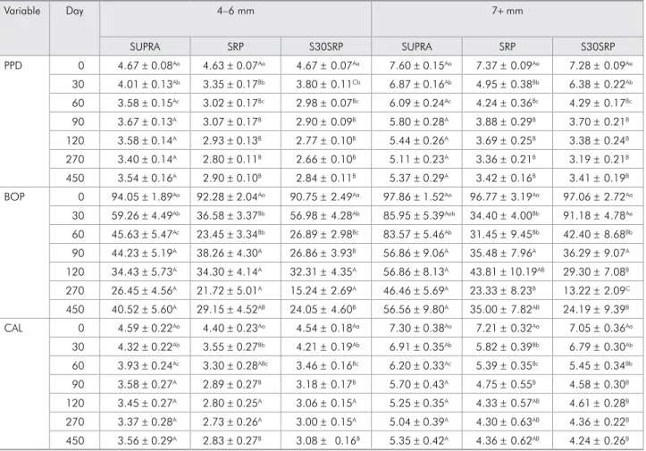
-table 2. Mean ± standard error for periodontal indicators according to PPD at baseline.
Variable Day 4–6 mm 7+ mm
SUPRA SRP S30SRP SUPRA SRP S30SRP
PPD 0 4.67 ± 0.08Aa 4.63 ± 0.07Aa 4.67 ± 0.07Aa 7.60 ± 0.15Aa 7.37 ± 0.09Aa 7.28 ± 0.09Aa 30 4.01 ± 0.13Ab 3.35 ± 0.17Bb 3.80 ± 0.11Cb 6.87 ± 0.16Ab 4.95 ± 0.38Bb 6.38 ± 0.22Ab 60 3.58 ± 0.15Ac 3.02 ± 0.17Bc 2.98 ± 0.07Bc 6.09 ± 0.24Ac 4.24 ± 0.36Bc 4.29 ± 0.17Bc 90 3.67 ± 0.13A 3.07 ± 0.17B 2.90 ± 0.09B 5.80 ± 0.28A 3.88 ± 0.29B 3.70 ± 0.21B 120 3.58 ± 0.14A 2.93 ± 0.13B 2.77 ± 0.10B 5.44 ± 0.26A 3.69 ± 0.25B 3.38 ± 0.24B 270 3.40 ± 0.14A 2.80 ± 0.11B 2.66 ± 0.10B 5.11 ± 0.23A 3.36 ± 0.21B 3.19 ± 0.21B 450 3.54 ± 0.16A 2.90 ± 0.10B 2.84 ± 0.11B 5.37 ± 0.29A 3.42 ± 0.16B 3.41 ± 0.19B BOP 0 94.05 ± 1.89Aa 92.28 ± 2.04Aa 90.75 ± 2.49Aa 97.86 ± 1.52Aa 96.77 ± 3.19Aa 97.06 ± 2.72Aa
30 59.26 ± 4.49Ab 36.58 ± 3.37Bb 56.98 ± 4.28Ab 85.95 ± 5.39Aab 34.40 ± 4.00Bb 91.18 ± 4.78Aa 60 45.63 ± 5.47Ac 23.45 ± 3.34Bb 26.89 ± 2.98Bc 83.57 ± 5.46Ab 31.45 ± 9.45Bb 42.40 ± 8.68Bb 90 44.23 ± 5.19A 38.26 ± 4.30A 26.86 ± 3.93B 56.86 ± 9.06A 35.48 ± 7.96A 36.29 ± 9.07A
120 34.43 ± 5.73A 34.30 ± 4.14A 32.31 ± 4.35A 56.86 ± 8.13A 43.81 ± 10.19AB 29.30 ± 7.08B 270 26.45 ± 4.56A 21.72 ± 5.01A 15.24 ± 2.69A 46.46 ± 5.69A 23.33 ± 8.23B 13.22 ± 2.09C 450 40.52 ± 5.60A 29.15 ± 4.52AB 24.05 ± 4.60B 56.56 ± 9.80A 35.00 ± 7.82AB 24.19 ± 9.39B CAL 0 4.59 ± 0.22Aa 4.40 ± 0.23Aa 4.54 ± 0.18Aa 7.30 ± 0.38Aa 7.21 ± 0.32Aa 7.05 ± 0.36Aa
30 4.32 ± 0.22Ab 3.55 ± 0.27Bb 4.21 ± 0.19Ab 6.91 ± 0.35Ab 5.82 ± 0.39Bb 6.79 ± 0.30Ab 60 3.93 ± 0.24Ac 3.30 ± 0.28ABc 3.46 ± 0.16Bc 6.20 ± 0.33Ac 5.39 ± 0.35Bc 5.45 ± 0.34Bb 90 3.58 ± 0.27A 2.89 ± 0.27B 3.18 ± 0.17B 5.70 ± 0.43A 4.75 ± 0.55B 4.58 ± 0.30B 120 3.45 ± 0.27A 2.80 ± 0.25A 3.06 ± 0.15A 5.25 ± 0.35A 4.33 ± 0.57AB 4.61 ± 0.28B 270 3.37 ± 0.28A 2.73 ± 0.26A 3.00 ± 0.15A 5.04 ± 0.39A 4.30 ± 0.63AB 4.36 ± 0.22B 450 3.56 ± 0.29A 2.83 ± 0.27B 3.08 ± 0.16B 5.35 ± 0.42A 4.36 ± 0.62AB 4.24 ± 0.26B
Different uppercase letters: significant differences between groups; different lowercase letters: significant differences within group.
tively). Again, the SUPRA quadrants showed obvi
-ous reductions, similar to those reported for subgin
-gival treatment.10,17,18
A signiicant improvement in BOP was observed in all groups. From days 0 to 60, all groups showed
table 3. Periodontal indicators considering the worst site of each tooth (day 450 minus day 90).
Variable* SUPRA SRP S30SRP
PPD -0.22 ± 0.16A -0.20 ± 0.14A -0.10 ± 0.13A (p = 0.17a) (p = 0.16a) (p = 0.76a) BOP -2.08 ± 5.58A -11.69 ± 6.81A -2.30 ± 5.17A
(p = 0.71b) (p = 0.10b) (p = 0.66b) CAL -0.19 ± 0.13A -0.01 ± 0.19A -0.07 ± 0.08A
(p = 0.15c) (p = 0.96c) (p = 0.40c)
a reduction in the percentage of sites with BOP (p < 0.05), although the best results were obtained in the SRP group (reductions from 56.8% to 76.8%). The latter results, which were maintained from day 90 forward, are similar to those reported in a study based on SRP plus antibiotics (reductions from 50% to 75%).18 Comparing the percentage of BOP at the 4–6 mm sites, the present indings showed a reduc
-tion from 51.48% to 74.58%. This pattern was also observed in the 7+ mm sites, where SRP and S30SRP together had a mean reduction of 51.95%, similar to data associated with subgingival control plus anti
-biotic therapies.18
Traditionally, it has been suggested that exclu -sive supragingival control is not effective for
clini-cal attachment maintenance.19 Nevertheless, Gomes et al.7 showed that an adequate supragingival regi
-men permitted clinical attach-ment gain. In the pres
-ent study, even sites with deep pockets (7+ mm at baseline) beneited from supragingival control and gained attachment throughout the 450 days.
Overall, considering the presence of BOP as the primary indicator for subgingival intervention,20 the treatment applied to the S30SRP group reduced the
number of sites requiring such intervention by 48.16% compared to the SRP group. At baseline, 72.55% of the sites in the SRP had BOP and received subgingi
-val SRP (i.e., periodontitis treatment). On the other hand, only 37.61% of sites from the S30SRP demon
-strated BOP after SUPRA and received the SRP inter
-vention. The same comparison in the 4–6 mm sites showed an important decrease of 38.25% in subgin
-gival treatment needs (Table 2). As at day 450, the SRP and S30SRP did not differ regarding BOP, but the SUPRA and SRP were similar. Thus, the S30SRP group may have had an even smaller percentage of sites with BOP. This inding underscores that prior well-performed SUPRA control not only reduces subgingival treatment needs, but also maintains this condition over time.
Conclusions
Adequate supragingival control permits long-term stability in the subgingival environment. If performed as a separate and prior step relative to subgingival intervention, SUPRA may substantially reduce the subgingival treatment needs in patients with gingivitis and periodontitis.
References
1. Jervoe-Storm PM, Semaan E, AlAhdab H, Engel S, Fimmers R, Jepsen S. Clinical outcomes of quadrant root planing versus full-mouth root planing. J Clin Periodontol. 2006 Mar;33(3):209-15.
2. Rhemrev GE, Timmerman MF, Veldkamp I, Van Winkelhoff AJ, Van der Velden U. Immediate effect of instrumentation on the subgingival microflora in deep inflamed pockets under strict plaque control. J Clin Periodontol. 2006 Jan;33(1):42-8. 3. Axelsson P, Nystrom B, Lindhe J. The long-term effect of a
plaque control program on tooth mortality, caries and peri
-odontal disease in adults. Results after 30 years of mainte
-nance. J Clin Periodontol. 2004 Sep;31(9):749-57.
4. Heasman PA, McCracken GI, Steen N. Supportive periodon
-tal care: the effect of periodic subgingival debridement compared with supragingival prophylaxis with respect to clinical outcomes. J Clin Periodontol. 2002;29 Suppl 3:163-72; discussion 195-6.
5. Haffajee AD, Teles RP, Socransky SS. The effect of periodontal therapy on the composition of the subgingival microbiota. Periodontol 2000. 2006 Oct;42(1):219-58.
6. Feres M, Gursky LC, Faveri M, Tsuzuki CO, Figueiredo LC. Clinical and microbiological benefits of strict supragingi
-val plaque control as part of the active phase of periodontal therapy. J Clin Periodontol. 2009 Oct;36(10):857-67.
7. Gomes SC, Piccinin FB, Susin C, Oppermann RV, Marcan
-tonio RA. Effect of supragingival plaque control in smok
-ers and never-smok-ers: 6-month evaluation of patients with periodontitis. J Periodontol. 2007 Aug;78(8):1515-21. 8. Angst PD, Piccinin FB, Oppermann RV, Marcantonio RA,
Gomes SC. Response of molars and non-molars to a strict supragingival control in periodontal patients. Braz Oral Res. 2013 Jan-Feb;27(1):55-60.
9. Darby IB, Mooney J, Kinane DF. Changes in subgingival microflora and humoral immune response following peri
-odontal therapy. J Clin Periodontol. 2001 Aug;28(8):796-805. 10. Silva MP, Feres M, Sirotto TA, Soares GM, Mendes JA, Faveri
M, et al. Clinical and microbiological benefits of metronida
11. Mombelli A. Clinical parameters: biological validity and clinical utility. Periodontol 2000. 2005 Oct;39(1):30-9. 12. Haffajee AD, Socransky SS, Goodson JM. Comparison of
different data analyses for detecting changes in attachment level. J Clin Periodontol. 1983 May;10(3):298-310.
13. Ainamo J, Bay I. Problems and proposals for recording gin
-givitis and plaque. Int Dent J. 1975 Dec;25(4):229-35. 14. Lesaffre E, Garcia Zattera MJ, Redmond C, Huber H, Needle
-man I. Reported methodological quality of split-mouth stud
-ies. J Clin Periodontol. 2007 Sep;34(9):756-61.
15. Hujoel PP. Design and analysis issues in split mouth clinical trials. Community Dent Oral Epidemiol. 1998 Apr;26(2):85-6. 16. Santos VR, Ribeiro FV, Lima JA, Miranda TS, Feres M, Bastos
MF, et al. Partial- and full-mouth scaling and root planing in type 2 diabetic subjects: a 12-mo follow-up of clinical param
-eters and levels of cytokines and osteoclastogenesis-related factors. J Periodontal Res. 2012 Feb;47(1):45-54.
17. Ioannou I, Dimitriadis N, Papadimitriou K, Sakellari D, Vouros I, Konstantinidis A. Hand instrumentation versus ultrassonic debridement in the treatment of chronic peri
-odontitis. A randomized clinical and microbiological trial. J Clin Periodontol. 2009 Feb;36(2):132-41.
18. Feres M, Haffajee AD, Allard K, Som S, Socransky SS. Change in subgingival microbial profiles in adult peri
-odontitis subjects receiving either systemically-admin
-istered amoxicillin or metronidazole. J Clin Periodontol. 2001 Jul;28(7):597-609.
19. Kaldahl WB, Kalkwarf KL, Patil KD, Molvar MP, Dyer JK. Long-term evaluation of periodontal therapy: I. Response to 4 therapeutic modalities. J Periodontol. 1996 Feb;67(2):93-102. 20. Brochut PF, Marin I, Baehni P, Mombelli A. Predictive value