Déborah Campos Telles Wagner Henriques Castro Ricardo Santiago Gomez Giovanna Ribeiro Souto Ricardo Alves Mesquita
Department of Oral Surgery and Pathology, School of Dentistry, Universidade Federal de Minas Gerais - UFMG, Belo Horizonte, MG, Brazil.
Corresponding Author: Giovanna Ribeiro Souto E-mail: [email protected]
Morphometric evaluation of keratocystic
odontogenic tumor before and after
marsupialization
Abstract: The aim of the present study was the morphometric evalua-tion of the epithelial lining and ibrous capsule in histological specimens of keratocystic odontogenic tumors (KOTs) before and after marsupial-ization. Histological sections from six KOTs that had undergone mar-supialization followed by enucleation were photographed. The thickness and features of the capsule and of the epithelial lining of the tumor were evaluated upon marsupialization and upon subsequent enucleation using Axion Vision software. The histological specimens taken upon marsu-pialization presented an epithelial lining that is typical of KOTs. After marsupialization, the enucleated specimens had a modiied epithelial lin-ing and a ibrous capsule that both presented a greater median thickness (p = 0.0277 and p = 0.0212, respectively), morphological changes, and signiicant enlargement. These modiications can facilitate full surgical treatment and may well be related to a low KOT recurrence rate.
Descriptors: Surgery, Oral; Odontogenic Tumors; Histology.
Introduction
A keratocystic odontogenic tumor (KOT) is a benign neoplasm of the jaws that originates either from a dental lamina or from the primordial odontogenic epithelium.1 KOTs were irst described by Phillipsen,2 who
named them “odontogenic keratocysts”. The aggressive clinical behav-ior and high recurrence rate of KOTs suggest a true neoplastic potential, which led the World Health Organization in 2005 to classify them as benign tumors containing an odontogenic epithelium with a mature and ibrous stroma and no odontogenic ectomesenchyme.1
KOTs are more commonly found in the mandible than in the max-illa,3,4 especially in the posterior maxillary region.3-6 This tumor affects
individuals of both genders,3 with a slight predilection for males,6,7 and
shows a higher incidence in the third decade of life, with an almost equal distribution in the other decades.3 Histopathologically, KOTs are
charac-terized by a connective tissue capsule lined with parakeratinized strati-ied squamous epithelium of 5–8 cell layers. The basal layer consists of columnar epithelial cells with hyperchromatic nuclei arranged in pali-sades. The epithelial-connective tissue junction is lat without epithelial projections.2
The treatment of KOTs includes decompression;8,9 marginal
resec-tion;5en bloc resection;10 and adjuvant therapy, such as cryotherapy,11
pe-Declaration of Interests: The authors certify that they have no commercial or associative interest that represents a conflict of interest in connection with the manuscript.
Submitted: May 10, 2013
Accepted for publication: Jul 31, 2013 Last revision: Aug 30, 2013
ripheral ostectomy,7,12,13 and Carnoy’s solution.3,13,14
The adjuvant therapies have been applied to reduce recurrences.3 The KOT recurrence rate varies from
5% to 62%.8 Several authors report that
marsupial-ization conducted to reduce the dimensions of ex-tensive KOTs promotes a second, less complex pro-cedure with low morbidity.4,8,9,15,16 In addition, prior
studies have reported that after marsupialization, the ibrous capsule becomes thicker and less friable, facilitating the surgical procedure of enucleation and reducing the recurrence rate.4,9,16 The
histologi-cal evaluation of the epithelial lining also showed metaplasia.17 However, to date, no study has
mor-phometrically evaluated the epithelial lining and i-brous capsule of KOTs at different times during their treatment with marsupialization. Therefore, the aim of the present study was to conduct a morphometric analysis of the epithelial lining and ibrous capsule in histological sections of KOTs before and after marsupialization.
Methodology
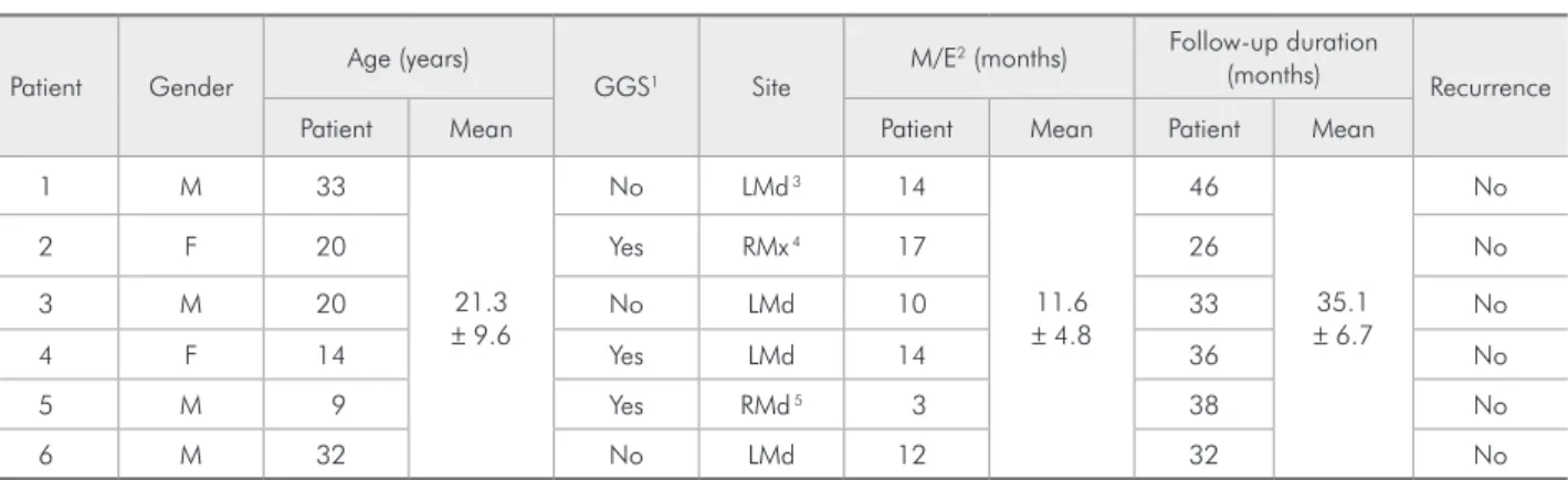
Six patients with KOTs who underwent marsupi-alization (incisional biopsy) followed by enucleation were included in this study. After marsupialization, the reduction in the size of their KOTs was exam-ined quarterly through clinical and radiographic examinations. The time between marsupialization and enucleation in each case is shown in Table 1. The enucleation was performed when a dimensional reduction of the tumor could not be radiographi-cally identiied within a 3-month period. After
enu-cleation, the surgical sites of all the patients were treated using peripheral ostectomy. Subsequently, Carnoy’s solution was applied (consisting of 60% ethanol, 30% chloroform, and 10% glacial acetic acid) for 5 minutes. This study was approved by the Research Ethics Committee of the Universidade
Federal de Minas Gerais - UFMG (COEP/UFMG
-15/08), and all of the participants signed an in-formed consent form.
The histological sections of selected KOT cases were retrieved from the iles of the Oral Pathology Service when the diagnosis proved to be in accor-dance with the indings of Barnes et al.1 All samples
were ixed in 10% buffered formalin and processed for hematoxylin-eosin staining (Sigma-Aldrich, Steinheim, Germany). The thickness of the epithelial lining and ibrous capsule at the time of marsupi-alization and after enucleation was measured using Axion Vision imagery analysis software, version 4.7.1 (Imaging Systems, Carl Zeiss, Oberkochen, Germany). The images of the specimens were viewed under a standard 25 microscope (Carl Zeiss, Oberkochen, Germany) with a inal magniication of 100× and photographed using a Canon Power Shot A-640 digital camera (Canon Inc., Tokyo, Japan). The photographs were transferred to the software, and the thickness measurements were conducted and recorded by a single examiner. The analyses of the images of each specimen were performed sepa-rately for the epithelial linings and the ibrous cap-sules, maintaining the surface of the epithelial lining parallel to the border of the picture frame so that Patient Gender
Age (years)
GGS1 Site
M/E2 (months) Follow-up duration
(months) Recurrence
Patient Mean Patient Mean Patient Mean
1 M 33
21.3 ± 9.6
No LMd 3 14
11.6 ± 4.8
46
35.1 ± 6.7
No
2 F 20 Yes RMx 4 17 26 No
3 M 20 No LMd 10 33 No
4 F 14 Yes LMd 14 36 No
5 M 9 Yes RMd 5 3 38 No
6 M 32 No LMd 12 32 No
per, middle, and lower) were performed on each im-age (Figures 1A, 1B, 2A, and 2B.) The mean values were recorded and expressed in µm. The BioEstat (BioEstat software version 4.0, Belém, Brazil) sta-tistical package was used to analyze the data, ap-plying the Wilcoxon test and Student’s t-test to the data from the epithelium and the ibrous capsule, the measurements could be taken perpendicularly to
the basal layer (Figures 1A and 1B).
When the total thickness of the ibrous capsule could not be determined from a single image, se-quential images were taken (Figures 1A, 1B, 2A, and 2B). The images were taken until all of the spec-imens had been included. Three measurements
(up-A B
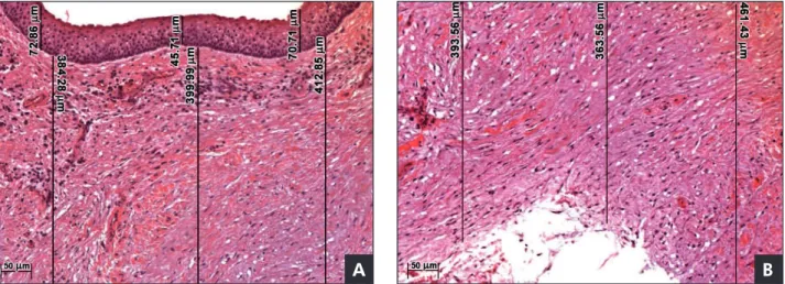
Figure 1 - Keratocystic odontogenic tumor specimen before marsupialization. A: The epithelial lining is represented by parake-ratinized stratified squamous epithelium with few layers and a basal palisade layer with cubic or columnar cells and hyperchro-matic nuclei. The epithelial-connective tissue junction is flat. B: Sequential measurements of the fibrous capsule shown in Figure 1A. The fibrous capsule consists of dense, sparsely cellularized, and slightly vascularized connective tissue. The three measure-ments (upper, middle, and lower) of the epithelial lining and fibrous capsule are represented by black lines (hematoxylin-eosin stain, 100× original magnification).
A B
Figure 2 - Keratocystic ontogenic tumor specimen after enucleation. A: The epithelial lining is represented by nonkeratinized, hyperplastic stratified squamous epithelium. The three measurements (upper, middle, and lower) of the epithelial lining and fi-brous capsule are represented by black lines (hematoxylin-eosin stain, 100× original magnification). B: Sequential measurements of the fibrous capsule shown in Figure 2A. The fibrous capsule consists of dense, sparsely cellularized, and slightly vascularized connective tissue with chronic inflammation (asterisk). The three measurements (upper, middle, and lower) are represented by black lines (hematoxylin-eosin stain, 100× original magnification).
A B
respectively. The probability value established for statistical signiicance was p < 0.05. The morphol-ogy of the epithelial lining and ibrous capsule was also observed and recorded both before and after the marsupialization.
Results
The data regarding the patients and the clinical features of their KOTs are described in Table 1.
In all cases, it was clinically obvious upon enucle-ation that the appearance of the tumor had changed from the time of marsupialization, with a consider-able thickening of the tumor’s ibrous capsule. This change facilitated the surgical procedure.
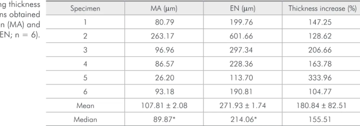
The histologically prepared specimens of the lesions were lined by parakeratinized stratiied squamous epithelium containing layers of only ap-proximately 5–6 cells before the marsupialization procedures. The basal layer consisted of cuboidal or columnar epithelial cells with hyperchromatic nu-clei arranged in palisades. The epithelial-connective tissue junction appeared lat with no epithelial pro-jections (Figure 1A). The specimens obtained after marsupialization showed stratiied squamous epi-thelium, which was predominantly nonkeratinized and hyperplastic, at times forming projections into the capsule. In addition, lymphocytic exocytosis and spongiosis could also be observed (Figure 2A). Four specimens presented focal areas of classic epithelial morphology associated with KOTs. The thickness measurements of the epithelium during marsupi-alization ranged from 26.2 to 263.17 µm (median of 89.87 µm), whereas the thickness in the KOT
specimens after enucleation ranged from 113.7 to 601.66 µm (median of 214.06 µm). This thickness difference was statistically signiicant (p = 0.0277, Wilcoxon test). Comparing the measurements taken during marsupialization and during enucleation, the epithelium presented a mean increase in thickness of 180% (Table 2).
The ibrous capsule in the specimens from the marsupialization procedure consisted of dense, cel-lularized, slightly vascularized connective tissue, at times with focal areas of inlammation. Microcysts could not be observed on the incisional or ation specimens. In the specimens from the enucle-ation procedure, the ibrous capsule consisted of dense and slightly cellularized connective tissue, with a variable amount of mononuclear inlamma-tory iniltrate (Figures 2A and 2B). The ibrous cap-sules from the incisional biopsy (marsupialization, 1395.01 µm ± 2.05) were not as thick as the capsules obtained from the inal enucleation (3562.15 ± 1.71,
p = 0.0212, Student’s t-test; Table 3). Therefore, the marsupialization procedure increased the thickness of the ibrous capsule by 294%.
Discussion
Previous studies have suggested that the lining of a keratocyst is only 5 or 6 cells thick and tears eas-ily during enucleation, in turn, causing a high recur-rence rate. However, after decompression or mar-supialization, the lining appears to become thicker and easier to enucleate; however, histologically, the lining appears to change and resembles that of the normal oral mucosa.4,8,9 Nevertheless, to date, only
Specimen MA (µm) EN (µm) Thickness increase (%)
1 80.79 199.76 147.25
2 263.17 601.66 128.62
3 96.96 297.34 206.66
4 86.57 228.36 163.78
5 26.20 113.70 333.96
6 93.18 190.81 104.77
Mean 107.81 ± 2.08 271.93 ± 1.74 180.84 ± 82.51
Median 89.87* 214.06* 155.51
* Statistically significant difference (p = 0.0277, Wilcoxon test). Table 2 - Epithelial lining thickness
descriptive studies have been performed to evaluate changes in the epithelial lining and ibrous capsule of KOTs at different times during marsupialization treatment. In the present study, a morphometric evaluation demonstrated that the epithelium and ibrous capsule in the specimens from enucleation were statistically signiicantly thicker than in the specimens from the incisional biopsy. This inding scientiically conirms the clinical observations.4,8,16
The histopathology of the incisional biopsies pre-sented the classic features of KOTs. After marsupi-alization, the features of the epithelium and ibrous capsule were modiied. Similar changes have also been observed in other studies.3,4,9,16 Pogrel9
report-ed that the coverreport-ed tumor was alterreport-ed with marsupi-alization and acquired the characteristics of normal oral mucosa in both routine histopathological and immunohistochemical analyses. The inlammatory iniltrate identiied by surgery can render the ap-pearance of KOTs similar to that of normal oral mu-cosa. In KOTs having some degree of inlammation, Stoelinga3 observed a loss of epithelial
keratiniza-tion that resembled that seen in radicular, inlamed dentigerous, and paradental cysts. De Paula et al.18
observed a predominantly nonkeratinized epithelial lining and an increased proliferative activity of in-lamed compared with noninin-lamed KOTs. August
et al.19 also noted a moderate hyperplasia and
in-lammation in many tumors that had been subjected to decompression. These authors also demonstrat-ed that the alterdemonstrat-ed expression of cytokeratin 10 is a marker of epithelial changes that occur in large
KOTs submitted to marsupialization. Marker et al.16 found changes in satellite cysts when the cysts
occurred in the ibrous capsule of KOTs subjected to marsupialization. This phenomenon was not ob-served in the tumors sampled in the present study. August et al.19 observed that the satellite cysts, upon
cystectomy, continued to express both cytokeratin 10 and KOT features, despite the differentiation of the main portion of the lesion.
One unexpected inding in the current study was the difference in the initial epithelial thickness of patient 5 compared with the thickness found for the other patients (Table 2). This patient had the great-est variation in thickness before and after marsu-pialization (333.96%, Table 2) over the short term (3 months, Table 1). Future studies with a larger sample of patients are warranted to examine the re-lationships of age at the diagnosis of KOT, physio-logical status, and individual genetic characteristics with the behavior of these lesions after marsupial-ization.
In the present study, the observations at the time of enucleation demonstrated that the appearance of the tumor changes after marsupialization. A thick-ening of the capsule covering the tumor could be observed, which facilitated the detachment of the entire lesion from the bone walls and its surgical removal. Marker et al.16 and Pogrel9 observed the
same alteration in a group of patients with KOTs treated by marsupialization. During the inspection of the surgical cavity in all cases, smooth bone walls but no remaining tumor could be observed. Despite Specimen MA (µm) EN (µm) Thickness increase (%)
1 1044.84 1478.92 41.54
2 3163.85 5446.77 72.15
3 1036.15 1884.64 81.88
4 526.09 3794.56 621.27
5 2063.86 3532.72 71.17
6 535.32 5235.32 877.97
Mean 1395.01 ± 2.05* 3562.15 ± 1.71* 294.33 ± 362.14
Median 1040.49 3663.64 77.01
* Statistically significant difference (p = 0.0212, Student’s t-test). Table 3 - Fibrous capsule thickness
apparent tumor removal, adjuvant treatment of sur-gical cavities with peripheral ostectomy and Car-noy’s solution was established as protocol.13
Alternatives have been proposed for the treat-ment of KOTs, and currently, the selection of more conservative procedures is advocated. The present study applied marsupialization under local anesthe-sia.6 This technique involves the cutting of an
open-ing in the wall of the lesion and the suturopen-ing of the tumor covering to the oral mucosa.8 A polyethylene
drain is routinely used to maintain communication between the inner tumor and the oral cavity. This drain is anchored by tying it with steel wire to the teeth adjacent to the surgical opening. Other stud-ies have described the use of an acrylic shutter as an alternative, especially in edentulous patients, as well as nasopharyngeal probes to maintain marsupializa-tion devices.4,9,16
Pogrel and Jordan8 distinguishes between
de-compression and marsupialization. However, the biological mechanism in both cases is the same, i.e., the reduction of the pressure within the lesion and establishment of contact between the surface of the tumor and the oral mucosa. The use or omission of a device to maintain the patency of the surgical open-ing depends on the anatomic features of the region.
Reducing the size of the KOT by marsupializa-tion facilitates its removal,4,9,16 as observed in the
current study. However, the option of marsupializa-tion generally requires the patient to undergo a sec-ond surgical procedure to enucleate the remaining lesion and to thus endure a longer treatment.4,5,16,19
A mean of 11.6 months elapsed between the inci-sional biopsy and the enucleation in the six evalu-ated patients. No problems could be identiied dur-ing this period. Therefore, it can be concluded that a longer treatment time is not a disadvantage of mar-supialization.
The maintenance period after marsupialization varied among the patients in this study from 3 to 17 months (mean of 11.6 months). During this time, the thickening of the ibrous capsule blocked the in-ner portions of the lesion and prevented its contin-ued remission. Therefore, the total regression of the lesion after marsupialization could not be identiied,
as reported by Pogrel and Jordan.8 Marker et al.,16
who also based their indings on radiographic evalu-ations, delayed approximately 10 months before performing enucleation and determined that longer delays promote more changes in the lining of tumor, which may be related to a decreased aggressiveness of the lesion. August et al.19 suggest a treatment
pe-riod of at least 9 months prior to enucleation to pro-duce epithelial differentiation. Brondum and Jen-ses,4 using radiographic evaluation, found that the
period from marsupialization to enucleation ranged from 1 to 14 months.
The literature reports that the recurrence rate of KOTs is between 0%5 and 62%.20 The capsule of thin
connective tissue and the plane junction between the epithelial and connective tissues cause the tumor wall to be friable. Moreover, cyst satellites can con-tribute to the recurrence of KOTs.5,10 Several authors
recommend a long period of postoperative follow-up.5,6,15 Woolgar et al.21 warn that the development
of a new tumor unrelated to the previous one in an adjacent region of the jaw can sometimes be inter-preted as a recurrence. Furthermore, the KOTs that affect patients with Gorlin-Goltz syndrome com-monly behave more aggressively,7 most likely due to
the higher rate of epithelial proliferation that occurs in these patients.22 In the present study, no
recur-rences could be observed among the evaluated pa-tients, including the syndromic patients.
Conclusion
In the present study, the epithelial lining and the ibrous capsule of KOTs were morphologically altered and signiicantly enlarged after marsupial-ization. These modiications facilitate full surgical treatment and may well be related to a low KOT re-currence rate.
Acknowledgements
The present study was supported by grants from the Conselho Nacional de Desenvolvimento
Cientíico e Tecnológico - CNPq
References
1. Barnes SL, Eveson JW, Reichart P, Sidransky D. World Health Organization classification of tumours. Pathology & genetics of Head and neck tumours. 1st ed. Lyon: IARC press; 2005. 306 p.
2. Phillipsen H. On keratocysts in the jaws. Tandleagebladet. 1956;60(1):963-71.
3. Stoelinga PJ. Long-term follow-up on keratocysts treated ac-cording to a defined protocol. Int J Oral Maxillofac Surg. 2001 Feb;30(1):14-25.
4. Brondum N, Jensen VJ. Recurrence of keratocysts and decom-pression treatment. A long-term follow-up of forty-four cases. Oral Surg Oral Med Oral Pathol. 1991 Sep;72(3):265-9. 5. Bataineh AB, al Qudah M. Treatment of mandibular
odon-togenic keratocysts. Oral Surg Oral Med Oral Pathol Oral Radiol Endod. 1998 Jul;86(1):42-7.
6. Vedtofte P, Praetorius F. Recurrence of the odontogenic kera-tocyst in relation to clinical and histological features. A 20-year follow-up study of 72 patients. Int J Oral Surg. 1979 Dec;8(6):412-20.
7. Forssell K, Forssell H, Kahnberg KE. Recurrence of kerato-cysts. A long-term follow-up study. Int J Oral Maxillofac Surg. 1988 Feb;17(1):25-8.
8. Pogrel MA, Jordan RC. Marsupialization as a definitive treat-ment for the odontogenic keratocyst. J Oral Maxillofac Surg. 2004 Jun;62(6):651-5; discussion 5-6.
9. Pogrel MA. Treatment of keratocysts: the case for decom-pression and marsupialization. J Oral Maxillofac Surg. 2005 Nov;63(11):1667-73.
10. Williams TP, Connor FA Jr. Surgical management of the odon-togenic keratocyst: aggressive approach. J Oral Maxillofac Surg. 1994 Sep;52(9):964-6.
11. Schmidt BL, Pogrel MA. The use of enucleation and liquid nitrogen cryotherapy in the management of odontogenic kera-tocysts. J Oral Maxillofac Surg. 2001 Jul;59(7):720-5; discus-sion 6-7.
12. Irvine GH, Bowerman JE. Mandibular keratocysts: surgical management. Br J Oral Maxillofac Surg. 1985 Jun;23(3):204-9.
13. Morgan TA, Burton CC, Qian F. A retrospective review of treatment of the odontogenic keratocyst. J Oral Maxillofac Surg. 2005 May;63(5):635-9.
14. Voorsmit RA, Stoelinga PJ, van Haelst UJ. The management of keratocysts. J Maxillofac Surg. 1981 Nov;9(4):228-36. 15. Pogrel MA, Jordan RC. Marsupialization as a definitive
treat-ment for the odontogenic keratocyst. J Oral Maxillofac Surg 2004 Jun;62(6):655-6.
16. Marker P, Brondum N, Clausen PP, Bastian HL. Treatment of large odontogenic keratocysts by decompression and later cystectomy: a long-term follow-up and a histologic study of 23 cases. Oral Surg Oral Med Oral Pathol Oral Radiol Endod. 1996 Aug;82(2):122-31.
17. Tabrizi R, Ozkan BT, Dehgani A, Langner NJ. Marsupializa-tion as a treatment opMarsupializa-tion for the odontogenic keratocyst. J Craniofac Surg. 2012 Sep;23(5):e459-61.
18. de Paula AM, Carvalhais JN, Domingues MG, Barreto DC, Mesquita RA. Cell proliferation markers in the odontogenic keratocyst: effect of inflammation. J Oral Pathol Med. 2000 Nov;29(10):477-82.
19. August M, Faquin WC, Troulis MJ, Kaban LB. Dedifferen-tiation of odontogenic keratocyst epithelium after cyst de-compression. J Oral Maxillofac Surg. 2003 Jun;61(6):678-83; discussion 683-4.
20. Pindborg JJ, Hansen J. Studies on odontogenic cyst epithe-lium. 2. Clinical and roentgenologic cspects of odontogenic keratocysts. Acta Pathol Microbiol Scand. 1963;58(1):283-94. 21. Woolgar JA, Rippin JW, Browne RM. A comparative study
of the clinical and histological features of recurrent and non-recurrent odontogenic keratocysts. J Oral Pathol. 1987 Mar;16(3):124-8.