Conservative Treatment Protocol for Keratocystic Odontogenic
Tumour: a Follow-up Study of 3 Cases
Gülsün Yildirim
1, Hanife Ataoglu
1, Abdullah Kalayci
1, Birkan Taha Özkan
1, Korhan Kucuk
1,
Alparslan Esen
11Department of Oral and Maxillofacial Surgery, Faculty of Dentistry, University of Selçuk, Konya, Turkey.
Corresponding Author: Birkan Taha Özkan
Yuzuncuyıl Üniversitesi, Dishekimligi Fakultesi Ağız, Dis, Cene Hastalıkları ve Cerrahisi Bölümü 65080, Kampus/Van
Turkey
Phone: + 90 532 616 91 63 Fax: + 90 432 225 17 47 E-mail: btozkan@hotmail.com
ABSTRACT
Background: The keratocystic odontogenic tumour is classiied as a developmental cyst derived from the enamel organ or from the dental lamina. The treatment of keratocystic odontogenic tumour of the jaw remains controversial. The aim of this study was to report the outcome of our conservative treatment protocol for keratocystic odontogenic tumour.
Methods: Three patients with different complaints referred to Oral and Maxillofacial Surgery Clinic, Faculty of Dentistry, Selçuk University. Initial biopsy was carried out in all patients and keratocystic odontogenic tumours were diagnosed subsequent to histopathological examination. The patients with keratocystic odontogenic tumours were treated by enucleation followed by open packing. This conservative treatment protocol was selected because of existing young aged patients. The average follow-up duration of the cases was 2 years.
Results: Out of 3 cases, 2 lesions were present in mandible and 1 lesion in maxilla. There was no evidence of recurrence during follow-up. All the cases were monitored continuously with panoramic radiographs, computed tomography and clinical evaluations.
Conclusions: This conservative treatment protocol for keratocystic odontogenic tumours, based on enucleation followed by open packing would be a possible choice with a view of offering low recurrence rate and low morbidity rate particularly in young patients.
Keywords: keratocysts; odontogenic cyst; jaw cysts; jaw neoplasms.
Accepted for publication: 12 June 2010 To cite this article:
Yildirim G, Ataoglu H, Kalayci A, Özkan BT, Kucuk K, Esen A. Conservative Treatment Protocol for Keratocystic Odontogenic Tumour: a Follow-up Study of 3 Cases.
J Oral Maxillofac Res 2010 (Jul-Sep);1(3):e7
INTRODUCTION
The odontogenic keratocyst (OKC) is classiied as a developmental cyst derived from the enamel organ or from the dental lamina [1-3]. The deinition “odontogenic keratocyst” irst was proposed by Philipsen in 1956, when he separated seven jaw cysts from cholesteatomas occurring in other cranial areas. Because of his thought that these were odontogenic cysts and not inlammatory in origin, he coined the term odontogenic keratocyst [4]. Later it was noted that other odontogenic cysts, such as radicular cysts, follicular cysts, and lateral periodontal cysts, are morphologically similar to OKCs [5]. In contrast there were observations which showed that the odontogenic keratocyst behaved more as a neoplasm and not like a cystic lesion [6]. Finally, in the latest World Health Organization classiication, the former odontogenic keratocyst was added to the benign odontogenic tumours category. The new term is “keratocystic odontogenic tumor” (KCOT). In contrast to other odontogenic cysts, KCOTs have a high recurrence rate, reportedly ranging from 13% to 80% based on the performed treatment [7-12]. The malignant transformation of KCOTs has also been reported [13]. Treatment of KCOTs remains a controversial subject [14]. Most cases recur within the irst 5 years after treatment [15,16]. Many attempts have been made to reduce the high recurrence rate of KCOTs by improving the operative technique. Advocates of conservative treatment suggest that marsupialization yields the results comparable to those obtained with more extensive surgery [16].
In this present study, 3 cases of keratocystic odontogenic tumour that were treated with enucleation followed by open packing through an intraoral approach and 2 year follow-up period will be presented.
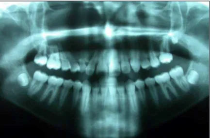
Figure 1. Preoperative panoramic radiograph shows the multilocular lucency extending from the neck of condyle to the right canine tooth (Case 1).
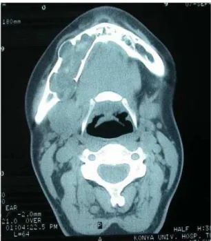
Figure 2. Preoperative computed tomography scan axial image shows the multilocular lucency extending from the neck of condyle to the right canine tooth (Case 1).
CASE DESCRIPTION AND RESULTS
Case 1
A 26 year old female was referred to Selcuk University, Faculty of Dentistry, Oral and Maxillofacial Surgery Clinic, for the evaluation of swelling and pain located at the posterior body and ascending ramus of the right mandible.
The patient’s medical history was unremarkable. In the clinical examination, there was no facial asymmetry. Intraorally, mild swelling was noticed on the right mandibular molar area. The overlying mucosa appeared normal on colour and texture. There was no
lympadenopathy.
The patient was evaluated radiographically by panoramic radiography and computed tomography (CT) imaging. The imaging revealed a multilocular lucency extending from the neck of the condyle to right canine tooth (Figures 1 and 2). All teeth in the lesion gave vital response to electric pulp testing.
Figure 3. Histopathologic view of the lesion shows the basal layer
of the epithelium composed of hyalinised ibrous connective tissue (hematoxylin and eosin stain, original magniication x100) (Case 1).
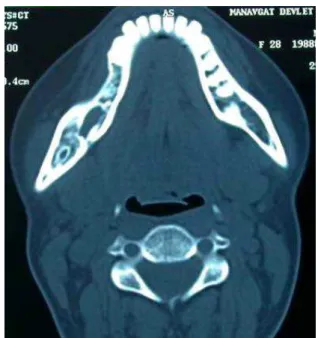
Figure 4. Computed tomography scan axial image at the end of 2 year follow-up period shows no signs of the recurrence (Case 1).
Figure 5. Panoramic radiograph at the end of 2 year follow-up period shows no signs of the recurrence (Case 1).
were either cuboidal or columnar and the basal cell nuclei were hyperchromatic and arranged in a “picket fence” coniguration. The ibrous connective tissue wall contained sparse chronic inlammatory cells which were composed of lymphocyte and plasma cells. On the basis of these indings, a diagnosis of a KCOT was made (Figure 3).
Due to the size of the lesion it was decided to treat it with enucleation followed by open packing. Under local anaesthesia, an incision extending from the mandibular ramus distal to the second molar was carried anteriorly around the crevices of the anteriomandibular teeth and a full thickness mucoperiosteal lap was raised. The cystic cavity was curetted from molar area to the neck of the condyle. The resulting cavity was packed with iodoform gauze impregnated with bacitracin ointment. The packing was replaced during the recall visits biweekly for six months following the initial surgery. The patient was reviewed radiologically every three months during follow-up period. At the end of 2 years follow-up period, no evidence of recurrence was noticed (Figures 4 and 5).
Case 2
A 14 year old female patient presented with complaint of a gradually swelling extending from the right maxillary canine to the third molar region. The swelling was slightly painful; it was irst noticed about 5 months previously. The patient’s medical history was unremarkable. Clinical examination revealed a buccal spherical and luctuant swelling involving the right lateral incisor, premolars and irst molar region. It was surfaced by normal oral mucosa. The maxillary right lateral incisor was displaced by the lesion (Figure 6). Evaluation radiographically by panoramic view and CT imaging revealed an expansible lesion extending from the primary canine tooth to the third molar
Figure 6. Preoperative intraoral view of the lesion shows a buccal
spherical and luctuant swelling involving the right lateral incisor, premolars and irst molar region (Case 2).
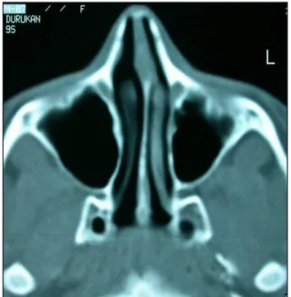
Figure 7. Preoperative computed tomography axial scan image shows an expansible lesion extending from the primary canine tooth to the
third molar region and superiorly to the loor of the orbit (Case 2).
Figure 8. Preoperative panoramic radiograph shows an expansible lesion extending from the primary canine tooth to the third molar region and superiorly
to the loor of the orbit (Case 2).
Figure 9. Postoperative intraoral view of the patient (Case 2). Figure 10. Photograph shows the totally excised lesion (Case 2).
diagnosis of KCOT.
Patient was seen for clinical and radiological evaluation regularly after treatment. The bony cavity has healed clinically and radiographically approximately 16 months after treatment (Figures 11 and 12). There was no evidence of residual disease on radiographic examination at 2 year clinical follow-up (Figure 13).
Case 3
Figure 11. Postoperative intraoral view of the patient shows no signs of the recurrence (Case 2).
Figure 12. Computed tomography scan axial image at the end of 2 year follow-up period shows no signs of the recurrence (Case 2).
Figure 13. Postoperative panoramic radiograph at the end of 2 year follow-up period shows no signs of the recurrence (Case 2).
treatment to our department. The radiographic examination revealed a lesion involving the entire mandible from second molar on the right to second molar on the left (Figure 14).
The incisional biopsy obtained by the general practitioner revealed presence of a KCOT.
His medical history was unremarkable. Extraoral examination revealed that there was a swelling and expansion at mandibular basis at both sides of the mandibular molar region. Radiologic examination revealed that the right mandibular canine tooth was impacted at the symphysis region, and there was a multilocular radiolucent cystic lesion related to this tooth.
A yellow serous liquid or semi-solid content was obtained as a ine needle aspiration material, matching the typical description of a cystic lesion. Biopsy was performed and impacted canine tooth was extracted at the same time. Biopsy result was KCOT.
Endodontic treatment was performed for all teeth related to a lesion. Enucleation followed by open packing treatment was performed. After the operation, the resulting bony cavity was packed with iodoform gauze impregnated with bacitracin ointment which again was changed biweekly for the following six months. There was no sign of recurrence at a postoperative 2 years period (Figure 15).
DISCUSSION
Figure 14. Preoperative panoramic radiograph shows a lesion involving the entire mandible from second molar on the right to second molar on the left (Case 3).
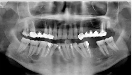
Figure 15. Panoramic radiograph at the end of 2 year follow-up period shows no signs of the recurrence (Case 3).
the production of collagenase, prostaglandins, and highly active oxidative enzymes. Piatelli et al. identiied bcl-2 protein overexpression in odontogenic keratocysts and concluded that it could produce an increase in the survival of the epithelial cells then, and this increased lifespan could, in turn, lead to the peculiar aggressive growth pattern of OKC. Moreover the bcl-2 staining can be useful to differentiate OKC from other types of odontogenic cysts [20].
Y. Kubota et al. [21] showed that Interleukin-1α (IL-1α) is strongly expressed in odontogenic keratocysts. IL-1α may up-regulate matrix metalloproteinase-2 activation by increasing the expression of membrane-type 1 matrix metalloproteinase in the ibroblasts isolated from odontogenic keratocysts synergistically with type I collagen. Ogata et al. [22] showed that in keratocystic odontogenic tumour ibroblasts, IL-1α may stimulate COX-2 expression. Andric et al. [23] investigated the expression of survivin, an inhibitor of apoptosis, in odontogenic keratocysts and compared it to the indings in non-neoplastic jaw cysts - periapical cysts,
they also studied a possible relationship between survivin expression and human cytomegalovirus presence within these cysts. It was found that survivin may contribute to the aggressive behavior of odontogenic keratocysts, and thus support the emerging opinion of their neoplastic nature.
However, the most important evidence that KCOT is a neoplasm comes from genetic studies demonstrating loss of heterozygosity of the tumour suppressor genes in KCOT cases [24-27]. The human homologue of the Drosophila segment polarity gene PTCH1 is a tumour suppressor gene within the Sonic hedgehog pathway. The Sonic hedgehog pathway has been shown to function in the patterning of limbs, the axial skeleton, the central nervous system, and tooth development. Mutations in the PTCH1 gene have been demonstrated in patients with nevoid basal cell carcinoma syndrome (NBCCS) 12 as well as sporadic neoplasms including the KCOT, basal cell carcinoma, and medulloblastoma [28,29]. Grachtchouk et al. [29] showed that epithelial expression of the Hh transcriptional effector Gli2 is suficient for highly penetrant keratocyst development in transgenic mice. Mouse and human keratocysts expressed similar markers, leading to tooth misalignment, bone remodeling, and craniofacial abnormalities. The frequent development of keratocysts in Gli2-expressing mice supports the idea that GLI transcription factor activity mediates pathological responses to deregulated Hh signaling in humans. Moreover, Gli2-mediated reactivation of quiescent epithelial rests to form keratocysts indicates that these cells retain the capacity to function as progenitor cells on activation by an appropriate developmental signal.
could support the theory that odontogenic keratocysts are neoplastic in origin, but other results clearly support that these lesions are developmental cysts with some neoplastic properties because of the high intrinsic growth potential.
A review of the biological KCOT behavior of this recognized aggressive pathological entity of the jaws and a contemporary outline of the molecular (growth factors, p53, PCNA and Ki-67, bcl-2) and genetic (PTCH, SHH) alterations associated with this odontogenic neoplasm, provides a better understanding of the mechanisms involved in its development and strengthen the current concept that the KCOT should, indeed, be regarded as a neoplasm. Furthermore, markers known to be rapidly induced in response to growth factors, tumour promoters, cytokines, bacterial endotoxins, oncogenes, hormones and shear stress, such as COX-2, may also shed a new light on biological mechanisms involved in the development of these benign but sometimes aggressive neoplasms of the jaws [31,32].
KCOT comprises approximately 11% of the all cysts of the jaws [33]. Approximately 65% of KCOTs occur in the mandible, with prevalence (73.2%) in the mandibular ramus and angle [34-37]. KCOTs usually occur in the second and third decades of life with an average patient age of 30.8 years [7,8,13]. An KCOT usually occurs as a single lesion. Multiple lesions are associated with the nevoid basal cell syndrome (Gorlin-Goltz syndrome); the lesions are also associated with younger patients [37]. In approximately 50% of patients, the lesion is asymptomatic and was found incidentally during routine radiographic examination. In others, pain, swelling, expansion, drainage, and bone perforation are reported [8,9].
On the radiography KCOTs appear as well-deined radiolucencies, which can be either unilocular or multilocular. Large unilocular KCOTs can be indistinguishable from cystic ameloblastoma [38]. The lesion’s proximity to the tooth may cause displacement and/or root resorption, although displacement (28.3%) is more commonly seen than resorption (5%) [39]. Conventional radiographic imaging, such as panoramic views and intraoral periapical ilms, in most cases are adequate to determine the location and estimate the size of KCOT. Advanced imaging techniques like computed tomography and magnetic resonance imaging can be useful in large cases involving the maxillary sinus and the rare cases that extend to the skull base [38].
Microscopically KCOT is characterized by epithelial lining of parakeratinized stratiied squamous epithelium. The basal layer of the epithelium is composed of hyalinised ibrous connective tissue. The basal cells are either cuboidal or columnar and the basal cell nuclei are hyperchromatic and arranged in a “picket fence”
coniguration. Furthermore, it has a corrugated parakeratinized luminal layer and a prominent basal cell layer [38].
Treatment of KCOTs remains a controversial subject [16]. The choice of treatment approach should be based on the size of the cyst, recurrence status, and radiographic evidence of cortical perforation [40]. This is general agreement that complete removal of large KCOTs of the mandibular ramus may be dificult [41,42]. Radical surgery, comprising resection with or without continuity defects, has been advocated for larger KCOTs and recurrent lesions. Resection may be indicated in the treatment of KCOTs in most cases; it completely removes the lesion, and recurrence is minimal. Bramley [43] recommended radical surgery with resection and bone transplantation. Irvine and Bowerman [44] recommended radical enucleation and bone grafting for large keratocysts. More aggressive treatment - resection or enucleation supplemented with Carnoy’s solution with or without peripheral ostectomy - results in a lower recurrence rate than enucleation alone or marsupialization. Notably, the recurrence rate after marsupialization followed by enucleation is not signiicantly higher than that following the so-called aggressive modalities [45].
Resection offers a high cure rate, but produces signiicant morbidity such as the loss of jaw continuity or facial disigurement. It should, therefore be reserved only for aggressive or recurrent lesions, or for the patients who cannot be closely followed-up after conservative treatments [46]. On the other hand, radical treatment would adversely affect the physcosocial condition of the young patient. The young patient would inhibit growth potential especially in the irst decade of life; therefore this condition of the patient represents a dificult decision for the surgeon for selecting conservative versus radical treatment protocol. Therefore, conservative treatment would also be offered in young patients with the view of posing low morbidity rate and conserving the most teeth adjacent to the lesion.
The one technique used in the treatment of the KCOT, as described in the reports of Brondum and Jensen [47] and Marker et al. [48], decompression is accomplished by opening into a cystic cavity followed by placement of a drainage tube to allow the opening to persist. The disadvantages of decompression and subsequent enucleation treatment - it requires two surgical procedures and the treatment time necessary is comparatively long.
production observed in 64% (n = 14) of patients treated by the cyst decompression/irrigation after a 9 month average treatment time. Otherwise, they mentioned that longitudinal follow-up of these patients should determine whether this change is associated with lower rates of recurrence than alternative KCOT therapy. Furthermore, Oka et al. [50] demonstrated that there may be a strong relationship among intracystic pressure, the expression of IL-1α, and bone resorption: positive pressure may play a crucial role in keratocystic odontogenic tumour growth via stimulating the expression of IL-1 in epithelial cells.
Currently, a total enucleation, with or without a “peripheral ostectomy” presents the most common surgical procedures used to treat most of the KCOTs [2,51]. Advocates of conservative treatment suggest that marsupialization yields the results comparable to those obtained with more extensive surgery [16]. In the strictest sense, marsupialization implies the exteriorization of cyst or an enclosed cavity by resecting a portion of its wall and suturing the cut edges of remaining wall to adjacent soft tissue, thereby creating a pouch. Marsupialization is not a newcomer to the armamentarium of possible KCOT treatment, and case series have appeared in the literature since early descriptions in the 1970s [52-54].
Enucleation of the KCOTs followed by open packing has been suggested as another conservative method of surgical treatment [16,55]. The current 3 patients were treated with enucleation followed by open packing. The resulting cavity was irrigated with mixture of normal saline and chlorhexidine gluconate for a full-of glass and also packed with iodoform gauze impregnated with bacitracin ointment to minimize the risk of recurrence in each recall visits. Regular recall visits are required to ensure cyst involution and the opportunity for appropriate treatment should be an evidence of recurrence. The beneit of this protocol lies in the minimal surgical morbidity. In addition, associated structures such as the inferior alveolar nerve and developing teeth are less vulnerable to the damage.
Different views on the recommended duration of radiologic and clinical follow-up are reported in the literature. According to Forssell [56], a 4 year period will sufice for the detection of most recurrences. As it was stated by the authors, the recurrence rate after the treatment of KCOTs is very broad and treatment dependent. At the end of 2 year follow-up period, no evidence of recurrence was registered in this report. Blanas et al. [57] conducted a systematic review of
literature and evaluated the collective recurrence rates reported with various types of the treatment. Forty-ive cases were treated by marsupialization with a recurrence rate of 24.4%. Enucleation alone was associated with a 28.7% recurrence [58]. Browne [36] has used three different methods of treatment: marsupialization, enucleation and primary closure, and enucleation and packing-open; he found approximately equal rates of recurrence with the three methods of treatment. This is in coincidence with Kolokythas et al. [40]: the aggressive nature of some KCOTs necessitates equally aggressive treatment, whereas long-term follow-up even for nonsyndromic patients with the single lesions is of paramount importance. Age of the patient and the site and histological characteristics of the treated lesions are not signiicantly associated with the incidence of recurrence.
Extraction of the teeth affected by the lesion, as well as a generous removal of partially eroded bone and overlying soft tissues, may contribute to lower recurrence rates. Bataineh and Al Qudah [59] advocated that resection without continuity, defects as a radical treatment, in which the removal of cyst, teeth, and overlying soft tissue is followed by packing of the resulting cavity to minimize the risk of recurrence. In order to minimizing the recurrence, eliminating the retention areas around the periapical portion of extracted teeth and the eradication of cyst, the teeth were removed during the surgical treatment in one case.
CONCLUSIONS
Enucleation followed by open packing can be suggested as a choice of conservative treatment with low recurrence rate for large keratocystic odontogenic tumours. However, due to the clinical behaviour of keratocystic odontogenic tumours, in case of possible risk of recurrence requirement for further operations should not be disregarded. Therefore, the patients should have control radiographic and clinical examinations for indeinite prognosis regardless of the treatment protocol.
ACKNOWLEDGMENTS AND DISCLOSURE STATEMENTS
REFERENCES
1. Rud J, Pindborg JJ. Odontogenic keratocysts: a follow-up study of 21 cases. J Oral Surg. 1969 May;27(5):323-30. [Medline: 5253512]
2. Panders AK, Haddlers HN. Solitary keratocysts of the jaws. J Oral Surg. 1969 Dec;27(12):931-8. [Medline: 5260718] 3. Shear M. Primordial cyst. Journal of the Dental Association of South Africa 1960;15(2):211-7.
4. Wright JM. The odontogenic keratocyst: orthokeratinized variant. Oral Surg Oral Med Oral Pathol. 1981 Jun;51(6):609-18. [Medline: 6166916] [doi: 10.1016/S0030-4220(81)80011-4]
5. el-Hajj G, Anneroth G. Odontogenic keratocysts--a retrospective clinical and histologic study. Int J Oral Maxillofac Surg. 1996 Apr;25(2):124-9. [Medline: 8727585] [doi: 10.1016/S0901-5027(96)80057-9]
6. Reichart PA, Philipsen HP, Sciubba JJ. The new classiication of Head and Neck Tumours (WHO)--any changes? Oral Oncol. 2006 Sep;42(8):757-8. Epub 2006 May 6. [Medline: 16679047] [doi: 10.1016/j.oraloncology.2005.10.011] 7. Brannon RB. The odontogenic keratocyst. A clinicopathologic study of 312 cases. Part II. Histologic features. Oral Surg
Oral Med Oral Pathol. 1977 Feb;43(2):233-55. [Medline: 264648] [doi: 10.1016/0030-4220(77)90161-X]
8. Haring JI, Van Dis ML. Odontogenic keratocysts: a clinical, radiographic, and histopathologic study. Oral Surg Oral Med Oral Pathol. 1988 Jul;66(1):145-53. [Medline: 2457195] [doi: 10.1016/0030-4220(88)90082-5]
9. Verbin RS, Barnes L. Cysts and cystlike lesions of the oral cavity, jaws, and neck. In: Barnes L, ed. Surgical Pathology of the Head and Neck, Vol. 2. New York, NY: Marcel Dekker; 1985. p. 1257–63.
10. Yoshiura K, Higuchi Y, Ariji Y, Shinohara M, Yuasa K, Nakayama E, Ban S, Kanda S. Increased attenuation in odontogenic keratocysts with computed tomography: a new inding. Dentomaxillofac Radiol. 1994 Aug;23(3):138-42. [Medline: 7530666]
11. DelBalso AM. An approach to the diagnostic imaging of jaw lesions, dental implants, and the temporomandibular joint. Radiol Clin North Am. 1998 Sep;36(5):855-90, vi. Review. [Medline: 9747192] [doi: 10.1016/S0033-8389(05)70067-1] 12. Som PM, Curtin HD. Chronic inlammatory sinonasal diseases including fungal infections. The role of imaging. Radiol
Clin North Am. 1993 Jan;31(1):33-44. [Medline: 8419978]
13. Myoung H, Hong SP, Hong SD, Lee JI, Lim CY, Choung PH, Lee JH, Choi JY, Seo BM, Kim MJ. Odontogenic keratocyst: Review of 256 cases for recurrence and clinicopathologic parameters. Oral Surg Oral Med Oral Pathol Oral Radiol Endod. 2001 Mar;91(3):328-33. [Medline: 11250631] [doi: 10.1067/moe.2001.113109]
14. Eyre J, Zakrzewska JM. The conservative management of large odontogenic keratocysts. Br J Oral Maxillofac Surg. 1985 Jun;23(3):195-203. [Medline: 3159419] [doi: 10.1016/0266-4356(85)90090-7]
15. Vedtofte P, Praetorius F. Recurrence of the odontogenic keratocyst in relation to clinical and histological features. A 20-year follow-up study of 72 patients. Int J Oral Surg. 1979 Dec;8(6):412-20. [Medline: 120338] [doi: 10.1016/S0300-9785(79)80079-4]
16. Forssell K, Forssell H, Kahnberg KE. Recurrence of keratocysts. A long-term follow-up study. Int J Oral Maxillofac Surg. 1988 Feb;17(1):25-8. [Medline: 3127485] [doi: 10.1016/S0901-5027(88)80224-8]
17. Scharffetter K, Balz-Herrmann C, Lagrange W, Koberg W, Mittermayer C. Proliferation kinetics-study of the growth of keratocysts. Morpho-functional explanation for recurrences. J Craniomaxillofac Surg. 1989 Jul;17(5):226-33. Review. [Medline: 2668340] [doi: 10.1016/S1010-5182(89)80074-5]
18. Formigli L, Orlandini SZ, Tonelli P, Giannelli M, Martini M, Brandi ML, Bergamini M, Orlandini GE. Osteolytic processes in human radicular cysts: morphological and biochemical results. J Oral Pathol Med. 1995 May;24(5):216-20. [Medline: 7616461] [doi: 10.1111/j.1600-0714.1995.tb01170.x]
19. Sorsa T, Ylipaavalniemi P, Suomalainen K, Vauhkonen M, Lindy S. Type-speciic degradation of interstitial collagens by human keratocyst wall collagenase. Med Sci Res 1988; 16: 1189-90.
20. Piattelli A, Fioroni M, Rubini C. Differentiation of odontogenic keratocysts from other odontogenic cysts by the expression of bcl-2 immunoreactivity. Oral Oncol. 1998 Sep;34(5):404-7. [Medline: 9861349] [doi: 10.1016/S1368-8375(98)00026-8]
21. Kubota Y, Oka S, Nakagawa S, Shirasuna K. Interleukin-1alpha enhances type I collagen-induced activation of matrix metalloproteinase-2 in odontogenic keratocyst ibroblasts. J Dent Res. 2002 Jan;81(1):23-7. [Medline: 11820363] [doi: 10.1177/154405910208100106] [FREE Full Text]
22. Ogata S, Kubota Y, Yamashiro T, Takeuchi H, Ninomiya T, Suyama Y, Shirasuna K. Signaling pathways regulating IL-1alpha-induced COX-2 expression. J Dent Res. 2007 Feb;86(2):186-91. [Medline: 17251521] [doi: 10.1177/154405910708600215]
23. Andric M, Dozic B, Popovic B, Stefanovic D, Basta-Jovanovic G, Djogo N, Andjus P, Milasin J. Survivin expression in odontogenic keratocysts and correlation with cytomegalovirus infection. Oral Dis. 2010 Mar;16(2):156-9. Epub 2009 Jul 29. [Medline: 19659890] [doi: 10.1111/j.1601-0825.2009.01612.x]
25. Shear M. The aggressive nature of the odontogenic keratocyst: is it a benign cystic neoplasm? Part 1. Clinical and early experimental evidence of aggressive behaviour. Oral Oncol. 2002 Apr;38(3):219-26. Review. [Medline: 11978543] [doi: 10.1016/S1368-8375(01)00065-3]
26. Shear M. The aggressive nature of the odontogenic keratocyst: is it a benign cystic neoplasm? Part 2. Proliferation and genetic studies. Oral Oncol. 2002 Jun;38(4):323-31. Review. Erratum in: Oral Oncol. 2004 Jan;40(1):107. [Medline: 12076694] [doi: 10.1016/S1368-8375(01)00066-5]
27. Shear M. The aggressive nature of the odontogenic keratocyst: is it a benign cystic neoplasm? Part 3. Immunocytochemistry of cytokeratin and other epithelial cell markers. Oral Oncol. 2002 Jul;38(5):407-15. Review. [Medline: 12110333] [doi: 10.1016/S1368-8375(01)00067-7]
28. Peacock ZS, Cox D, Schmidt BL. Involvement of PTCH1 mutations in the calcifying epithelial odontogenic tumor. Oral Oncol. 2010 May;46(5):387-92. Epub 2010 Apr 3. [Medline: 20371205] [doi: 10.1016/j.oraloncology.2010.02.023] 29. Grachtchouk M, Liu J, Wang A, Wei L, Bichakjian CK, Garlick J, Paulino AF, Giordano T, Dlugosz AA. Odontogenic
keratocysts arise from quiescent epithelial rests and are associated with deregulated hedgehog signaling in mice and humans. Am J Pathol. 2006 Sep;169(3):806-14. [Medline: 16936257] [doi: 10.2353/ajpath.2006.060054] [FREE Full Text]
30. de Vicente JC, Torre A, Gutiérrez AM, Lequerica P. Immunohistochemical comparative study of the odontogenic keratocysts and other odontogenic lesions. Med Oral Patol Oral Cir Bucal. 2010 Apr 11. [Epub ahead of print]. [Medline: 20383104] [FREE Full Text]
31. Mendes RA, Carvalho JF, van der Waal I. Characterization and management of the keratocystic odontogenic tumor in relation to its histopathological and biological features. Oral Oncol. 2010 Apr;46(4):219-25. Epub 2010 Feb 26. [Medline: 20189443] [doi: 10.1016/j.oraloncology.2010.01.012]
32. Mendes RA, Carvalho JF, van der Waal I. Biological pathways involved in the aggressive behavior of the keratocystic odontogenic tumor and possible implications for molecular oriented treatment - an overview. Oral Oncol. 2010 Jan;46(1):19-24. Epub 2009 Dec 9. [Medline: 20004133] [doi: 10.1016/j.oraloncology.2009.10.009]
33. Shear M. Developmental odontogenic cysts. An update. J Oral Pathol Med. 1994 Jan;23(1):1-11. Review. [Medline: 8138974] [doi: 10.1111/j.1600-0714.1994.tb00246.x]
34. Ahlfors E, Larsson A, Sjögren S. The odontogenic keratocyst: a benign cystic tumor? J Oral Maxillofac Surg. 1984 Jan;42(1):10-9. [Medline: 6199488] [doi: 10.1016/0278-2391(84)90390-2]
35. Forssell K, Sorvari TE, Oksala E. A clinical and radiographic study of odontogenic keratocysts in jaws. Proc Finn Dent Soc. 1974 Aug;70(4):121-34. [Medline: 4411585]
36. Browne RM. The odontogenic keratocyst. Clinical aspects. Br Dent J. 1970 Mar 3;128(5):225-31. [Medline: 5270271] [doi: 10.1038/sj.bdj.4802449]
37. Brannon RB. The odontogenic keratocyst. A clinicopathologic study of 312 cases. Part I. Clinical features. Oral Surg Oral Med Oral Pathol. 1976 Jul;42(1):54-72. [Medline: 1065842] [doi: 10.1016/0030-4220(76)90031-1]
38. Ali M, Baughman RA. Maxillary odontogenic keratocyst: a common and serious clinical misdiagnosis. J Am Dent Assoc. 2003 Jul;134(7):877-83. [Medline: 12892445] [FREE Full Text]
39. Haring JI, Van Dis ML. Odontogenic keratocysts: a clinical, radiographic, and histopathologic study. Oral Surg Oral Med Oral Pathol. 1988 Jul;66(1):145-53. [Medline: 2457195]
40. Kolokythas A, Fernandes RP, Pazoki A, Ord RA. Odontogenic keratocyst: to decompress or not to decompress? A comparative study of decompression and enucleation versus resection/peripheral ostectomy. J Oral Maxillofac Surg. 2007 Apr;65(4):640-4. [Medline: 17368357] [doi: 10.1016/j.joms.2006.06.284]
41. Zachariades N, Papanicolaou S, Triantafyllou D. Odontogenic keratocysts: review of the literature and report of sixteen cases. J Oral Maxillofac Surg. 1985 Mar;43(3):177-82. [Medline: 2579223] [doi: 10.1016/0278-2391(85)90156-9] 42. Webb DJ, Brockbank J. Treatment of the odontogenic keratocyst by combined enucleation and cryosurgery. Int J Oral
Surg. 1984 Dec;13(6):506-10. [Medline: 6210260] [doi: 10.1016/S0300-9785(84)80021-6]
43. Bramley P. The odontogenic keratocyst--an approach to treatment. Int J Oral Surg. 1974;3(5):337-41. Review. [Medline: 4214174] [doi: 10.1016/S0300-9785(74)80074-8]
44. Irvine GH, Bowerman JE. Mandibular keratocysts: surgical management. Br J Oral Maxillofac Surg. 1985 Jun;23(3):204-9. [Medline: 3159420] [doi: 10.1016/0266-4356(85)90091-9]
45. Madras J, Lapointe H. Keratocystic odontogenic tumour: reclassiication of the odontogenic keratocyst from cyst to tumour. J Can Dent Assoc. 2008 Mar;74(2):165-165h. [Medline: 18353202] [FREE Full Text]
46. Pitak-Arnnop P, Chaine A, Oprean N, Dhanuthai K, Bertrand JC, Bertolus C. Management of odontogenic keratocysts of the jaws: a ten-year experience with 120 consecutive lesions. J Craniomaxillofac Surg. 2010 Jul;38(5):358-64. Epub 2009 Nov 7. [Medline: 19897381] [doi: 10.1016/j.jcms.2009.10.006]
47. Worrall SF. Recurrent odontogenic keratocyst within the temporalis muscle. Br J Oral Maxillofac Surg. 1992 Feb;30(1):59-62. [Medline: 1550808] [doi: 10.1016/0266-4356(92)90139-A]
49. August M, Faquin WC, Troulis MJ, Kaban LB. Dedifferentiation of odontogenic keratocyst epithelium after cyst decompression. J Oral Maxillofac Surg. 2003 Jun;61(6):678-83; discussion 683-4. [Medline: 12796876] [doi: 10.1053/joms.2003.50137]
50. Oka S, Kubota Y, Yamashiro T, Ogata S, Ninomiya T, Ito S, Shirasuna K. Effects of positive pressure in odontogenic keratocysts. J Dent Res. 2005 Oct;84(10):913-8. [Medline: 16183790] [doi: 10.1177/154405910508401008] [FREE Full Text]
51. Killy HC, Kay LW. Benign cystic lesions of the jaws: their diagnosis and treatment. 2nd ed. Edinburgh: Churchill Livingstone; 1972. p. 94-107.
52. Emerson TG, Whitlock RI, Jones JH. Involvement of soft tissue by odontogenic keratocysts (primordial cysts). Br J Oral Surg. 1972 Mar;9(3):181-5. [Medline: 4505406] [doi: 10.1016/S0007-117X(71)80032-X]
53. Roser SM, Davee JS. An iniltrating odontogenic keratocyst: a clinical pathological correlation. J Oral Med. 1978 Jan-Mar;33(1):28-30. [Medline: 278825]
54. van der Wal KG. Development of a keratocyst in the facial soft tissues. J Oral Maxillofac Surg. 1985 Aug;43(8):614-6. [Medline: 3859612] [doi: 10.1016/0278-2391(85)90131-4]
55. Donoff RB, Guralnick WC, Clayman L. Keratocysts of the jaws. J Oral Surg. 1972 Nov;30(11):880-4. [Medline: 4507244] 56. Forssell K. [Primordial cyst. Clinical and radiographic study]. Proc Finn Dent Soc. 1981;77(5):301-4. Finnish.
[Medline: 7301831]
57. Omura S, Kawabe R, Kobayashi S, Mizuki N. Odontogenic keratocyst appearing as a soap-bubble or honeycomb radiolucency: report of a case. J Oral Maxillofac Surg. 1997 Feb;55(2):185-9. [Medline: 9024360] [doi: 10.1016/S0278-2391(97)90242-1]
58. Jackson IT, Potparic Z, Fasching M, Schievink WI, Tidstrom K, Hussain K. Penetration of the skull base by dissecting keratocyst. J Craniomaxillofac Surg. 1993 Dec;21(8):319-25. [Medline: 8113423] [doi: 10.1016/S1010-5182(05)80490-1]
59. Bataineh AB, al Qudah M. Treatment of mandibular odontogenic keratocysts. Oral Surg Oral Med Oral Pathol Oral Radiol Endod. 1998 Jul;86(1):42-7. Review. [Medline: 9690244] [doi: 10.1016/S1079-2104(98)90148-2]
To cite this article:
Yildirim G, Ataoglu H, Kalayci A, Özkan BT, Kucuk K, Esen A. Conservative Treatment Protocol for Keratocystic Odontogenic Tumour: a Follow-up Study of 3 Cases.
J Oral Maxillofac Res 2010 (Jul-Sep);1(3):e7
URL: http://www.ejomr.org/JOMR/archives/2010/3/e7/e7ht.pdf doi: 10.5037/jomr.2010.1307
Copyright © Yildirim G, Ataoglu H, Kalayci A, Özkan BT, Kucuk K, Esen A. Accepted for publication in the JOURNAL OF ORAL & MAXILLOFACIAL RESEARCH (http://www.ejomr.org), 12 June 2010.