Keratocystic odontogenic tumor
Tumor odontogênico ceratocístico
Brendade Souza Moura1; Maria aparecida cavalcante1; Wagner HeSpanHol1.
INTRODUCTION
O
dontogenic tumors are neoplasias that develop exclusively in the gnatic bones, originating from odontogenic tissues by proliferation of epithelial tissue, mesenchymal one, or both. The term keratocyst odontogenic (KO) was introduced by Philipsen, in 1956, and referred to any maxillarycyst that presented keratin formation1,2. In 1962, the histological criteria and the specific clinical behavior were established for this lesion, which was different from the other jaw cysts2,3. The last World Health Organization (WHO) classification of odontogenic tumors called odontogenic keratocyst a keratocystic odontogenic tumor, based on the presence of genetic and molecular alterations, which would also be present in some neoplasias4. Despite this change in the KOclassification, Neville et al.4 and Regezi et al.5 continued to classify this lesion as an odontogenic cyst.The keratocystic odontogenic tumor is an injury that requires special considerations because of its aggressive appearance and its potential for recurrence and malignization. It has slow and painless growth. There are twotheories for its development: the first from remnants of the dental lamina and the
other from the proliferation of cells of the basal layer of the oral epithelium of the mandible and maxilla3,6.
In the literature, there are few published studies that evaluate and correlate the presence or absence of recurrence among cases diagnosed as KOT with age, gender and location of the odontogenic lesion.
The objective of the present study was to evaluate the frequency of keratocystic odontogenic tumors in the Oral Surgery Service (SCO) of the Clementino Fraga Filho University Hospital, Federal University of Rio de Janeiro (HUCFF/UFRJ), regarding recurrence rate, gender, age of recurrence and lesion location.
METHODS
We conducted a retrospective study of data obtained from clinical records of the HUCFF/UFRJ Oral Surgery Service patients and histopathological reports issued by the HUCFF/UFRJ Pathological Anatomy Service from 2002 to 2012. The study included complete patient information on age, lesion location, gender, recurrence end treatmentemployed, besides the diagnosis of the lesions by histopathology according to the classification of the World Health Organization
1 - Federal University of Rio de Janeiro, Oral Surgery Service, Rio de Janeiro, Rio de Janeiro State, Brazil.
A B S T R A C T
Objective: to evaluate the frequency of keratocystic odontogenic tumor(KOT) in the Oral Surgery Service (OSS) of the University Hospital Clementino Fraga Filho of the Federal University of Rio de Janeiro (HUCFF / UFRJ), with respect to recurrence rate, gender, age of recurrence and location of the injury. Methods: clinical records were reviewed and histopathological reports of KOT patients of the HUCFF/UFRJ between 2002 and 2012. Patients diagnosed with KOT were divided into two groups for the occurrence of relapse: positive (n=6) and negative (n=19). Results: regarding the location, there was a predilection for the mandible. In the average age of patients in the positive group was 40.5 and the negative group, 35.53. In the distribution by gender, positive group showed equal distribution, different from that observed in the negative group, which showed a predilection for males. Conclusion: KOT was the second most frequent injury in our patients, recurrence was lower among males and had the jaw as most affected location
(WHO, 2005). The exclusion criterion was the absence of three or more relevant data in the medical record.
We tabulated the data in a database and analyzed them descriptively with the SPSS 20.0 program.
Surgeries were performed at the hospital and, depending on the extent and locality of the lesion, local or general anesthesia was chosen. The treatment of choice was curettage enucleation of the cyst. In cases of relapse, a second surgery was performed to remove the remaining lesion.
This work was approved by the Ethics in Research Committee (CEP) of HUCFF/UFRJ under opinion No. 993,649.
RESULTS
There were 96 cases of odontogenic lesions. Of these, 25 (26.04%) were diagnosed as KOT, these being more frequent in the age range of 10-20 years. Other observed odontogenic lesions thatare differential diagnosis with KOT were: ameloblastoma, dentigerous cyst, radicular cyst, central giant cell granuloma, traumatic bone cyst, Gorlin’s cyst, residual cyst and odontogenic myxoma.
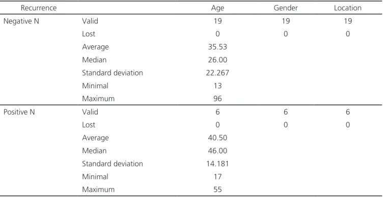
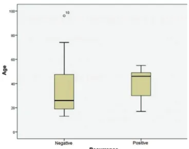
Of the total number of patients with KOT, 24% presented recurrence. Among those who relapsed, the predominant age group was from 41 to 50 years. Weassessed the relationship between age and relapse (Figure 1) with the Mann Whitney test, which did not reveal statistical significance (p>0.05). The age distribution of patients with odontogenic keratocysts can be seen in Table 1.
The patients’mean age in the positive group was 40.5, and in the negative, 35.53. In the positive group, the minimum age was 17 years and the maximum, 55. In the negative relapse group, the minimum age was 13 years and the maximum, 96 (Table 1).
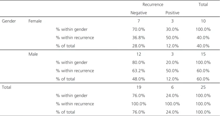
The positive group had an equal gender distribution, differently from the negative group, which presented a male preference (Table 2). However, the assessment of relapse in relation to gender, which we performed through the Fisher’s exact test, was not statistically significant (p>0.05).
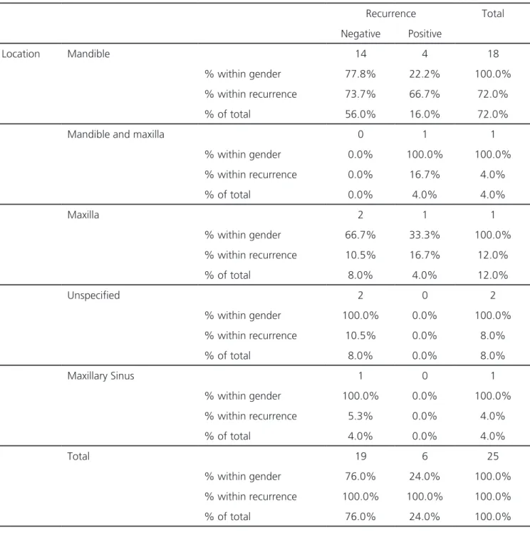
As for location, there was predilection for the mandible, with 56% of the cases negative for relapse; Among the relapsingcases, the mandible was also the most frequent location (Table 3).
Table 1. Average, median, standard deviation, minimum and maximum age between groups.
Recurrence Age Gender Location
Negative N Valid 19 19 19
Lost 0 0 0
Average 35.53
Median 26.00
Standard deviation 22.267
Minimal 13
Maximum 96
Positive N Valid 6 6 6
Lost 0 0 0
Average 40.50
Median 46.00
Standard deviation 14.181
Minimal 17
DISCUSSION
The keratocystic odontogenic tumor is a maxillary odontogenic lesion of epithelial development, affecting mainly the maxilla and the mandible. Few published studies evaluated KOT regarding gender, age and location in a given region or country based on the 2005 WHO classification7-12. In our study, KOT was the second most common lesion, differing from the studies by Chrysomali et al.13 and Johnson et al.14, in which KOT was more prevalent.
In the present study of the 96 cases, KOT represented 26.04% of them, presenting a higher frequency when compared with the epidemiological data described by Meningaud et al.12, who analyzed 695 cases diagnosed as odontogenic cysts and observed odontogenic keratocysts in 19,1%. Siriwardena et al.15 investigated the frequency of odontogenic tumors in a given population in Sri Lanka, showing a KOT incidence of 25.7%. Tawfik et al.16 reported an incidence of 19.5%.
In 2012, Servato et al.17 reported the cases diagnosed at the Federal University of Uberlândia, Brazil, and described KOT as one of the most frequent odontogenic tumors, with a rate of 31.7%. Luo et al.18
reported 1309 cases between 1985 and 2006, and Avelar et al.19 observed a higher frequency of KOT than in the present study; the two rates were, respectively, 38.73% and 30%.
Chirapathomsakul et al.8 analyzed KOT recurrence and observed seven recurrences (22.6%) in their study, which corroborates the data seen in the present study, in which six cases recurred (24%); Of these, 50% appeared in the age group of 41 to 50 years. Madras and Lapointe et al.7 studied 21 KOT patients, and the proportion of recurrence of these lesions was 29%. Regezi et al.5 reported a recurrence rate of 10 to 30%. This explains the importance of the patient’s prolonged clinical and radiographic follow-up after removal of the odontogenic keratocystic tumor.
According to Katase et al.20, KOT is a benign cystic neoplasm that may be associated with basal cell nevus carcinoma syndrome, characterized by multiple cystic lesions. Of the 25 cases of KOT considered in the present study, one case had the described syndrome. Ramaglia et al.21 reported um case of an eight-year-old girl affected by basal cell nevus carcinoma syndrome and Habibi et al.10 reported 8.1% of bearers of this syndrome.
Table 2. Distribution of patients by gender between groups.
Recurrence Total
Negative Positive
Gender Female 7 3 10
% within gender 70.0% 30.0% 100.0%
% within recurrence 36.8% 50.0% 40.0%
% of total 28.0% 12.0% 40.0%
Male 12 3 15
% within gender 80.0% 20.0% 100.0%
% within recurrence 63.2% 50.0% 60.0%
% of total 48.0% 12.0% 60.0%
Total 19 6 25
% within gender 76.0% 24.0% 100.0%
% within recurrence 100.0% 100.0% 100.0%
According to Lopes et al.6, KOT is a differential diagnosis of odontogenic cysts or tumors, such as ameloblastoma, giant cell central granuloma, dentigerous cyst, adenomatoid odontogenic tumor, ameloblastic fibroma, traumatic bone cyst, central giant cell granuloma, lateral periodontal cyst and Gorlin’s cyst. Regezi et al.5 point out, as odontogenic lesions that are KOT’s differential diagnosis, dentigerous cyst, ameloblastoma, odontogenic myxoma, adenomatoid odontogenic tumor and ameloblastic fibroma. Neville et
al.4 emphasize that the absence of KO bone expansion helps the differential diagnosis with root cyst and dentigerous cyst. In the present study, the differential diagnosis of KOT were: ameloblastoma, dentigerous cyst, radicular cyst, central giant cell granuloma, traumatic bone cyst, Gorlin’s cyst, residual cyst and odontogenic myxoma, which corroborates the literature’s findings.
The mandible is the most frequent site of odontogenic keratocyst1,4,7,9,10,11,13,22-24. According to Neville
et al.4, the mandible is affected in 60% or 80% of cases. Table 3. Anatomical location of the KOT between groups.
Recurrence Total
Negative Positive
Location Mandible 14 4 18
% within gender 77.8% 22.2% 100.0%
% within recurrence 73.7% 66.7% 72.0%
% of total 56.0% 16.0% 72.0%
Mandible and maxilla 0 1 1
% within gender 0.0% 100.0% 100.0%
% within recurrence 0.0% 16.7% 4.0%
% of total 0.0% 4.0% 4.0%
Maxilla 2 1 1
% within gender 66.7% 33.3% 100.0%
% within recurrence 10.5% 16.7% 12.0%
% of total 8.0% 4.0% 12.0%
Unspecified 2 0 2
% within gender 100.0% 0.0% 100.0%
% within recurrence 10.5% 0.0% 8.0%
% of total 8.0% 0.0% 8.0%
Maxillary Sinus 1 0 1
% within gender 100.0% 0.0% 100.0%
% within recurrence 5.3% 0.0% 4.0%
% of total 4.0% 0.0% 4.0%
Total 19 6 25
% within gender 76.0% 24.0% 100.0%
% within recurrence 100.0% 100.0% 100.0%
The present study confirms the data from the literature. Among the cases studied, a simultaneous occurrence was found in the maxilla and mandible, as reported by Auluck
et al.25. In the present study there was a case in which the KOT was present in the maxillary sinus.
Our study showed that KOT was more frequent in males. This is similar to that reported in other studies1,4,11,13,15,16,22. Avelar et al.19 and Mallman
et al.11 contradict the literature data, presenting a higher frequency in the female gender.
The mean age was 36.72, higher than that observed in two previous studies, by Habibi et al.10 and Avelar et al.19. Chirapathomsakul et al.8 showed that the most frequent age group is 11-40 years. The current study found that the age group with the highest occurrence of KOT was 10-20 years, which is confirmed by other papers11,13,17. Sekerci et al.2 observed a higher frequency of KOT in the age group between 20 and 29 years.
Madras and Lapointe7 performed more aggressive treatment in 21 patients with KOT, with resection or enucleation complemented with Carnoy’s solution, with or without peripheral ostectomy. Regezi et al.5, on their turn, cited enucleation with peripheral bone curettage or ostectomy as a preferred management method. Habibi et al.10 demonstrated that marsupialization followed by enucleation was more efficient, with no recurrences. The treatment performed in the cases of the present study was enucleation with cyst curettage.
Our results and the literature review expose the need for new scientific work on KOT and its characteristics, which we consider to be of utmost importance for the institution of the most adequate treatment to minimize recurrence of this odontogenic lesion.
REFERENCES
1. Amorim RFB, Godoy GP, Figueiredo CRL, Pinto LP. Ceratocisto odontogênico: estudo epidemiológico de 26 casos. Rev Odonto Ciên. 2003;18(39):23-30. 2. Santo AMB, Yurgel LS. Ceratocisto odontogênico: ava-liação das variantes histológicas paraceratinizada e or-toceratinizada. Rev Odonto Ciên. 1999;14(27):61-86.
3. Marques JAF, Neves JL, Alencar DA, Lemos IM, Mar-ques LC. Ceratocisto odontogênico: relato de caso. Siti-entibus. Rev Univ Est Feira de Santana. 2006;34:59-69. 4. Neville BW, Damm DD, Allen CM, Bouquot JE, edi-tores. Patologia oral e maxilofacial. 3rd ed. Philadel-phia: Elsevier; 2009.
5. Regezi JA, Sciubba JJ, Jordan R, editores. Patologia oral: correlações clinicopatológicas. 5ª ed.
Philadel-Figure 1. Age of patients according recurring or non recurring KOT.
Objetivo: avaliar a frequência do tumor odontogênico ceratocístico(TOC) no Serviço de Cirurgia Oral (SCO) do Hospital Universitário Clementino Fraga Filho da Universidade Federal do Rio de Janeiro (HUCFF/UFRJ), no que diz respeito à taxa de recidiva, ao sexo, à idade de recorrência e à localização da lesão. Métodos: foram examinados os prontuários clínicos e laudos histopatológicos de pacientes do SCO do HUCFF/UFRJ no período de 2002 a 2012. Os pacientes diagnosticados com TOC foram divididos em dois grupos quanto à ocorrência de recidiva: positivo (n=6) e negativo (n=19). Resultados: com relação à localização, houve predileção pela mandíbula. Em relação à média de idade dos pacientes, no grupo positivo foi 40,5, e no grupo negativo, de 35,53. Na distribuição por sexo, o grupo positivo apresentou distribuição igualitária, diferentemente do observado no grupo negativo, em que predominou o sexo masculino.
Conclusões: o TOC representou a segunda lesão mais frequente em nossos pacientes, tem menor recidiva no sexo masculino e tem a mandíbula como localização mais acometida.
Descritores: Tumores Odontogênicos. Recidiva. Diagnóstico Diferencial.
phia: Elsevier; 2008.
6. Lopes MWF, Souza GFM, Carvalho EJA, Gondola AO. Aspectos clínico-morfológicos do queratocisto odontogênico: relato de caso. Odontol Clín Cient. 2004;3(1):61-5.
7. Madras J, Lapointe H. Keratocystic odontogenic tu-mour: reclassification of the odontogenic keratocyst from cyst to tumour. J Can Dent Assoc. 2008;74(2):165. 8. Chirapathomsakul D, Sastravaha P, Jansisyanont P.
A review of odontogenic keratocysts and the be-haviour of recurrences. Oral Surg Oral Med Oral Pa-thol Oral Radiol Endod. 2006;101(1):5-9.
9. Lima GM, Nogueira RLM, Rabenhorst SHB. Consi-derações atuais sobre o comportamento biológico dos queratocistos odontogênicos. Rev Cir Trauma-tol Buco-Maxilo-Fac. 2006;6(2):9-16.
10. Habibi A, Saghravanian N, Habibi M, Mellati E, Habibi M. Keratocystic odontogenic tumor: a 10-year retrospective study of 83 cases in a Iranian population. J Oral Sci. 2007;49(3):229-35.
11. Mallmann CT, Vieira RR, Silva SO, De Carli BMG, De Carli JP. Tumor odontogênico ceratocístico - levantamento de casos e revisão de literatura. Odonto. 2012;20(40):67-72.
12. Meningaud JP, Oprean N, Pitak-Amnop P, Ber-trand JC. Odontogenic cysts: a clinical study of 695 cases. J Oral Sci. 2006;48(2):59-62.
13. Chrysomali E, Leventis M, Titsinides S, Kyriakopou-los V, Sklavounou A. Odontogenic tumors. J Cra-niofac Surg. 2013;24(5):1521-25.
14. Johnson NR, Savage NW, Kazoullis S, Batstone MD. A prospective epidemiological study for odonto-genic and non-odontoodonto-genic lesions of the maxilla and mandible in Queensland. Oral Surg Oral Med Oral Pathol Oral Radiol. 2013;115(4):515-22. 15. Siriwardena BS, Tennakoon TM, Tilakaratne WM.
Relative frequency of odontogenic tumors in Sri Lanka: analysis of 1677 cases. Pathol Res Pract. 2012;20(4):225-30.
16. Tawfik MA, Zyada MM. Odontogenic tumors in Da-kahlia, Egypt: analysis of 82 cases. Oral Surg Oral Med Pathol Oral Radiol Endod. 2010;109(2): e67-73. 17. Servato JP, Prieto-Oliveira P, de Faria PR, Loyola
AM, Cardoso SV. Odontogenic tumours: 240 cas-es diagnosed over 31 years at a Brazilian
univer-sity and a review of internacional literature. Int J Oral Maxillofac Surg. 2012;42(2):288-93.
18. Luo HY, Li TJ. Odontogenic tumors: a study of 1309 cases in a Chinese population. Oral Oncol. 2009;45(8):706-11.
19. Avelar RL, Antunes AA, Santos TS, Andrade ESS, Dourado E. Tumores odontogênicos: estudo clíni-co-patológico de 238 casos. Rev Bras Otorrinola-ringol. 2008;74(5):668-73.
20. Katase N, Nagatsuka H, Tsujigiwa H, Gunduz M, Tamamura R, Pwint HP et al. Analysis of the neo-plastic nature and biological potential of sporadic and nevoid basal cell carcinoma syndrome-as-sociated keratocystic odontogenic tumor. J Oral Pathol Med. 2007;36(9):550-4.
21. Ramaglia L, Morgese F, Pighetti M, Saviano R. Odontogenic keratocyst and uterus bicornis in nevoid basal cell carcinoma syndrome: case report and literature review. Oral Surg Oral Med Oral Pathol Oral Radiol Endod. 2006;102(2):217-9. 22. Sekerci AE, Nazlim S, Etoz M, Deniz K, Yasa Y.
Odontogenic tumors: a collaborative study of 218 cases diagnosed over 12 years and comprehen-sive review of the literature. Med Oral Patol Oral Cir Bucal. 2015;20(1):e34-44.
23. Ali M, Baughman RA. Maxillary odontogenic ker-atocyst: a common and serious clinical misdiagno-sis. J Am Dent Assoc. 2003;134(7):877-83. 24. Waldron CA. Cistos e tumores odontogênicos. In:
Neville BW, Damm DD, Allen CM, Bouquot JE. Pa-tologia oral e maxilofacial. 2ª ed. Rio de Janeiro: Guanabara Koogan; 1998. p.485-90.
25. Auluck A, Suhas S, Pai KM. Multiple odontogenic keratocysts: report of a case. J Can Dent Assoc. 2006;72(7):651-6.
Received in: 23/08/2016
Accepted for publication: 15/11/2016 Conflict of interest: none.
Source of funding: none.
Mailing address:
Brenda de Souza Moura