the assessment itself. The usual lips position can be classiied as lips closed (normal), half-open, closed with tension, closed in dental contact, sometimes open, sometimes closed, and open1.
On the other hand, the assessment of usual tongue position is not easy to diagnose, since very often it is not possible to observe the tongue positioning inside the oral cavity. In view of this, one may classify the usual tongue position as non-observable. When it is possible to visualize it, as in cases of mouth breathing, the tongue can be classiied as in the palatine papilla, on the loor of the mouth, with high dorsum or interdental1.
The literature also suggests that the patient is asked about the site the tongue occupies inside the oral cavity2, and this information depends on the
USUAL TONGUE AND LIPS POSITION IN
ANTEROPOSTERIOR AND VERTICAL GROWTH PATTERNS
Posição habitual da língua e dos lábios nos padrões
de crescimento anteroposterior e vertical
Luana Cristina Berwig(1), Rodrigo Agne Ritzel(2) Ana Maria Toniolo da Silva(3),
Carolina Lisbôa Mezzomo(3), Eliane Castilhos Rodrigues Côrrea(4) Eliane Oliveira Serpa(2)
(1) Programa de Residência Multiproissional Integrada em
Gestão e Atenção Hospitalar no Sistema Público de Saúde da Universidade Federal de Santa Maria - UFSM, Santa Maria, RS, Brasil.
(2) Universidade Federal de Santa Maria - UFSM, Santa Maria,
RS, Brasil.
(3) Departamento de Fonoaudiologia da Universidade Federal de Santa Maria – UFSM, Santa Maria, RS, Brasil.
(4) Departamento de Fisioterapia da Universidade Federal de Santa Maria – UFSM, Santa Maria, RS, Brasil.
Conlict of interest: non-existent ABSTRACT
Purpose: to study the usual tongue and lips position in anteroposterior and vertical growth patterns
in children with mixed dentition. Methods: the sample comprised 54 children, aged seven to 11 years
old. The selected children were referred for radiographic evaluation and cephalometric analysis, which made it possible to obtain the SNA, SNB and AND angles (anteroposterior growth pattern) and the classiication of the facial type:brachyfacial, mesofacial and dilocofacial (vertical growth pattern). The tongue and lips position was determined from the observation of cephalometric radiographs made by two speech therapists experienced in orofacial motricity. The usual tongue position was classiied as in the papilla, high dorsum or on the loor of the mouth, and the usual lips position, as closed or half-open/open. In order to verify the relationship between the usual tongue and lips position with anteroposterior and vertical growth patterns, statistical tests like Analysis of variance, Student’s t test, Mann-Whitney U and chi-square test at a signiicance level of 5% was used. Results: a statistically signiicant relationship between the tongue position and the SNB angle was identiied, children with tongue position on the loor of the mouth showed signiicantly lower SNB angle than children with tongue position in the papilla. SNB angle was a statistically signiicant lower in children with open or half open lips than children with closed lips. There was no difference between the normal position of the tongue and lips in other growth patterns anteroposterior and vertical growth. Conclusion: The usual position of lips and tongue were related to mandibular growth pattern and hasn’t been inluenced by facial type.
KEYWORDS: Radiography; Dental; Evaluation; Tongue; Lip; Face
INTRODUCTION
Considering the scarcity of studies found on this matter and trying to contribute to the clinical practice in the area of orofacial motricity, this research was carried out aiming to study the usual tongue and lips position in the anteroposterior and vertical growth patterns in children during the mixed dentition phase.
METHODS
This study was registered and approved by the Research Ethics Committee of the institution of origin under the protocol number 220.0.243.000-8. It presents an analytic transversal character. The sample comprised children from four schools in the state education network from a municipality in the state of Rio Grande do Sul - RS. The children agreed with the participation in the study and had the Informed Consent Term signed by their respective guardians.
The inclusion criteria were: children aged between seven and 11, to be in the mixed dentition phase and to be caucasian. The children whose respective cephalometric radiography didn’t allow the visualization of lips and tongue position, who presented history of phonologic and/or orthodontic and/or orthopedic treatment, evident signs of neuro -logical involvement and/or craniofacial syndromes and malformations were excluded.
The children selected according to the study criteria were referred for cephalometric assessment. This assessment was carried out from the teleradi -ography in lateral norm, with the use of the Kodak® T-Mat 18x24 radiographic ilm, placed in metallic chassis, covered with screen Kodak® lanex regular, in Soredex Cranex Tome Ceph. The ilm devel -opment was performed in automatic dental ilm processor Revel with Kodak® luids (developer and ixer). The image obtained through teleradiography was digitalized and inserted in the CDT program.
From the cephalometric measures obtained, the following angles regarding the anteroposterior growth pattern11 were considered in this study:
1. SNA: denotes a sagital relation of the maxilla in relation to the skull base. Its increase denotes maxillary protusion and the decrease points to a maxillary retrusion. Average clinical norm: 820.
2. SNB: denotes a sagital relation of the mandible in relation to the skull base. Its increase denotes mandibular protusion and the decrease points to mandibular retrusion. Average clinical norm: 800.
3. ANB: difference between the angles SNA and SNB. It deines the maxilla and mandible degree of perception of the patient’s oral
strucu-tures. In this context, a recent study showed that the coniability of the information provided by the individuals in a sample regarding the usual tongue position was low, either in children or in adults, even after stimulating intraoral perception with a spatula3.
In view of the dificulty assessing the usual tongue position, some instrumental resources can be found in the literature that have been used aiming to overcome this dificulty, and the use of cepha -lometric teleradiography is the most commonly reported, serving as the basis for cephalometric tracing4,5.
The observation of the tongue through teleradi -ography brings important information to deine the feasibility of the adequacy of tongue positioning with the existing fuctional space. The tongue position observed will depend on the tongue size and tension, the palatine tonsils size, the possibility of nasal airlow, the bony basis position and size, the hard palate morphology, the dento-occlusal condition and the facial typology6.
From the cephalometric teleradiography, the usual lips position can also be classiied. This should be analysed taking into account the respi -ratory mode displayed by the patient, the tension of the lips, the overjet, the size of the lower third of the face in relation to the middle third and the maxil -lomandibular bony bases size6.
As can be observed, according to the literature the lips and tongue position may vary according to the size of the skull bony bases (anteroposterior pattern) and the facial typology (vertical pattern) the individual presents, and theses aspects can be obtained from cephalometric tracing.
The anteroposterior bony bases relation allows the facial proile classiication. The individuals with skeletal pattern Class I display straight proile, and the mandible is directly below the maxilla. The individuals with skeletal pattern Class II display a convex proile, associated with a mandibular reduction, maxillary projection, or both, in relation to the cranial base. The skeletal Class III determines the concave proile due to the mandibular increase, maxillary reduction or both in relation to the cranial base7,8.
constricted, or against the inferior incisors, or interdentalized.
• Classiication of lips position:
1. Closed: lower lip in contact with the superior lip. 2. Half-open or open: when the lower lip was not
in contact with the upper lip.
In order to verify the relation between the tongue position and the the mean values of the horizontal angles clinical norm (SNA, SNB e ANB) the Analysis of Variance (ANOVA) was used, and when a signiicant statistical difference was veriied, multiple comparisons were made. In order to verify the relation between the lips position and the mean values of the horizontal angles clinical norm, the Student’s t-test was used for comparing the values of SNA and ANB horizontal angles, and the Mann-Whitney U test for comparison between the values of SNB, since the latter didn’t show a normal distribution. Ir order to verify the relation between the tongue position and the lips position with the horizontal angles (SNA, SNB and ANB) and with the vertical growth pattern (facial type) the Chi-square test was applied. In the statistical analyses, the level of signiicance 5% (p<0,05) was used.
The analyses were made with the use of the SPSS 17.0 software.
RESULTS
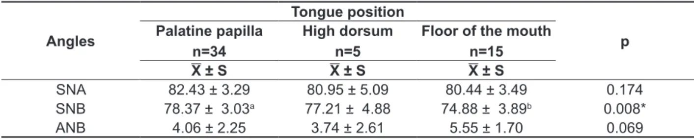
Table 1 shows the study of the relation between the tongue position and the mean values of SNA, SNB and ANB angles. It was veriied that the children with tongue position on the loor of the mouth display a SNB angle signiicantly smaller than the children with the usual tongue position on the palatine papilla.
anteroposterior relation. Average clinical norm: 20.
The vertical growth pattern was also determined from the classiication of the facial type, by calcu -lating the VERT index of the Ricketts cephalometric analysis12. The cephallometric points in this analysis
are based on ive cephalometric measures: the facial axis angle, facial depth, mandibular plane angle, lower facial height and mandibular arc. According to this index value, the facial types are as follows: brachifacial (VERT index higher than 0,5), mesofacial (VERT index between -0,5 and +0,5), dolicofacial (VERT index less than -0,5).
In order to contemplate the objectives of this study, the tongue and lips position was determined from the observation of the cephalometric radio -graphs made by two speech therapists experienced in orofacial motricity, whose evaluation was made individually. In the cases where the responses between the speech therapists were different, a new evaluation was made in conjunction. Thus, of the 55 cephalometric radiographs, only one was excluded by de evaluators for not reaching a consensus regarding the tongue position.
After a careful evaluation of the tongue and lips position, a classiication of the veriied positions was made, wich made it possible the analysis of data as follows.
• Classiication of tongue position:
1. In the palatine papilla: Apex of the tongue elevated with high dorsum or lowered;
2. High dorsum: high dorsum with the apex lowered and/or constrained.
3. Floor of the mouth: apex and lowered dorsum on the loor of the mouth, with the tongue either
Table 1 – Relation between the tongue position and the mean values of the anteroposterior angles (SNA, SNB and ANB)
Angles
Tongue position
p Palatine papilla
n=34
High dorsum n=5
Floor of the mouth n=15
X ± S X ± S X ± S
SNA 82.43 ± 3.29 80.95 ± 5.09 80.44 ± 3.49 0.174
SNB 78.37 ± 3.03a 77.21 ± 4.88 74.88 ± 3.89b 0.008*
ANB 4.06 ± 2.25 3.74 ± 2.61 5.55 ± 1.70 0.069
smaller in children with lips position half-open or open when compared to children with lips position closed.
Table 2 shows the relation between the lips position and the mean values of the SNA, SNB and ANB angles. It was veriied a SNB angle signiicantly
Table 2 – Relation between lips position and mean values of the anteroposterior angles (SNA, SNB and ANB)
Angles
Lips position
p Closed
n=45
Half-open or open N=9
X ± S X ± S
SNA 81.98 ± 3.58 80.54 ± 3.48 0.273
SNB 77.69 ± 3.69 75.34 ± 3.51 0.036*
ANB 4.30 ± 2.29 5.20 ± 1.76 0.271
X=average; S=standard deviation; p=signiicance value; *signiicance by Mann-Whitney U test (p <0.05).
Figure 1 shows the relation of lips position with the facial types. No difference was veriied between the usual lips position in the vertical growth patterns.
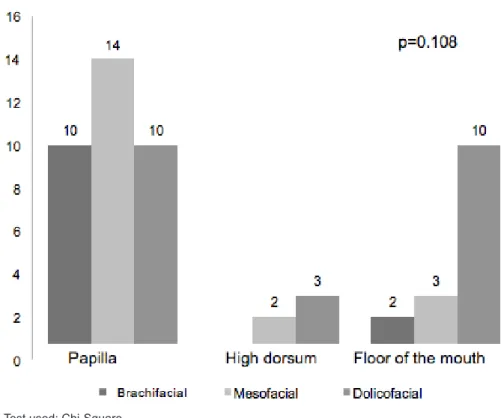
Figure 2 shows the relation of tongue position with the facial types. No difference was veriied between the usual tongue position in the vertical growth patterns.
Test used: Chi-Square
the tongue position and the SNB angle. From the multiple comparisons, it was determined that the children with the tongue on the loor of the mouth displayed the mean SNB angle value smaller than the children with the tongue position on the papilla. It is believed that the mean SNB value closer to normality in the children with tongue position on the palatine papilla has favored the correct positioning of the tongue because of the suficient intraoral space.
The literature reports that in the cases of mandibular prognathism, the tongue occupies the loor of the mouth, and can be bulky, hypotensive and projected13,14. On the other hand, when mandibular retrognathism occurs with reduction in the anteroposterior space, the usual tongue position shows lowered apex with dorsum more elevated, which can be located between the dental arcs in case there is a concomitant maxillary retrusion14,15.
From the analysis of the results on Table 2, it was observed that children with half-open or open lips present the mean SNB angle value smaller than that of children with closed lips.
The mandibular retrognathism does not favor the labial sealing, once the bony bases must be in balance in order to have the apropriate labial oclusion, with an ANB angle in a relation of two degrees. In the cases of mandibular retrognathism, it can be veriied the anterior sealing with the lower
DISCUSSION
Taking into account the dificulty of assessing the usual tongue positioning due to the frequent unvia -bility of visualization of this structure inside the oral cavity, radiological techniques have been used to complement the insuficient clinical assessment4,5.
The use of barium sulfate on the mouth to perform cephalometric radiographs has salso been proposed, as it facilitates the visualization of several regions of the tongue4,5.
In this study, the option was to perform a telera -diography without the use of contrast on the tongue, aiming a better illustration of the clinical practice, where cephalometric radiographs are requested by dentists, and sometimes the speech therapist has access to cephalometric documentation previously requested during the orthodontic treatment.
It should be noted that in this study, of the 55 cephalometric radiographs analyzed, only one radiograph was escluded by the evaluators due to a lack of consensus regarding the tongue position because of the dificulty of visualizing the entire structure. No studies were found that have analyzed the agreement between evaluators regarding the classiication of the tongue and lips positioning.
Concerning the results, it can be observed on Table 1 the statistically signiicant relation between
Test used: Chi-Square
in Figure 2 makes it possibel the observation that the tongue positioning on the loor of the mouth seems to have been favored by the dolicofacial pattern, being in accordance with the literature9.
In the craniofacial growth pattern predominantly vertical, due to the increase of the lower third of the face and the tension decrease in the orofacial musculature, the mandible and tongue lowered position is favored, many times favoring the mouth breathing instalation, which may be the cause or consequence of the dolicofacial pattern6,10,18,19.
CONCLUSION
One may conclude with this study that the usual lips and tongue position in children in the mixed dentition phase showed relation with the mandibular growth pattern, and was not inluenced by the facial type.
lip occluding on the upper incisors, half-open lips and the upper lip hypofunction14.
From the descriptive analysis of data in Figure 1, it was also possible to observe that the lips often present sealed in mesofacial and brachifacial children, because the growth pattern in these facial types is respectively balanced and horizontal.6,10 In dolicofacial children, there was an increase in the frequency of half-open and open lips, because in this case there is a prevalence of vertical growth6,9,10,16, mainly in the lower third, which makes it dificult for the lower lip to reach in the direction of the upper lip. Besides, in the dolicocefacials the mandible levator muscles are more stretched out and less powerful, resulting in a lowered mandibular position, which also compromises the proper usual lips position10,17.
No statiscally signiicant difference was veriied between the tongue position and the facial type (Figure 2). However, the descriptive analysis of data
RESUMO
Objetivo: estudar a posição habitual da língua e dos lábios nos padrões de crescimento
anteroposte-rior e vertical de crianças em fase de dentição mista. Métodos: a amostra foi constituída por 54
crian-ças, na faixa etária entre sete e 11 anos. As crianças selecionadas foram encaminhadas para ava-liação radiográica e análise cefalométrica, que possibilitou a obtenção de ângulos SNA, SNB e ANB (padrão de crescimento anteroposterior) e da classiicação do tipo facial entre braquifacial, mesofacial e dolicofacial (padrão de crescimento vertical). A posição da língua e dos lábios foi determinada a par-tir da observação das radiograias cefalométricas por duas fonoaudiólogas com experiência na área de motricidade orofacial. A posição habitual da língua foi classiicada como na papila palatina, com dorso elevado ou no assoalho oral, e a posição habitual dos lábios, como fechados ou entreabertos/ abertos. Para veriicar a relação entre a posição habitual da língua e dos lábios com os padrões de crescimento anteroposterior e vertical foram utilizados os testes estatísticos Análise de Variância, t de Student, U de Mann-Whitney e Qui-Quadrado, ao nível de signiicância de 5%. Resultados:
veriicou--se relação estatisticamente signiicante entre a posição da língua e o ângulo SNB, sendo que as crianças com posição de língua no assoalho oral apresentaram ângulo SNB signiicantemente menor do que as crianças com posição habitual de língua na papila palatina. Veriicou-se ângulo SNB signi-icantemente menor nas crianças com posição de lábios entreabetos ou abertos quando comparadas às crianças com posição de lábios fechados. Não houve diferença entre a posição habitual da língua e dos lábios nos demais padrões de crescimento anteroposterior e de crescimento vertical. Conclusão:
a posição habitual de lábios e de língua apresentou relação com o padrão de crescimento mandibular, não tendo sido inluenciada pelo tipo facial.
do palato duro em diferentes tipologias faciais de respiradores nasais e orais. Rev CEFAC. 2011;ahead of print:0.
11. Rahal A, Pierotti S. Eletromiograia e cefalometria na fonoaudiologia. In.: Ferreira LP, Bei-Lopes DM, Limongi SCO. Tratado de Fonoaudiologia. São Paulo: Roca; 2004, p.237-53.
12. Ricketts RM, Roth RH, Chaconas SJ, Schulhof RJ, Engel GA. Orthodontic diagnosis and planning their roles in preventive and rehabilitative dentristy. 1 ed. Denver: Rocky Mountain; 1982.
13. Aléssio CV, Mezzomo CL, Körbes D. Intervenção Fonoaudiológica nos casos de pacientes classe III com indicação à Cirurgia Ortognática. Arq odontol. 2007;43(3):102-10.
14. Coutinho TA, Abath MB, Campos GJL, Antunes AA, Carvalho RWF. Características cefalométricas do padrão face longa. Adaptações do sistema estomatognático em indivíduos com desproporções maxilo-mandibulares: revisão da literatura. Rev Soc Bras Fonoaudiol. 2009;14(2):275-9.
15. Mory M, Baroni L, Tessitore A, Assencio-Ferreira V. Análise radiográfca da posição habitual da língua nos portadores de distoclusão. Rev CEFAC. 2003;5(6):231-4.
16. Cardoso MA, Bertoz FA, Capelozza Filho L, Reis SAB. Características cefalométricas do padrão face longa. Rev Dental Press Ortodon Ortopedi. 2005;10(2):29-43.
17. Pereira AC, Jorge TM, Ribeiro Júnior PD, Berretin-Felix G. Características das funções orais de indivíduos com má oclusão Classe III e diferentes tipos faciais. Rev Dental Press Ortodon Ortopedi. 2005;10(6):111-9.
18. Castro AMA, Vasconcelos MHF. Avaliação da infuência do tipo facial nos tamanhos dos espaços aéreos nasofaríngeo e bucofaríngeo. R Dental Press Ortodon Ortop Facial. 2008;13(6):43-50. 19. Castro AMA, Teles RP. Infuência do tipo facial no tamanho do espaço aéreo nasofaríngeo. Orto SPO. 2008;41(4):393-8.
REFERENCES
1. Genaro KF, Berretin-Felix G, Rehder MI, Marchesan IQ. Avaliação miofuncional orofacial: protocolo MBGR. Rev CEFAC. 2009;11(2):237-55. 2. Junqueira P. Avaliação miofuncional. In.: Marchesan IQ. Fundamentos em Fonoaudiologia: Aspectos clínicos da motricidade orofacial. Rio de Janeiro: Guanabara Koogan;1998. p. 13-21.
3. Cardoso AFR, Bommarito S, Chiari BM, Motta AR. A coniabilidade da informação fornecida pelo indivíduo a respeito de seu posicionamento habitual de língua. Rev. CEFAC. 2011;13(2):236-44.
4. Tessitore A. Análise radiográica da posição habitual da língua [dissertação]. Campinas (SP): Universidade Estadual de Campinas. Faculdade de Ciências Médicas; 2001.
5. Tessitore A, Crespo AN. Análise radiográica da posição habitual de repouso da língua. Pró-Fono R. Atual. Cient. 2002;14(1):7-16.
6. Bianchini EMG. Avaliação fonoaudiológica da motricidade oral – distúrbios miofuncionais orofaciais ou situações adaptativas. Rev Dental Press Ortodon Ortop Facial. 2001;6(3):73-82. 7. Cardoso KR, Giellow I; Mattos MC. Posicionamento habitual de língua nos padrões faciais anteroposteriores. In.: Marchesan IQ, Zorzi JL, Gomes IC. Tópicos em fonoaudiologia 1997/1998. São Paulo: Editora Lovise; 1998. p.233-59.
8. Silva Filho OG, Queiroz APC, Herkrath FJ, Silva GFB. Correlação entre padrão facial e relação sagital entre os arcos dentários no estágio de dentadura decídua: considerações epidemiológicas. R Dental Press Ortodon Ortop Facial. 2008;13(1):101-12. 9. Ramires RR, Ferreira LP, Marchesan IQ, Cattoni DM, Andrada e Silva MA. Tipologia facial aplicada à Fonoaudiologia: revisão de literatura. Rev Soc Bras Fonoaudiol. 2010;15(1):140-5.
10. Berwig LC, Silva AMT, Côrre ECR, Moraes AB, Montenegro MM, Ritzel RA. Análise quantitativa
Received on: May 17, 2012 Accepted on: December 06, 2012
Mailing address: Luana Cristina Berwig
Rua Coronel Niederauer, número 792, apartamento 202
Bonim - Santa Maria - RS CEP: 97015-120