Dengue
E
RICM
ARTÍNEZT
ORRESDefinition
D
ENGUE is a viral illness, of an endemic-epidemic nature, transmittedby mosquitoes of the genus Aedes, especially by Aedes aegypti. It
constitutes nowadays, at the global level, the most important arbovirosis in terms of morbidity, lethality and economic implications (Guzmán et al., 2004; Kindhauser, 2003), and presents several forms of clinical expression: from an undifferentiated fever (frequent in children) and fever with headache, intense general malaise, ostheomyoarticular pain with or without rash, leucopenia and some kind of bleeding, until severe forms that – having started with the previous symptoms – evolve to hypovolemic shock due to leakage of plasma, with moderate or intense thrombocytopenia, and with large hemorrhages in the gastrointestinal tract and other locations. Dengue can also express itself by means of the so-called “atypical” forms, which are relatively infrequent and result from the particularly intense damage to an organ or system: encephalopathy, myocardiopathy or hepatopathy by dengue, among others (Martínez, 1995; Martínez, 1997).
Etiology
Dengue is constituted by four serologically differentiable viral serotypes (Dengue 1, 2, 3 and 4) sharing structural and pathogenic analogies, which means that any of them can cause the severe forms of the illness - although the serotypes 2 and 3 have been associated to the higher number of severe cases and deaths. Those viruses are constituted by spherical particles with 40 to 50 nm in diameter, present in the structural proteins of the envelope (E), membrane (M) and capsid (C), as well as a ribonucleic acid (RNA) genome. They also contain other non-structural (NS) proteins: NS1, NS2A, NS2B, NS3, NS4A, NS4B, and NS5-3. The dengue viruses belong to the genus Flavivirus of the Flaviviridae family (Gubler, 1998).
The viruses of dengue and the host’s response
antibody-dependent amplification (ADA) that is expressed in an enhanced viral replication and an increase in the viremia, which promotes and favors the development of the severe form of the illness (Guzmán et al., 1992; Halstead, 2002).
Epidemiology
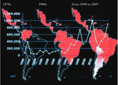
Nearly half the world population is in risk of suffering this infection for living in tropical and subtropical areas, as well as over 400 million European and North-American travelers that, each year, cross the borders and return to their countries from Asia, Africa and Latin-America (Wichmann et al., 2007; Pinazo et al., 2008). The global prevalence of dengue has increased dramatically along recent years. Fifty million infections, half a million patients in hospitals and over 25 thousand deceased per year are estimated. Approximately 100 countries have reported cases of dengue and / or hemorrhagic dengue, and over 60 of them do it regularly every year (WHO, 1997; Jacobs, 2000), reason why the World Health Organization (WHO) regards this condition as one of the main health issues of mankind, in addition to the fact that it causes serious social and economic implications. In the region of the Americas, a progressive increase in the number of dengue cases has occurred along the last three decades (Kouri, 2006), and the illness has reached almost the total number of countries (Figure 1).
Source: OPS/OMS.
Figure 1 – Evolution of the situation of dengue and DHF (dengue hemorrhagic fever) in the Americas, 1980-2007.
In order to enable the transmission of the illness in a given town, region or country, the virus, the vector and the susceptible host have to be present there simultaneously. When infected and going through the viremia stage (five to seven days), the host is the reservoir of the disease. All known vectors that can transmit
the four serotypes of the dengue virus belong to the genus Aedes, of which the
Aedes aegypti is the most important. This species follows the human being inside his
/ her residence and at its surroundings, since the female prefers the human blood and stings, mainly during daylight, one or several individuals in order to produce each laying of minute eggs, which is performed in natural or artificial reservoirs of water, until these eggs become larvae, pupae and adult mosquitoes. The other epidemiologically important species is the Aedes albopictus, imported from Asia in tires brought to the United States and currently present in most countries of the Americas region.
The dengue viruses can only infect man and higher primates if introduced by means of the vector mosquito’s sting. This is the only path of clinical-epidemiological importance, since dengue is not transmitted by the oral, respiratory or sexual paths, like other viruses. However, there is also the infrequent and still poorly documented vertical transmission (Maroun et al., 2008) and the recently reported infection by the transfusional way, apparently with a very scarce occurrence (Blanco, 2008; Tambyah et al., 2008).
Why is dengue a re-emergent disease at global level?
Dengue is a re-emergent illness at global level due to the unusual increase in the vector along the last decades (Calisher, 2005). The Aedes aegypti is a domestic or peridomestic mosquito, of which the female needs human blood in order to maintain its reproduction, and lays its eggs in reservoirs of clean or cleanish water. The eggs become larvae and later pupae, until emerging in adult form. The infecting female can live up to two months and sting several times a day. Other mosquitoes have also demonstrated their vectorial competence, such as the Aedes albopictus, known as “the Asian tiger”, which was taken to America two decades ago and presently infects several countries in Europe.
There are the so-called macro-determinant factors that can explain this increase in dengue in a global scale: those of the climatic kind – such as the global warming – and those of the social kind, such as the increase in the world population, the trend towards a disorderly urbanization, the international trips, and poverty expressed in issues associated to dwelling, education, water supply, collection of solid refuses and others, as well as the lack of effective national and international programs against this disease and its vector (Gubler, 2005). Presently, the control of the vector constitutes the only strategy for the prevention of dengue.
Disease burden
predominance of adults, similarly to what was occurring in Brazil and other South-American countries. In recent years, however, there was a change in the age in which dengue sets in and becomes severe, and its frequency in the pediatric age has increased (Teixeira et al., 2008). The negative effects on the economy result from the high cost of the control of epidemics, the occupational and school absenteeism, and indirect implications on some countries of which the income depends on tourism, among others.
Physiopathology
There are a number of pathogenic theories to explain the severe forms of dengue. According to the sequential theory, a second infection caused by another serotype results in an amplification of the antibodies-mediated infection, or inmuno-amplification, with an extensive viral replication and an increase in the viremia, which determines the severity of the illness (Cummings et al., 2005). Other theories consider that the differences in the pathogenicity of the viral strains explain the severe forms of dengue (Anantapreecha et al., 2005). In practical terms, in a same epidemic of dengue there is a coexistence of factors associated to the host and factors associated to the virus, as well as of epidemiologic or environmental factors.
When the virus is introduced into the skin, the first target cell is the dendritic cell present in the epidermis (Palucka, 2000; Kwan et al., 2005), especially the Langerhans cells, which are activated and present the virus to the lymphocyte T. Similarly, the viruses that have broken into the blood are identified by the monocyte and endothelial cells, which also perform the presenting function. The first lymphocytes to be activated are the CD4 and then the CD8, with the release of cytokines (Cardier et al., 2005).
The immune response of the host may be protective (and lead to the healing) or pathogenic, expressed by a “deregulation” that is characterized by an excessive production of cytokines, as well as by a change in the response from the type TH1 to TH2 (Mabalirajan et al., 2005) and an inversion of the index CD4 / CD8. The excessive effusion of cytokines causes an increase in the vascular permeability that is expressed in a leakage of plasma, which is the fundamental physiopathological alteration of dengue, by means of which water and proteins escape to the extravascular space – resulting in hemoconcentration and, sometimes, hypovolemic shock (Basu, 2008).
The viral infection induces the apoptosis of lymphocytes T along the first days of the infection, which, according to its severity, may exert a favorable influence in the disappearance of the virus or may cause the lysis of extensive amounts of these cells and transitorily decrease the patient’s immune competence, as well as cause damage to other cells and tissues of the host, such as the endothelia, hepatocytes, myocardiocytes, neurons, tubular cells of the kidneys, and others, which could explain the damage to many organs during this infection (Limonta
the peripheral blood by means of an immune-mediated mechanism. The bleedings that occur during the dengue are not directly associated to the intensity of the thrombocytopenia (Gomber et al., s. d.), since they are produced by a whole set of factors (Schexneider & Reedy, 2005). The causes of the bleedings in the dengue are multiple (Srichaikul & Nimmannitya, 2000), including the vascular ones and some alterations in clotting by means of the cross-action of some antiviral antibodies against the plasminogen and other proteins, as well as an unbalance between the mechanisms of coagulation and those of fibrinolysis.
Dengue is a sole disease
The dengue infection may be clinically unapparent and cause an illness with varied intensity, including from febrile forms with body pains and higher or lesser encumbrance to the body, to severe pictures of shock and large hemorrhages. It has been accepted so far that the main difference between the classical dengue or dengue fever (DF) and the dengue hemorrhagic fever (DHF) is not precisely expressed by the bleedings but by the leaking of plasma, particularly when it has a clinical expression and impact by causing a significant elevation in the hematocrit and an accumulation of fluid in serous cavities, such as pleural leakage, ascites and pericardial leakage.
The so varied clinical spectrum of dengue explains the diversity of clinical pictures that may be detected within a same family or population during an epidemic outbreak, since some patients (maybe most of them) are only slightly affected and – mistakenly – do not even look for medical assistance; others present scarce symptoms (oligosymptomatic), and others are intensely affected, presenting a great prostration and perhaps an unfavorable evolution, clinical deterioration and death, sometimes in a couple of hours. Each one of the four viruses of dengue may cause any clinical picture of the aforementioned spectrum.
There are also the clinical forms that, for not being so frequent, are known
as “atypical”, and result from the especially intense damage to an organ or system:
encephalopathy, myocardiopathy or hepatopathy by dengue, as well as kidney dysfunction with acute kidney insufficiency and others that are also associated to mortality (Martínez, 2005).
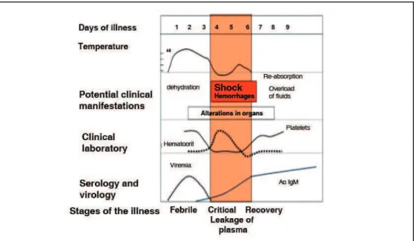
Dengue is a very dynamic illness, in spite of having a very short duration (no more than one week in nearly 90% of the cases). Its expression can change as the days go by and can also worsen suddenly, reason why the patient needs a repeated assistance by the doctor, preferably on a daily basis. The course of the dengue disease goes through three clinical stages: the febrile stage – the single one for the immense majority of patients –, the critical stage and the recovery stage (Figure 2).
towards recovery. Other times, the decrease in the fever is associated to the moment in which the patient’s state worsens, and the period during which the fever subsides indicates, therefore, the start of the critical stage of the illness.
Source: Formulated by Eric M. Torres.
Figure 2 – Progress of the dengue illness.
This is characteristic of dengue: the first day with no fever is the day in which there is a greater risk of the occurrence of complications. The critical stage coincides with the leakage of plasma (escape of fluids from the intravascular to the extravascular space), and its most feared expression is the shock, with coldness in the teguments, weak pulse, tachycardia and hypotension – sometimes associated to large digestive hemorrhages, as well as to damage to the liver and perhaps to other organs. In this stage, the hematocrit is elevated and the platelets – which were decreasing already – reach their lowest amounts. In the recovery stage, the patient’s improvement is usually evident, but on occasion a state of fluid overload occurs, as well as some additional bacterial infection.
Clinical picture
A slight abdominal pain and diarrhea may occur, and the latter is more frequent in those patients with less than two years of age and in adults.
Sequence of the clinical signs in the diagnosis of the clinical forms of dengue
Identifying the sequence of the clinical and laboratory manifestations is very important to differentiate the dengue from another disease that may present similar alterations but with a different order of appearance (leptospirosis, meningococcemia, influenza, sepsis, acute abdomen, and others); and, furthermore, constitutes the only possibility to precociously detect the dengue patient that can evolve or is already evolving towards the severe clinical form of hemorrhagic dengue and shock by dengue. During the first days a rash appears in a variable percentage of the patients; it has not been demonstrated, though, that the rash is a prognostic factor.
The aforementioned manifestations are predominant at least along the first 48 hours of the disease, and can remain for an additional couple of days in the stage that can be regarded as its FEBRILE STAGE, during which it is not possible to know if the patient is going to remain with symptoms and signs of classical dengue all the time and is going to evolve toward a spontaneous healing, or if that is but the
beginning of a severe dengue, with shock and extensive bleedings.
Between day 3 and day 6 for the children, and day 4 and day 6 for the adults (as a more frequent period but not exclusive of the patients that evolve to the severe dengue), the fever decreases, the abdominal pain becomes intense and continuous, a plural or ascitic leakage is detected, vomiting becomes more frequent, and the
CRITICAL STAGE of the illness starts - moment in which the installation of
shock is more frequent. In this stage the hepatomegalia also becomes evident. The presence of signs of alarm is very characteristic of the beginning of this stage, indicating complications such as the shock (Rigau & Laufer, 2006).
The hematocrit is normal at the beginning but starts to get higher and higher, while the radiological studies of the thorax or the abdominal ultrasonography show ascites or right-sided or bilateral pleural leakage. The maximum elevation of the hematocrit coincides with the shock. The platelet count shows a progressive decrease
and reaches its lowest levels during the day of the shock, then starts to increase quickly
and reaches a normal level in a couple of days. The shock occurs with a frequency 4- or 5-fold higher at the moment in which the fever decreases or along the first 24 hours after the fever has completely subsided than during the febrile stage of the disease.
of alarm indicate the moment in which the patient can be saved by receiving a treatment with hydro-electrolytic solutions in amounts sufficient to replace the losses caused by the leakage of plasma, sometimes exacerbated by exterior losses of fluids (sweating, vomiting, diarrhea).
At the beginning, not all clinical signs of shock have to be present. It is enough to detect the narrowing of the differential arterial tension (AT) or pulse pressure (difference of 20 mmHg or less between the maximum or systolic AT and the minimum or diastolic AT), which, in general, has been preceded by signs of hemodynamic instability (tachycardia, coldness, delayed capillary filling, among others). Therefore, it is not necessary to wait for hypotension in order to diagnose shock (Martínez & Velázquez, 2002).
As a general rule, the signs of shock last for a couple of hours. When the shock becomes prolonged or recurrent, that is, lasts for more than 12 or 24 hours, and exceptionally for over 48 hours, the lungs show radiological images of interstitial edema, sometimes similar to pneumonic lesions. Later, a syndrome of difficulty in breathing due to non-cardiogenic pulmonary edema may occur, leading to a darker prognosis.
Following the critical stage, the patient remains, for a variable period of time, in the RECOVERY STAGE, which also requires medical assistance, since during this period the patient must physiologically eliminate the surplus of fluids that had leaked until normalizing all of his / her vital functions; in children and healthy adults, this increased diuresis is well tolerated, but patients suffering of cardiopathies or nephropathies and the elderly should receive special attention. The doctors must also be attentive to a possible bacterial co-infection, usually pulmonary, as well as to the occurrence of the so-called late rash (10 days and later). Some adult patients present asthenia for several days and others report bradypsychia for weeks.
Tests in clinical laboratories and tests by images
The physician assisting a DF patient will probably prescribe a count of leucocytes for detecting the frequent leucopenia, which can be as intense as showing less than 1,000 leucocytes x mm3. The differential formula will evidence
the neutropenia typical of the early stage of the disease, some band cells and atypical lymphocytes. The hematocrit and the platelet count will be the indispensable clinical laboratory tests for the patient suspected of possibly evolving to the severe dengue, with leaking of fluids, shock and hemorrhages, although their execution is not strictly required during the follow-up of the febrile case suspected of dengue in the absence of spontaneous bleedings or – at least – of a positive loop test. The patients requiring hematocrits and platelet counts, in general, require them in a series along several days.
In spite of this, the leucocytes count > 6000 cells / mm3 has been a factor
prognostic indicators due to the frequency of these alterations in patients that had evolved to death, as well as the increase in the transaminases, mainly the GOT (glutamic-oxaloacetic transaminase) (Azevedo et al., 2002).
The study of the patient must be completed according to the clinical picture, the possibilities of the place and the kind of medical attention that he / she is receiving – ambulatory or with hospitalization; the latter may include the execution of a comprehensive coagulogram, erythrosedimentation, total proteins, ionogram, gasometry, urea, creatinine, transaminases or other enzymes in the blood that indicate liver cytolysis (Villar-Centeno et al., 2008), as well as medullogram, as required.
For the differential diagnosis (Bruce et al., 2005; Wilder-Amith et al., 2004), the physician – in given cases – may request a hemoculture, thick film, study of the cerebrospinal fluid (cytochemical and bacteriological), and other more specific tests. The radiological studies of the thorax and the abdominal ultrasonography are very useful in the hemorrhagic dengue, as well as the electrocardiogram and the echocardiogram if a myocardial dysfunction is suspected. With the echocardiogram a pericardial leakage can be identified, but also something that is more important: a decreased myocardial contractility that may be the expression of a myocarditis by dengue.
The radiological study of the thorax (anteroposterior and lateral views) allows the detection of the presence of a pleural leakage, as well as of a cardiomegalia or other thoracic alteration. Along the last decade, the utilization of sonographic studies made possible the early identification of ascites, pleural and pericardial leakage, and the thickening of the gallbladder wall due to edema of the wall, all of which are signs of fluid leakage; and allowed also the diagnosis of fluid accumulation in the perirenal areas, which has been associated to the shock by dengue and cannot be explained but by the capillary escape itself, this time to the retroperitoneal space (Setiawan et al., 1998; Venkata et al., 2005).
How is the infection by dengue confirmed?
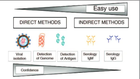
The possibility of performing the culture and isolation of the dengue virus from the patients’ blood during the febrile stage is available. This method is still the golden rule but is expensive and difficult, reason why it is not applicable to most patients. Furthermore, the virology laboratories qualified to perform the culture and isolation are scarce, and the application of techniques of molecular biology for the detection of the viral genome is more feasible. The polymerase chain reaction (PCR) is used to identify the viral serotype and the viral burden as well, in this case by means of the so-called PCR in real time (Guzmán & Kourí, 2004). So far these are the most reliable methods, although they are not the most frequently utilized (Figure 3).
means of the method ELISA or others. The serological study for IgM should not be prescribed before day 5, or should preferably be prescribed as of day 6. This study, therefore, does not constitute an aid for the assisting physician to decide about procedures, since the patient’s state may worsen as of day 3 or day 4. Nevertheless, it is important to prescribe those serological studies, for the laboratory results
complete the diagnostic tripod along with the clinic and the epidemiology. Laboratory
tests to identify viral antigens, particularly to identify some of the non-structural proteins of the dengue virus, exist already (determination of antigens NS1) and are in process of validation and implementation. They are especially useful during the first four days of the febrile stage of the illness.
Laboratory criteria for the confirmation of the diagnosis (WHO, 1997)
The laboratory criteria for the diagnosis confirmation are the following (at least one of them must be present):
• Isolation of the dengue virus from the serum, the plasma, the leucocytes or the autopsy samples;
• Ascertainment of a 4-fold increase in the reciprocal titles of antibodies IgG or IgM against one or several antigens of the dengue virus in matching serum samples;
• Demonstration of the antigen of the dengue virus in autopsy tissues by means of immunochemistry or immunofluorescence tests, or in serum samples by means of immunoassay techniques.
• Detection of viral genomic sequences in the autopsy tissue, the serum or the samples of cephalorachidian fluid by the polymerase chain reaction
Source: Adapted with permission of J. Cardosa.
(PCR).
In spite of not being regarded as a confirmation diagnosis, the elevation of the IgM specific of the dengue, as of the day 6 of the disease, contributes to the diagnosis of the clinical case and the epidemiological monitoring.
Dengue classification
During three decades, the World Health Organization (WHO) has recognized and recommended the classification of dengue in: dengue fever (DF) and dengue hemorrhagic fever (DHF) with or without dengue shock syndrome (DSS). In order to be regarded as a DF (or classical dengue) case, the patient must present fever and two symptoms out of the following: headache, retroocular pain, ostheomyoarticular pains, rash, leucopenia, and some kind of bleeding (WHO, 1997).
The dengue hemorrhagic fever requires the presence of the four following criteria: a) fever (or the patient having presented fever along last week), some kind of spontaneous bleeding – usually petechias, or other – or, at least, having a positive loop test; c) thrombocytopenia lower than 100,000 per mm3; and d) plasma
leakage, evidenced by a 20% elevation of the hematocrit, or by a 20% decrease of the hematocrit after the critical stage, or by the verification of pleural leakage, ascites or pericardial leakage by means of image studies, usually sonography (Pan-American Health Organization, 1995).
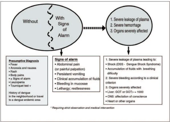
In recent years, articles have been published (Balmaseda et al., 2005; Setiati et al., 2007) that bring into question the usefulness of this classification for regarding it as too stern, much too dependent on laboratory results, and for not including dengue patients with other severe forms of the illness, such as the particular damage to the Central Nervous System (encephalitis), to the heart (myocarditis) or to the liver (severe hepatitis). It was not useful for the clinical management of the patients, either. For this reason, the TDR/WHO (Program of Training and Research on Transmissible Diseases of the World Health Organization) has sponsored an international study, named DENCO (Dengue Control), of which one of the components was of clinic, and which main purpose was to obtain information from a high number of patients with confirmed dengue and find out a better way to classify them, as well as to identify those signs of alarm that could be useful to improve the protocol of management of dengue cases.
Clinical information was obtained from nearly 2,000 patients with confirmed dengue, proceeding from seven countries in two continents. The study concluded that 18 to 40% of the cases could not be classified by means of the current WHO Classification, and over 15% of cases with shock could not be classified as severe cases of dengue either, since they did not comply with some of the criteria to be regarded as a case of DHF/DSS. The study had also another consistent result in the proposal of a binary classification of the disease: DENGUE and SEVERE DENGUE.
leakage, expressed by hypovolemic shock, and / or by breathing difficulty due to the excess of fluids accumulated within the lungs; b) severe hemorrhages, according to the criterion of the assisting physician; and c) damage to organs: severe hepatitis by dengue (transaminases higher than 1000 units), encephalitis by dengue, or a serious damage to other organs, such as the myocarditis by dengue. These severity criteria had 95% of sensitivity and 97% of specificity (Figure 4).
Source: Formulated by Eric M. Torres.
Figure 4 – Classification of dengue.
The DENCO study allowed also the identification of some signs and symptoms that were present in the patients one day before the worsening of their state. Those signs of alarm allow the early identification of the dengue patient that is going to evolve to Severe Dengue and – mainly – provides the physician with the opportunity of starting an early treatment with the replacement of fluids intravenously and, by doing so, improving the patient’s prognosis. The abdominal pain or the pain reported with the abdomen palpation was a significant factor of risk for adults and children, as well as the bleeding in mucosae and the thrombocytopenia lower than 10,000 x mm3. In adults, other signs of alarm
B. Results from the DENCO study. TDR/WHO Expert Meeting on Dengue Classification and Case Management. Implications of the DENCO study. WHO, Geneve, Sep 30-Oct 1/2008).
This new classification counts on a strict scientific support and coincides, in broad outlines, with the criteria of the clinicians that are experts in dengue in our region; but it still needs to be validated in practice, reason why the agreement signed in the headquarters of the World Health Organization was about applying it during a new period in a greater number of countries, in order to assess its usefulness and feasibility in situations of endemicity of dengue and during epidemics of the disease, both in hospitals and in units of Primary Assistance to Health.
Crucial data for the treatment of dengue patients
It is not correct to say that dengue and severe dengue cannot be treated. The lack of an antiviral drug or another specific medicine may be successfully replaced by the application of a set of pieces of knowledge that allows the classification of the
patients according to their symptoms and the stage of the illness, as well as the early
acknowledgement of the signs of alarm that announce the imminence of the shock,
allowing the physician to anticipate the complications and decide about the most
adequate therapeutic procedures (Martínez, 2006).
The doctor should question every feverish patient with a clinical and epidemiological mind, asking him / her to go into detail on the duration of the symptoms as of the first day of fever; in addition to that, the physician should subject the patient to a physical examination, in order to diagnose other causes of fever that also concur during the dengue epidemics. The questions that a doctor should ask himself / herself in face of a patient suspected of dengue are four: A) Does he / she have dengue?, B) Does he / she present any bleeding, any comorbidity or signs of alarm?, C) Is he / she in shock?
The answers to those questions allow the doctor to classify the patient into one of three groups (A, B and C) and decide about procedures:
• To send him / her home, providing him / her with guidance and ambulatory treatment (group A);
• Hospitalization for a close observation and medical treatment (group B); • Urgent intensive treatment (group C).
Group A – patients that can be sent home
associated to sweating, vomiting, or other losses. In order to mitigate the body pains and decrease the fever, the doctor may prescribe paracetamol (never more than 4 g a day for adults, and in the dosage of 10-15 mg x kg of body weight x day in children), as well as the application of water-soaked sponges on the skin to make the temperature drop. Aspirin or non-steroidal anti-inflammatory drugs should not be administered. The patient and his / her family should be instructed about the signs of alarm that, if occurring, will be a reason for seeking immediate medical assistance, especially at the moment in which the fever subsides (Azevedo
et al., 2002), such as abdominal pain, frequent vomiting, and sleepiness, as well as
bleedings in mucosae, including an excessive bleeding during menstruation.
Group B – patients that should be taken to a hospital for a better observation and treatment
Those are the patients presenting any of the following manifestations:
Signs of alarm
Co-existing medical conditions – conditions that can make the disease or its handling more complicated, such as: pregnancy, extreme ages in life (children with less than one year of age and the elderly), obesity, diabetes mellitus, chronic hemolytic illnesses and any chronic illness, or patients being treated with anticoagulants or corticoids, as well as social circumstances such as living alone, or living too far away from the unit of assistance with no reliable means of transportation.
Action plan for the patients presenting signs of alarm: Start the replacement of fluids intravenously (IV) utilizing crystalloid solutions, such as an isotonic saline solution at 0.9% or other (Dung et al., 1999; Wills et al., 2005). Begin with 5-7 ml x kg x hour, and then maintain the dosage or decrease it in accordance with the patient’s clinical response. If possible, collect a blood sample for the hematocrit before starting the IV fluid replacement, and then repeat the hematocrit periodically. Administer the minimum amount required to maintain the adequate perfusion and diuresis (0.5 ml x kg x hour). Usually this IV fluid administration needs to be executed for 48 hours. In the event of a clinical worsening or an elevated hematocrit, increase the dose of IV crystalloids to 10 ml x kg of body weight x hour, until the patient stabilizes or has to be taken to an Intensive Care Unit (ICU).
Action plan for the patients not presenting signs of alarm
Group C – Patients requiring emergency treatment and intensive care for suffering of severe dengue
The action plan comprises the treatment of the shock by means of resuscitation with the IV administration of crystalloid solutions at 10-20 ml x kg x hour in the first hour and the re-evaluation of the patient’s condition (vital signs, time of capillary filling, hematocrit, diuresis). Then it is necessary to decide – depending on the situation – to decrease progressively the amount of fluids if the patient shows signs of recovery, or to administer a second dosage of crystalloids if the vital signs are still unstable and the hematocrit has increased – which indicates that the shock persists. Now the amount of the transfused crystalloid solution may be of 20 ml x kg x hour. If an improvement in the patient’s state is obtained, the amount of fluids should be progressively reduced. If not, the doctor should consider the possibility of utilizing a dose of colloidal solution. If the hematocrit decreases and the patient maintains the state of shock, the doctor should consider that a hemorrhage (usually digestive) has occurred, and prescribe a transfusion of red blood cells. The patients in shock by dengue should be frequently monitored until the danger period has passed. A careful balance between the fluids that the patient receives and loses should be kept. The patients with severe dengue should be assisted in a place where they can receive intensive care (Ranjit et al., 2005; Shann, 2005).
Complications and severe and unusual forms of dengue
The shock by dengue is present in the great majority of the patients that worsen and die, as the direct cause of death or by causing complications such as massive hemorrhages, disseminated intravascular coagulation, non-cardiogenic pulmonary edema, failure of multiple organs (syndrome of hypoperfusion-reperfusion). More than complications of dengue, those are complications resulting from a prolonged or recurrent shock. Preventing the shock or treating it in a precocious and effective way means preventing the other complications of the DHF and avoiding death.
Dengue patients frequently present some kind of liver damage, usually recoverable. They may present also some myocardial damage – particularly in adults –, with little electrocardiographic expression. The damage to the kidneys and the neurological dysfunctions are less frequent. However, some dengue patients may
manifest a special damage to an organ or system, reason why these occurrences have
that may lead to an acute kidney failure or selectively affect the function of re-absorption of the distal renal tubules, and, thus, contribute to an increased amount of fluid in the extravascular space.
Prevention
It is necessary to provide the population with sanitary education and environmental reordering, with communitary and multisector participation (Pérez-Guerra et al., 2005; Sánchez et al., 2008). The measures of prevention are associated to the vector control: avoiding the breeding spots by destroying unserviceable water receptacles (discarded tires, tin cans, bottles, etc.), as well as covering and protecting the containers of consumable water (tanks and other vessels), changing the cultivation of plants in recipients with water, to which sand or dirt can be added, and avoiding peridomiciliary stagnant water. Chemical larvicides (temephos) or biologic larvicides may be used in tanks and other containers with water. The insecticides against adult mosquitoes (adulticides) are justifiable only during epidemics or to block the transmission in face of high levels of infestation, but should be always associated to the aforementioned educational measures (Espinoza-Gómez et al., 2002).
Vaccines against dengue?
So far there is no effective, safe and cost-saving vaccine against dengue. The complexity of its development is due to the fact that there are four different viruses, with some evidence of cross-protection but…, the interference between the vaccinal viruses when co-administered, the complex character of the illness itself with its wide range of severity, the inexistence of an animal model, the insufficient knowledge about the severe disease in a previously infected individual, and the lack of knowledge of the molecular markers of virulence (Hatch et al., 2008).
The vaccine should be quadrivalent, reason why the first great problem is of identifying four immunogens that are apt to provide a balanced immune response that can protect against the four viruses simultaneously. Furthermore, the theoretical danger exists that a vaccine against dengue could potentially cause severe dengue in the vaccinated individuals, due to the immuno-amplification phenomenon known as ADA.
obtained (Hombach J. Revised dengue classifications: implications for vaccine trials. TDR/WHO Expert Meeting on Dengue Classification and Case Management. Implications of the DENCO study. WHO, Geneve, Sep 30-Oct 1/2008).
Bibliography
ANANTAPREECHA, S. et al. Serological and virological features of dengue fever and dengue hemorrhagic fever in Thailand from 1999 to 2002. Epidemiol. Infect., v.133, n.3, p.503-7, 2005.
AZEVEDO, M. B. et al. O previsível e o prevenível: mortes por dengue na epidemia carioca. Saúde em Foco/Informe Epidemiol Saúde Colectiva, Rio de Janeiro, v.24, p.65-80, 2002.
BALMASEDA, A. et al. Assessment of the World Health Organization scheme for classification of dengue severity in Nicaragua. Am. J. Trop. Med. Hyg., v.73, n.6, p.1059-62, 2005.
BASU, U. C. Vascular endothelium: the battlefield of dengue virus. FEEMS Immunol. Med. Microbiol., p.1-13, 2008.
BLANCO, C. Dengue and Chikungunya viruses in blood donations: risks to the blood supply? Transfusin, v.48, p.1279-81, 2008.
BRUCE, M. G. et al. Leptospirosis among patients presenting dengue-like illness in Puerto Rico. Acta Trop., v.96, n.1, p.36-46, 2005.
CALISHER, C. H. Persistent emergence of dengue. Emerg. Infect. Dis., v.11, n.5, p.738-9, 2005.
CARDIER, J. E. et al. Proinflammatory factors present in sera from patients with acute dengue infection induce activation and apoptosis of human microvascular endothelial cells: possible role of TNF-alpha in endothelial cell damage in dengue. Cytokine, v.30, n.6, p.359-65, 2005.
CUMMINGS, D. A. et al. Dynamic effects of antibody dependent enhancement on the fitness of viruses. Proc. Natl. Sci. USA, v.102, n.42, p.15259-64, 2005.
DUNG, N. M. et al. Fluid replacement in dengue shock syndrome: a randomized double-blind comparison of four intravenous-fluid regimens. Clin. Infect. Dis., v.29, n.4, p.787-94, 1999.
ESPINOZA-GÓMEZ, F. et al. Educational campaign virus malathion spraying for the control of Aedes aegypti in Colima, Mexico. J. Epidemiol. Comm. Health, v.56, n.2, p.148-52, 2002.
GOMBER, S. et al. Hematological observations as diagnostic markers in dengue hemorrhagic fever: a reappraisal. Indian Pediatr., v.38, n.5, p.477-81, s. d.
GUBLER, D. J. Dengue and dengue hemorrhagic fever. Clin. Microbiol. Rev., v.11, n.3, p.480-96, 1998.
GUZMÁN, M. G.; KOURÍ, G. Dengue diagnosis, advances and challenges. Int. J. Infect. Dis., v.8, p.69-80, 2004.
GUZMÁN, M. G, et al. Sequential infection as risk factor for DHF/SSD during the 1981 Dengue Hemorrhagic cuban epidemic. Mem. Inst. Oswaldo Cruz, v.86, n.3, p.367, 1992. _______. Dengue, one of the greatest emerging health challenges of the 21st century. Expert Rev. Vaccines, v.3, n.5, p.511-20, 2004.
HALSTEAD, S. B. Dengue hemorrhagic fever: two infections and antibody dependent enhancement, a brief history and personal memoir. Rev. Cubana Med. Trop., v.54, n.3, p.171-9, 2002.
HARRIS, E. et al. Fluid intake and decreased risk for hospitalization for dengue fever, Nicaragua. Emerg. Infect. Dis., v.9, n.8, p.1003-6, 2003.
HATCH, S. et al. Dengue vaccines: opportunities and challenges. Drugs, v.11, n.1, p.42-5, 2008.
JACOBS, M. Dengue: emergence as a global public health problem and prospects for control. Trans. R. Soc. Trop. Med. Hyg., v.94, n.1, p.7-8, 2000.
KINDHAUSER, M. K. Dengue y fiebre hemorrágica dengue. In: Defensa Global ante la amenaza de Enfermedades Infecciosas. Ginebra: Organización Mundial de la Salud, 2003. p.140-3.
KOURI, G. El dengue, un problema creciente de salud en las Americas. Rev. Panam. Salud Publica, v.19, n.3, p.143-5, 2006.
KWAN, W. H. et al. Dendritic cell precursors are permissive to dengue virus and Human Immunodeficiency Virus infection. J. Virol., v.79, n.12, p.7291-99, 2005.
LIMONTA, D. et al. Apoptosis in tissues from fatal dengue shock syndrome. J. Clin. Virol., v.40, p.50-4, 2007.
MABALIR AJAN, U. et al. The immune response in patients with dengue during defervescence: preliminary evidence. Am. J. Trop. Med. Hyg., v.72, n.6, p.783-5, 2005. MAROUN, S. L. C. et al. Case report: vertical dengue infection. J. Pediatr., Rio de Janeiro, v.84, n.6, 2008.
MARTÍNEZ, E. Dengue y dengue hemorrágico. Aspectos clínicos. Salud Pública Mex., v.37, p.29-44, 1995.
_______. Dengue y dengue hemorrágico. Buenos Aires: Ed. Univ. Quilmes, 1998.
MARTÍNEZ, E. Dengue. In: GONZÁLEZ-SALDAÑA, N. et al. (Ed.) Infectología clínica pediátrica. México, DF: Editorial Trillas, 1997. p.589-95.
_______. Dengue. Rio de Janeiro: Fiocruz, 2005.
_______. La prevención de la mortalidad por dengue: un espacio y un reto para la atención primaria de salud. Rev. Panam. Salud Pública, v.20, n.1, p.60-74, 2006.
MARTÍNEZ, E.; VELÁZQUEZ, J. C. Dengue. In: RUZA, F. (Ed.) Tratado de cuidados intensivos pediátricos. 3.ed. Madrid: Capitel–Norma Ediciones, 2002. p.1760-4.
PALUCK A, A. K. Dengue virus and dendritic cells. Nature Med., v.6, n.7, p.748-9, 2000.
PÉREZ-GUERR A, C. et al. Knowledges and attitudes in Puerto Rico concerning dengue prevention. Rev. Pan. Salud Publica, v.17, n.84, p.243-53, 2005.
PINAZO, M. J. et al. Imported dengue hemorrhagic fever, Europe. Emerg. Infect. Dis., v.14, n.8, p.1329-30, 2008.
R ANJIT, S. et al. Aggressive management of dengue shock syndrome may decrease mortality rate : a suggested protocol. Pediatr. Crit. Care Med., v.6, n.4, p.412-9, 2005. RIGAU, J. G.; LAUFER, M. K. Dengue-related deaths in Puerto Rico, 1992-1996: diagnosis and clinical alarm signals. Clin. Infect. Dis., v.42, p.1241-6, 2006.
SÁNCHEZ, L. et al. Estrategia de educación popular para promover la participación comunitaria en la prevención del dengue en Cuba. Rev. Panam. Salud Publica, v.24, n.1, p.61-9, 2008.
SCHEXNEIDER, K. I.; REEDY, E. A. Thrombocytopenia in dengue fever. Curr. Hemaol. Rep., v.4, n.2, p.145-8, 2005.
SETIATI, T. E. et al. Dengue disease severity in Indonesian children: an evaluation of the World Health Organization classification system. BMC Infect Dis., v.7, n.22, 2007.
Available in: <http://www.biomedcentral.com/1471-2334/7/22>.
SETIAWAN, M. W. et al. Dengue hemorrhagic fever: ultrasound as an aid to predict the severity of the disease. Pediatr. Radiol., v.28, n.1, p.1-4, 1998.
SHAH, I. Dengue and liver disease. Scand J Infect Dis., v.1-2, iFirst article, 2008.
SHANN, F. Severe dengue: coming soon to a pediatric intensive care unit near you?
Pediatr. Crit. Care Med., v.6, n.4, p.490-2, 2005.
SRICHAIKUL, T.; NIMMANNITYA, S. Haematology in dengue and dengue haemorrhagic fever. Baillieres Best Pract. Res Clin. Haematol., v.13, n.2, p.261-76, 2000. TAMBYAH, P. A. et al. Dengue Hemorrhagic Fever transmitted by blood transfusion. N Engl J Med 359, 14. www NEJM. Org, October 2, 2008. Downloaded from www.nejm. org on October 7, 2008.
TEIXEIR A, M. G. et al. Recent shift in age pattern of dengue hemorrhagic fever, Brazil.
Emerg. Infect. Dis., v.14, n.10, p.1663, 2008.
VENK ATA, P. M. et al. Role of ultrasound in dengue fever. Br. J. Radiol., v.78, n.929, p.416-8, 2005.
VILLAR-CENTENO, L. A. et al. Biochemical alterations as markers of Dengue Hemorrhagic Fever. Am. J. Trop. Med. Hyg., v.78, n.3, p.370-4, 2008.
WICHMANN, O. et al. Severe dengue virus infection in travelers: risk factors and laboratory indicators. J. Infect. Dis., v.195, p.1089-96, 2007.
WILDER-AMITH, A. et al. Use of simple laboratory features to distinguish the early stage of severe acute respiratory syndrome from dengue fever. Clin. Infect. Dis., v.39, n.12, p.1818-23, 2004.
WORLD HEALTH ORGANIZATION. Dengue Haemorrhagic Fever. Diagnosis, treatment, prevention and control. Geneva, 1997. p.1-84.
A
BSTRACT – Dengue is the most important arbovirosis in the world, with a huge burdenof disease and social implications. It is transmitted by mosquitoes of the genus Aedes, particularly Aedes aegypti, which live in the domestic and peridomestic habitat. The clinical picture includes fever, headache, retroorbital pain, body pains, rash and malaise. Sometimes patients have a sudden worsening with hypovolemic shock and hemorrhages, with an elevated lethality. Not an antiviral drug is available, but death can be prevented by early intravenous infusion of crystalloid solutions. Some vaccine candidates are being now evaluated.
Prevention depends on vector control by health education and environmental reordering.
K
EYWORDS: Dengue, Severe dengue, Dengue shock, Vector control, Health education,Environmental reordering.
Eric Martínez Torres is a Scientiae Doctor and physician, second grade specialist in Pediatrics. Titular member of the Cuban Academy of Sciences (Academia de Ciencias de Cuba), of the Pan-American Association of Infectology (API – Asociación Panamericana de Infectología) and of the Cuban Association of Educators in Health (Asociación Cubana de Educadores de la Salud). He is a member of the Group of Experts in Dengue of the Pan-American Organization of Health [Grupo de Expertos de Dengue de la Organización Panamericana de la Salud (OPS)] and of the World Health Organization (WHO) as well. @ – emartinez@ infomed.sld.cu / [email protected]
Received on 11.3.2008 and accepted on 11.4.2008.