Metabolism through Activation of the Constitutive
Androstane Receptor and the Estrogen Receptor Alpha
Mei-Fei Yueh1, Tao Li3, Ronald M. Evans3, Bruce Hammock4, Robert H. Tukey1,2*
1Laboratory of Environmental Toxicology, Department of Pharmacology, University of California San Diego, La Jolla, California, United States of America,2Laboratory of Environmental Toxicology, Department of Chemistry and Biochemistry, University of California San Diego, La Jolla, California, United States of America,3Gene Expression Laboratory, Howard Hughes Medical Institute, The Salk Institute for Biological Studies, La Jolla, California, United States of America,4Department of Entomology University of California Davis Cancer Center, University of California Davis, Davis California, United States of America
Abstract
Triclocarban (3,4,49-trichlorocarbanilide, TCC) is used as a broad-based antimicrobial agent that is commonly added to personal hygiene products. Because of its extensive use in the health care industry and resistance to degradation in sewage treatment processes, TCC has become a significant waste product that is found in numerous environmental compartments where humans and wildlife can be exposed. While TCC has been linked to a range of health and environmental effects, few studies have been conducted linking exposure to TCC and induction of xenobiotic metabolism through regulation by environmental sensors such as the nuclear xenobiotic receptors (XenoRs). To identify the ability of TCC to activate xenobiotic sensors, we monitored XenoR activities in response to TCC treatment using luciferase-based reporter assays. Among the XenoRs in the reporter screening assay, TCC promotes both constitutive androstane receptor (CAR) and estrogen receptor alpha (ERa) activities. TCC treatment tohUGT1mice resulted in induction of theUGT1Agenes in liver. This induction was dependent upon the constitutive active/androstane receptor (CAR) because no induction occurred in
hUGT1Car2/2 mice. Induction of theUGT1Agenes by TCC corresponded with induction ofCyp2b10, another CAR target gene. TCC was demonstrated to be a phenobarbital-like activator of CAR in receptor-based assays. While it has been suggested that TCC be classified as an endocrine disruptor, it activates ERaleading to induction ofCyp1b1in female ovaries as well as in promoter activity. Activation of ERa by TCC in receptor-based assays also promotes induction of human
CYP2B6. These observations demonstrate that TCC activates nuclear xenobiotic receptors CAR and ERabothin vivoand
in vitro and might have the potential to alter normal physiological homeostasis. Activation of these xenobiotic-sensing receptors amplifies gene expression profiles that might represent a mechanistic base for potential human health effects from exposure to TCC.
Citation:Yueh M-F, Li T, Evans RM, Hammock B, Tukey RH (2012) Triclocarban Mediates Induction of Xenobiotic Metabolism through Activation of the Constitutive Androstane Receptor and the Estrogen Receptor Alpha. PLoS ONE 7(6): e37705. doi:10.1371/journal.pone.0037705
Editor:Vasu D. Appanna, Laurentian University, Canada
ReceivedFebruary 23, 2012;AcceptedApril 23, 2012;PublishedJune 15, 2012
Copyright:ß2012 Yueh et al. This is an open-access article distributed under the terms of the Creative Commons Attribution License, which permits unrestricted use, distribution, and reproduction in any medium, provided the original author and source are credited.
Funding:Funding for this work was provided by United States Public Health Service Grants P42ES010337 (RE and RHT), a 2009 ARRA Supplement to Superfund grant P42ES010337 (RHT and MFY) and P42ES004699 (BDH), and GM086713 (RHT). BDH is a George and Judy Marcus Senior Fellow of the American Asthma Foundation. The funders had no role in study design, data collection and analysis, decision to publish, or preparation of the manuscript.
Competing Interests:The authors have declared that no competing interests exist.
* E-mail: rtukey@ucsd.edu
Introduction
Triclocarban (3,4,49-trichlorocarbanilide, TCC), containing the diphenyl urea moiety, is generally classified as a halogenated aromatic hydrocarbon. Along with triclosan [5-chloro-2-(2,4-dichlorophenoxy)phenol; TCS], TCC is used as a broad based antimicrobial agent [1] that is commonly added to personal hygiene products such as soaps and deodorants [2]. It has been estimated that the antibacterial agent is present in 76% of all liquid soaps, while TCC is the predominant antibacterial compound in all soaps [3]. Up to 454,000 kg of TCC is used in the United States each year [4]. As a result of its use in the health care industry and its resistance to degradation in sewage treatment processes, TCC has become a significant waste product that is found in numerous environmental compartments, such as water resources, sewage and sludge [4–6]. Evidence indicates that TCC passes freely through wastewater treatment facilities into the effluent and accumulates in
the processed sludge, which is often used as a soil amendment or fertilizer, thus allowing TCC to be reintroduced into the environment. TCC has been detected at microgram per liter levels in water compartments in the environment [7], and it is among the top 10 most commonly detected organic waste water compounds for frequency and concentration indicating extensive contamination of aquatic systems [8,9]. Its ubiquitous distribution in the environment has led to identification of TCC in biological fluids both in wild animals and humans [10]. The use of TCC containing soap products has demonstrated that TCC is readily absorbed through the skin and can be detected in urine samples following a single shower [11].
receptors [10]. For example, studies with cell culture bioassays have demonstrated that TCC can interfere with the Ah receptor ligand binding by acting as an antagonist, and TCC can synergistically enhance testosterone action through interaction with the androgen receptor [10,13,14]. TCC evokes a modest synergistic response with a cell-based estrogen receptor (ER)-mediated bioassay in ERa positive human ovarian cancer cells [10], but does not serve as a direct ligand. It has been suggested that TCC be classified as an endocrine disruptor since it was shown to synergize the enlargement of male sex accessory organs in mice [13]. More recently, TCC has been shown to inhibit soluble epoxide hydrolase (sEH), which could potentially lead to biological regulation of inflammation, pain and blood pressure [15]. These findings suggest that TCC has the potential to influence human health.
Xenobiotic nuclear receptors (XenoRs) belong to a superfamily of transcription factors that regulate genes involved in drug metabolism and hormone homeostasis. Members of the XenoR superfamily each contain a ligand-binding domain (LBD) and a DNA binding domain (DBD) that targets the receptors to specific DNA sequence in the regulatory regions of potential target genes [16]. Upon ligand binding, the receptors translocate from the cytoplasm to the nucleus and shift to a transcriptionally active state, where they heterodimerize with the retinoic acid receptor (RXR) and become functional transcription factors that activate their target genes [17,18]. Considering their modulation by small lipophilic molecules, we monitor activities of XenoRs, including pregnane X receptor (PXR), constitutive active/androstane receptor (CAR), liver X receptor alpha (LXRa), farnesoid X receptor (FXR), vitamin D nuclear receptor (VDR), peroxisome proliferator-activated receptor (PPARa), PPARb, PPARc, ERa, and ERb, in response to TCC treatment. The initial study was to screen this wide variety of XenoRs for interaction with TCC, thus providing the framework for understanding the underlying mechanism through which TCC modulates gene regulation. To examine if TCC displayed CAR or PXR ligand specificity, we elected to initially evaluate the impact of TCC treatment in humanized UGT1 (hUGT1) mice [19]. The human UDP-glucur-onosyltransferase 1 (UGT1) locus and the nine UGT1A genes are expressed as a transgenic gene construct [20] in a Ugt1-null background [21] and express each of theUGT1Agenes in a tissue specific fashion that is comparable to their expression patterns in human tissues [22–25]. In liver, it has been demonstrated that activation of PXR [20] and CAR [26] leads to induction of each of theUGT1Agenes. In addition, murine target genes activated by PXR, such as Cyp3a11 and CAR which target induction of
Cyp2b10, can also be evaluated. Although TCC was shown previously to have little ER agonist activity in receptor-based bioassay screens [10], we elected to examine the impact of TCC on Cyp1b1expression in vivoin ERasensitive tissues. Combined with techniques employing reverse genetics in mice and mecha-nistic studies in tissue culture, these findings add additional support to the current body of literature that TCC is capable of altering programmed gene expression, which may ultimately impact human health.
Materials and Methods
Cells, Transfections, and Culture Conditions
a) XenoR screening assay: Nuclear receptors (i.e., PXR, CAR, ERa, LXRa, VDR, FXR, PPARa, PPARd, PPARc, GR, and RXR), expression plasmids, and the luciferase reporter constructs containing their corresponding response elements were generated as described previously [18,27–32]. CV-1 cells were maintained in
phenol-red free DMEM (Life Technologies) supplemented with 10% super-stripped FBS, seeded in 96-well plates, and transfected with expression plasmids to supply a specific nuclear receptor (i.e., PXR, CAR, LXRa, VDR, FXR, PPARa, PPARd, PPARc, or GR) and RXR along with a luciferase reporter containing the appropriate DNA response element. The assay was conducted with TCC (10mm) and various ligands as positive controls including pregnenolone 16a-carbonitrile (PCN, 10mM; Sigma) for PXR, 1,4-Bis-[2-(3,5-dichloropyridyloxy)]benzene (TCPO-BOP, 250 nM; Sigma) for CAR,b-estradiol (10 nM; Sigma) for ER, T0901317 (1mM; Cayman Chemical) for LXRa, calcipotriol (10mM; Sigma) for VDR, GW4064 (1mM; Sigma) for FXR, WY14643 (30mM; Sigma) for PPARa, GW501516 (100 nM) for PPARd, rosiglitazone (1mM; Cayman Chemical) for PPARc, dexamethasone (100 nM; Sigma) for GR to validate the process of screening. b) ERa-mediated promoter activation: CV-1 cells were transfected with luciferase vectors containing the 2-kb promoter regions upstream of the start codon forCYP1B1orCYP2B6genes (Switch Genomics, CA) and the pcDNA3.1 expression vector alone or pcDNA3.1 expression vector for ERa. c) CAR ligand binding assays: CV-1 cells were transiently transfected with the expression vector containing the Gal4 DNA binding domain fused with the ligand binding domain of murine or human CAR, and co-transfected with the luciferase reporter plasmid, mh 100-luc [16]. All the transfection experiments were carried out using transient transfection reagent FugeneHD (Life Technologies) and transfection efficiency was measured by co-transfection of ab-gal expression vector. The day after the transfection, fresh phenol-red free medium with super-stripped serum containing DMSO, the positive compound of each nuclear receptor, or TCC (10mM) was added, and the cells were incubated for an additional 24 hours. The luciferase activities were measured and normalized byb-gal activity [33]. The human breast adenocarcinoma cell lines MCF-7 and MDA-MB-231 were cultured in DMEM (4.5 mg/L glucose) supplemented with 10% fetal bovine serum (Invitrogen). Following TCC treatments, cells were incubated for 24 hours and harvested for total RNA isolation (Trizol, Invitrogen). Cell lines CV-1, MCF7, and MDA-231 were purchased from American Type Culture Collection (Manassas, VA).
Animals
Humanized UGT1 mice (hUGT1*28) were generated as described previously [19]. TheCar-null (Car2/2) mouse line was a generous gift from Dr. Negishi (National Institute of Environ-mental Health Sciences, Research Triangle Park, NC). Genotyp-ing forCar-null mice was described previously [34]. Humanized
UGT1*28mice [19] were bred with Car2/2 mice, and
hUGT1*28/Car+/2 were backcrossed to produce hUGT1*28/
Car2/2 mice. Ethics Statement: All animals received food and water ad libitum, and mouse handling and experimental procedures were conducted in accordance with our protocol (approval ID: No. S99100), previously approved by the University of California San Diego (UCSD) Institutional Animal Care and Use Committee (IACUC). The IACUC oversees the UCSD animal care and use program and is responsible for reviewing all animal use protocols, ensuring compliance with the federal regulations, inspecting animal facilities and laboratories and overseeing training and educational programs.
In vivo Studies with hUGT1*28 and Car-null Mice
from each treatment group, were pulverized and used for preparation of total RNA. For experiments to detectCyp1b1and
Cyp2b10gene products in mouse ovary tissues, 10-day old female mice were treated with TCC (20 mg/kg) or corn oil by i.p. injection every other day for three weeks. Following the treatment, ovary tissues were used to prepare total RNA.
Transfection of ERa-target Specific siRNA
Small interfering RNA (siRNA) duplexes were prepared by Bioneer (Alameda, CA). Targeted coding regions of the ERa
oligonucleotides sequences were as follows iERa1, 59- CTG-TCTTCTGTTGGGAACA -39, iERa2, 59 -GTCACTACTCAG-GCTGACT -39, and iERa3, 59- CACTGAAATGGCCATT-GAT -39. MCF7 cells were seeded in a 6-well plate and transfected in the presence of 10 nM of either siRNA or negative-control RNA in a final volume of 1 ml OPTI-MEM with Lipofectamine 2000 (Invitrogen). After 5 hours, cells were replenished with fresh medium containing 10% fetal bovine serum. The next day after the transfection, cells were treated with either TCC or DMSO and incubated for an additional 24 hours followed by total RNA extraction. Real time RT-PCR results confirmed that transfection with iERas reduced mRNA levels of ERa.
Real-time Reverse Transcription-PCR
Mouse CYP2B10, human UGT1A1, 1A3, 1A4, 1A6 and 1A9, human CYP1B1 and human CYP2B6 mRNA levels were quantitated by real-time RT-PCR. Total RNA was isolated and cDNA was synthesized as described previously [35]. Following the cDNA synthesis, real-time PCRs were conducted with a pair of gene-specific primers and 2X MESA GREEN qPCR MasterMix (Eurogentec, San Diego, CA) to determine a Ct value of the corresponding gene using the MX4000 Multiplex Quantitative PCR (Stratagene, La Jolla, CA). Briefly, one micro liter of the cDNA template from the RT-PCR reaction was used in a 20ml of reaction mixture containing 10ml of 2X MESA Green qPCR MasterMix and 0.4mM of a pair of corresponding primers. The forward and reverse primers for human CYP1B1 are (forward, 59 -TGACTGCCGTGTGTTTCGG -39, and reverse, 59 -GTG-CCTCAAGAACTTGTCCAG -39); for human CYP2B6 the primers are (forward, 59- AGACGCCTTCAATCCTGACC, and reverse, 59- CCTTCACCAAGACAAATCCGC -39), and those for mouseCyp2b10and humanUGTgenes were described previously [20]. Each sample was performed in triplicate, normalized to the internal control genes mouse cyclophilin (forward, 59- CAGACGCCACTGTCGCTTT -39, reverse, 59 -TGTCTTTGGAACTTTGTCTGCAA) or human b-actin (for-ward, 59- GGCGGCACCACCATGTACCCT -39, and reverse, 59- AGGGGCCGGACTCGTCATACT -39), and quantified based on the formulaDCt = Ct(tested gene)– Ct(cyclophilin).
Microsomal Protein Preparation and Immunoblot Analysis
Age-matched mice were treated with either DMSO or TCC by i.p. injection for 48 hours. Following exposure by chemical treatment, mice were sacrificed and the microsomal fraction from mouse liver tissues was prepared as described previously [36]. The extracted microsomal protein (30mg) was loaded on pre-cast Bis-Tris gel (NuPAGE, Novex) and electrophoresis was performed following determination of protein concentrations. The resolved protein was transferred onto a nitrocellulose membrane, and the membrane blocking, incubation with the primary antibody CYP2B10 (a kind gift from Dr. Negishi, NIEHS) and horseradish peroxidase-conjugated secondary antibody (Cell Signaling) were
described previously [36]. Protein was detected by Renaissance Western Blot chemiluminescence reagent (PerkinElmer Life Sciences) and visualized using Bio-Rad ChemiDoc imager.
Reagents
TCC, 1,4-bis[2-(3,5-dichloropyridyloxy)]benzene (TCPOBOP), and DMSO were from Sigma-Aldrich. (6-(4-Chlorophenyl)imi-dazo[2,1-b] [1,3]thiazole-5-carbaldehyde-O -(3,4-dichlorobenzy-l)oxime) (CITCO) was from Tocris Bioscience (St. Louis, USA). The Bradford assay for protein concentration analysis was from Bio-Rad Laboratories. The dual-luciferase reporter assay system and reporter plasmid, pGL3-basic vector and pRL-SV40 vector were from Promega. The plasmids for CAR ligand binding assay and CAR-mediated promoter activation was described previously [37]. The expression vector for ERa (pcDNA-ERa) was constructed in house (Evans Lab).
Statistics
The values in the table and figures correspond to the mean6
standard deviation (SD) of at least three samples. Student’s t-test was used to assess differences between groups. Statistically significant differences are indicated with *, p,0.05; **, p,0.005; ***, p,0.0005.
Results
TCC Activates CAR and ERa
a) XenoR screening:Of the 11 XenoRs screened with TCC
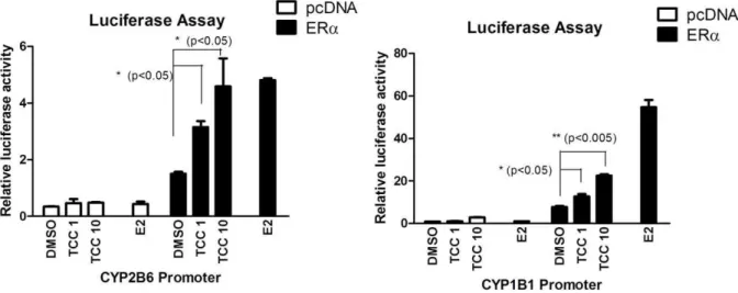
at the concentration of 10mM, ERa and CAR were effectively activated by TCC (Table 1). CAR was moderately activated by TCC with 1.75-fold induction of luciferase activity. TCC also promotes ERaactivity with similar potency as estradiol (Table 1). All other nuclear receptors produce statistically insignificant induction with VDR, FXR, PPARc, and GR being mostly unaffected, thus TCC likely only has minimal effects on these receptors.b) ERa-mediated promoter activation:It has been
shown previously that the humanCYP1B1 gene is regulated by estradiol through the ERa[38]. The ERareceptor has also been reported to regulate the expression of the humanCYP2B6 gene [39]. To identify the potential role of ERa in gene induction for
CYP1B1andCYP2B6by exposure to TCC, transient transfections with a luciferase vector containing the 2kb fragment upstream from the CYP2B6 start site (pGL3–2B6) with or without co-transfection of ERa were performed. The reporter activity of pGL3–2B6 increased with TCC treatment in a dose-dependent manner to a level compatible to that of estradiol treatment in the presence of ERa(Figure 1A). To further support the role of ERa
in TCC-mediated gene regulation, gene expression of human
CYP1B1, a well-characterized enzyme that is linked to expression in estrogen-regulated tissue through ERa [38] was examined. Similar to the CYP2B6 response to TCC, CYP1B1 promoter activity was significantly induced by TCC in a dose-dependent fashion. Mimicking the estradiol action, ERa is required for the
CYP1B1promoter activation by TCC (Figure 1B).c) CAR ligand
binding assay: CAR has been shown to display a species
response to TCC with CITCO as a potent ligand (Figure 2A and 2B) suggesting that TCC is a CAR activator, but not an agonist ligand for either mouse or human CAR.
TCC Induction of Xenobiotic Metabolism by CAR
TheUGT1Agenes expressed inhUGT1*28mice are susceptible to regulation by CAR [26], PXR [20], peroxisome proliferator-activated receptor-alpha (PPARa) [41] and the Ah receptor [20]. Initial experiments were conducted by treating hUGT1*28mice with 16 mg/kg TCC by the i.p. route and evaluatingUGT1Agene expression patterns in liver after 48 hours. In comparison to DMSO treated mice,UGT1A1,21A3, 21A4,21A6, and21A9
gene products were each induced in hUGT1*28mice following TCC administration (Figure 3). When gene targets that are regulated by CAR (Cyp2b10), PXR (Cyp3a11), PPARa (Cyp4a11) and the Ah receptor (Cyp1a1) were evaluated by real time RT-PCR analysis, only Cyp2b10 gene expression was found to be induced (Figure 4), indicating that CAR activation by TCC was leading to the induction of theUGT1Agenes. To examine if CAR was underlying the induction pattern, we treated hUGT1*28/ Car2/2 mice with TCC and examined expression of theUGT1A
genes. In comparison tohUGT1*28mice, there was no induction of theUGT1Agenes inhUGT1*28/Car2/2mice. This correlated with a nearly complete absence of Cyp2b10 gene induction following TCC treatment in hUGT1*28/Car2/2 mice (Figure 4A). To access the expression of Cyp2b10 protein in microsomal preparation from treated mouse livers, Western blot analysis was performed with antibody against the CYP2B6 protein. Consistent with the transcript levels, Cyp2b10 protein expression was induced by TCC treatment, and the induction was blocked in Car2/2livers (Figure 4B).
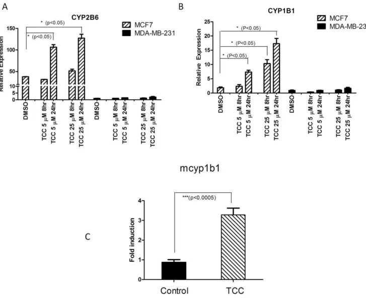
CYP2B6 and CYP1B1 Gene Expression Regulated by TCC
To investigate the effect of TCC on CYP2B6 mRNA expression through a ERa-dependent mechanism, ERapositive MCF7 and ERa negative MDA-MB-231 human breast cancer cells were treated with TCC. Results of real time RT-PCR revealed dose-and time-dependent effects of TCC treatment on induction of
CYP2B6 mRNA expression in ERa positive MCF7 cells (Figure 5A). In contrast, CYP2B6 expression was unaffected in TCC-treated ERa negative MDA-MB-231 cells. To further support the role of ERain TCC-mediated gene regulation, gene expression of humanCYP1B1, a well-characterized enzyme that is linked to expression in estrogen-regulated tissue through ERa[38] was examined. Similar toCYP2B6response to TCC, MCF7 cells exhibited a TCC-dependent increase in CYP1B1 mRNA levels that was not observed in MDA-MB-231 cells, further assuring the involvement of ERain TCC-mediated gene induction (Figure 5B). Furthermore, we examined mouse Cyp1b1 induction in ERa
sensitive tissue such as the ovaries. TCC treatment to female
hUGT1*28mice resulted in the induction ofCyp1b1(Figure 5C). These results clearly link TCC exposure to activation of ERa
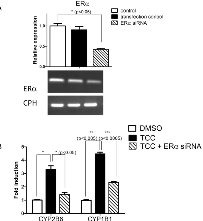
target genes, Cyp1b1/CYP1B1 and Cyp2b10/CYP2B6 in both cultured human breast cancer cells andin vivo estrogen-sensitive tissues. Finally, transfection experiments with siRNAs targeting ERawere performed to interrupt ERagene expression in MCF7 cells. Gene expression levels forCYP1B1,CYP2B6, and ERawere analyzed by real time PCR following siRNA transfections and TCC treatment. SiRNA knockdown of ERawith 58% effective-ness significantly reducedCYP1B1andCYP2B6gene expression in MCF7 cells (Figure 6), further assuring the involvement of ERain TCC-mediated gene induction.
Discussion
With the XenoR screening assay, our results show divergent receptor binding activities of TCC as indicated by fold induction of luciferase activity. This system appears to be a suitable qualitative model for evaluating XenoR activation toward tested compounds. CAR and ERaappeared most susceptible to TCC activation and exhibited moderate luciferase activity. We then assessed potential biological effects arising from TCC exposure in the perspective of drug and steroid metabolism and discussed the molecular mechanism through which the key phase I and phase II xenobiotic metabolizing enzymes are induced. The use ofin vitro
cell-based assays to predict and define the underlying mechanisms
Table 1.Xenobiotic receptors screening assay.
Xenobiotic receptors Reporter constructs
Fold induction in the absence of ligand
Fold induction with the
positive ligand Fold induction with TCC
Era ERE 160.1 2.8960.26 2.1360.14**
Erb ERE 160.06 1.8560.26 1.4760.15
CAR PBRE 160.07 2.0560.33 1.7560.30***
PXR CYP3A promoter 160.27 4.2260.73 1.4460.44
LXRa LXRE 160.4 2.3461.49 1.5160.73
VDR CYP24A1 promoter 160.45 9.6763.94 1.0660.07
FXR FXRE 160.14 29.363.79 0.8860.18
PPARa PPRE 160.13 8.662.89 1.5360.27
PPARd PPRE 160.32 10.3660.64 1.5760.55
PPARc PPRE 160.32 5.8960.64 1.0360.55
GR GRE 160.1 5.8362.17 1.160.18
TCC was tested for the ability to activate PXR, CAR, LXRa, FXR, VDR, PPARa, PPARb, PPARc, ERa, and ERbby binding to the corresponding response element. CV1 cells were transfected with an expression plasmid to supply a human nuclear receptor, a luciferase reporter plasmid containing the appropriate DNA response element, and ab-galactosidase expression vector to control for transfection efficiency. The positive ligand for each receptor is described in Material and Method. Luciferase activity was represented as fold induction relative to DMSO-treated cells. The results are expressed as the mean6S.D.
**p,0.005.
combined with TCC exposure within in vivo animal models is a useful approach to understand xenobiotic interactions caused by TCC. An indirect interaction of TCC with CAR that enables TCC to induce the humanUGT1Agenes inhUGT1*28mice was revealed in our animal models. The specificity of CAR-mediated regulation by TCC is supported by observations that hepatic
Cyp2b10is also induced, while theUGT1Agenes and theCyp2b10
gene are not induced in hUGT1*28/Car2/2 mice. We also eliminated any interaction with PXR, since PXR was insensitive to TCC in the luciferase reporter assay, and TCC has no effect on expression of the PXR-target gene Cyp3a11 (data not shown). CAR is well recognized to regulate the induction of theCYP2B
genes by PB and a group of diverse agents referred to as ‘‘PB-like’’ agents including the pesticide contaminant TCPOBOP, the most potent PB-like inducers known in rodents [42]. To date, only a few CAR agonists, such as TCPOBOP for murine CAR and CITCO for human CAR are identified. Our results with the GAL4-human/mouse CAR LBD fusion proteins, representing the ligand specificity of CAR, indicate that TCC, mimicking PB, acts as an
indirect CAR activator. Although the luciferase activation by TCC was at a moderate level, this induction was significant compared to that of the control and 2-fold induction by TCPOBOP. The moderate scale CAR activation in the luciferase screening assay by TCC may be due to the unique feature of CAR being a constitutive active receptor even in the absence of an exogenous ligand as well as being a CAR activator through an indirect mechanism that facilitates translocation of CAR to the nucleus and transactivation of its target genes.
The ability of TCC to stimulate CAR target genes may be of significance. The current study has demonstrated cross-regulation withCYP2B and human UGT1A genes by TCC through CAR activation. This is not surprising because a PB response element flanking the gene promoter has been identified in both human
CYP2B6andUGT1A1genes [37,39]. It is well characterized that the CAR-mediated inducible expression of xenobiotic-metaboliz-ing enzymes and accelerated metabolic elimination of correspond-ing substrates is associated with drug-drug interaction clinically. In addition, CAR can act as a negative regulator controlling
Figure 1. Promoter Activation by TCC.Transfection of a Luc reporter containing the 2 kbCYP2B6(A) orCYP1B1(B) promoter region with or without cotransfection of an ERaexpression vector to CV-1 cells was performed. Following transfection, cells were treated for 24 hours with DMSO or different concentrations of TCC as indicated. Firefly luciferase activity was measured and normalized by using the level ofb-galactosidase activity. doi:10.1371/journal.pone.0037705.g001
Figure 2. CAR ligand binding assay.HepG2 cells were transfected with the expression vector containing the Gal4 DNA binding domain fused with the ligand binding domain of murine CAR (mCAR, A) or human CAR (hCAR, B), and cotransfected with the Luciferase reporter plasmid, mh 100-luc. Following transfection, cells were treated for 24 hours with phenobarbital (1 mM), TCC (10mM), TCPOBOP (25mM), or CITGO (10mM). Firefly Luciferase activity was measured and normalized by using the level ofrenillaluciferase activity.
inhibition of biotransformation genes, e.g.,UGT2B7andCYP7A1
[35,43]. This down regulation might lead to changes in steady-state dynamics of steroids and bile acid homeostasis. Thus, TCC contamination in biological systems may be critically relevant by interfering with metabolism of endogenous and exogenous substrates by CAR-target xenobiotic genes. For example, a recent study has linked CAR pre-activation and alcohol infusion to a synergistic decrease in the expression of enzymes that metabolize the alcohol in liver including alcohol dehydrogenase 1, aldehyde dehyrogenase (ALDH) 1A1, ALDH3A2, and CYP2E1, which supports the role of CAR in modulating alcoholic liver injury [44]. Secondly, it has been characterized that PB-like agents possess CAR-dependent liver enlargement properties that are evidenced by increases in cell proliferation and suppression of apoptosis [45]. This CAR-dependent action is thought to be a prerequisite and underlying mechanism for nongenotoxic PB-mediated liver tumorigenesis [46]. Therefore, TCC might deserve close scrutiny to determine if its long-term effects on liver disease and tumorigenesis through a CAR dependent mechanism, similar to the actions of PB.
While transcriptional regulation of theCYP2Bgene is evident through TCC-mediated CAR activation, the slight induction of
Cyp2b10in TCC-treated CAR null livers (1.77–3.26 fold induction compared with DMSO-treated ones) implies the
CAR-indepen-dent mechanism. Recently, a study has confirmed ERa-dependent regulation ofCYP2B6, in which a functional ER response element was identified in theCYP2B6regulatory region [39]. In this study, we provide compelling evidence showing CYP2B6 induction by TCC is also mediated through ERa. In addition to CAR, TCC enables induction of CYP2B6 promoter activity in an ERa -dependent fashion. Moreover, ERa activation by TCC affects other ERatarget genes;CYP1B1transcription is subjected to ERa -dependent TCC regulation as evidenced by activation of the
CYP1B1promoter region, which was previously characterized as harboring a functional ERa binding site [38]. TCC-mediated induction of CYP2B6 and CYP1B1 was observed in estrogen sensitive cultured cells and tissues further supporting the observa-tion that TCC exposure can regulate target genes of ERathrough an ERa-dependent mechanism.
Human TCC metabolism involves direct N2 and N9 -glucuronidation and ring hydroxylation to 2-OH-TCC and 6-OH-TCC, which can further undergo sulfate and glucuronide conjugation [47]. TCC metabolites are detected in plasma and urine in subjects who have showered with TCC-containing soap with TCC-N-glucuronides as the major route of renal excretion [11]. The robust TCC-mediatedCYP2Bgene induction through both CAR and ERaactivation may be relevant considering that contact to TCC maybe occurring consistently over time through
Figure 3. The comparative expression of hepatic UGT1A genes between hUGT1 and hUGT1/Car2/2mice.RT-PCR analysis using liver RNA from DMSO- or TCC-treated (16 mg/kg, dissolved in 50ml DMSO) mice and isoform specific-primer pairs was performed to detect the expression
ofUGT1Agenes as indicated.
the use of personal care products. CYP2B6 is one of the major enzymes to metabolize clinical drugs (e.g., cyclophosphamide and tamoxifen), and 202 to 250-fold inter-individual variation in CYP2B6 expression has been demonstrated [48], presumably due to transcriptional regulation and polymorphisms. Environmental exposure to agents like TCC may be contributing towards variation inCYP2B6since both CAR and ERaare regulators of this gene and potential targets following TCC exposure.
With environmentally relevant concentrations, a recent report indicated that TCC increased embryo production in the freshwater mud snail suggesting that TCC may be causing reproductive effects in a manner similar to that seen with some known environmental estrogens [49]. Although TCC is not an agonist for the AR (androgen receptor), TCC has the ability to amplify the effect of testosterone on the androgenic activity in the AR-mediated luciferase assay [13]. To study ER interactions, Ahn
et al. [10,14] performed a cell-based ER-mediated bioassay to study TCC-ERa interaction. At 1 and 10mM, TCC evoked a modest response equivalent to about 30% of that produced by 1 nM estradiol. Although it is a mild agonist for the ERain the reporter gene assay, TCC enhances the estrogen action in the presence of estrogen [10]. Combined with our results, it can be anticipated that TCC has a significant influence on modulating transcriptional control of ERatarget genes, such as those involved in xenobiotic and steroid metabolism. For example, regulating
CYP1B1gene expression by TCC reinforces its ability to disrupt estrogen homeostasis. Estrogen has long been associated with breast cancer. CYP1B1 is highly expressed in estrogen target tissues and changes in the expression of proteins such as CYP1B1 are known to metabolize estrogens, which can potentially alter physiological levels and the intensity of estrogen action. In addition, CYP1B1 catalyzes the 4-hydroxylation of estradiol to
Figure 4. Cyp2b10 gene expression following the TCC treatment.Age-matched heterozygous (CAR+/2) andCar-null mice (Car2/2) (n = 3) were treated with either DMSO or TCC (16 mg/kg) by intraperitoneal injection for 48 hours. A. The liver tissues were used for preparation of total RNA. After the reverse transcription for cDNA synthesis, real-time PCR was conducted to determine the Ct value with cyclophilin as an internal control gene. B. The microsomal proteins were prepared from liver tissues and subject to Western Blot analysis using Cyp2b10 antibody as the primary antibody.
generate 4-hydroxy estradiol [50], a catechol metabolite that produces free radicals, causes cellular damage, and is associated with carcinogenic activity of estrogen and the development of breast cancer. The continual use of personal care products that contain antimicrobial agents like TCC may enhance over time the generation of endogenously produced metabolites capable of eliciting a toxic or carcinogenic episode. In addition, CAR activation has recently been shown to inhibit human UGT2B7
gene expression [35]; indicating that continued exposure to TCC would have a negative influence on normal drug metabolism and clearance. Combined with evidence that there is considerable cross-talk between the ER and CAR [51,52], TCC accumulation may surely influence the many target genes involved in xenobiotic metabolism. Therefore, the perceived benefits of using TCC as an antimicrobial agent in personal care products should be weighed against possible risks.
In Conclusion
These studies demonstrate that acute exposure to TCC results in the activation of important regulatory pathways dictated by CAR and ERathat can potentially impact the steady-state levels of hormones, as well as altering routes of drug metabolism. One of the important check points in steroid homeostasis is biological inactivation by glucuronidation, which is shown in this study to be induced in a CAR dependent fashion following TCC treatment. Induction of cytochrome P450 genes, whose products are steroid hydroxylases, is also regulated by TCC both through CAR as well as ERa. Combined with findings that TCC is capable of synergizing the actions of testosterone through the AR [13], these studies suggest that long term exposure to TCC, from daily use of personal hygiene products, has the potential of altering normal steroid biogenesis. Thus, long term alterations in hormone homeostasis initiated by chronic exposure to TCC could potentially lead to human health problems.
Figure 5. Activation of gene expression by TCC.Human breast cancer cell lines, MCF7 and MDA-MB-231, were cultured and treated with either DMSO or 5 or 10mM TCC. Following total RNA isolation and reverse transcription reaction,CYP2B6(A) orCYP1B1(B) mRNA levels were measured by real-time PCR using a gene specific pair of primers. (C) Induction of cyp1b1 gene in mouse ovary and fallopian tube by TCC. 10-day old
hUGT1*28mice were treated with corn oil or TCC (20 mg/kg) by i.p. injection every 48 hours until 30 days of age. The tissues of ovaries and fallopian
tubes were used to prepare total RNA. Following the reverse transcription for cDNA synthesis, real-time PCR was conducted to determine the Ct value
forcyp1b1with cyclophilin as an internal control gene.
Author Contributions
Conceived and designed the experiments: MFY TL RHT. Performed the experiments: MFY TL. Analyzed the data: MFY TL. Contributed
reagents/materials/analysis tools: RME BH RHT. Wrote the paper: MFY TL RHT.
References
1. Walsh SE, Maillard JY, Russell AD, Catrenich CE, Charbonneau DL, et al. (2003) Activity and mechanisms of action of selected biocidal agents on Gram-positive and -negative bacteria. J Appl Microbiol 94: 240–247.
2. Scientific Commitee on Consumer Products (2005) SCCP. Opinion on triclocarban for other uses as a preservative. Calipa n degree P29. European Union. SCCP/0851/04.
Figure 6. TCC-mediated gene induction is blocked by ERasiRNAs.siRNAs for ERa. were designed and synthesized by Bioneer (Alameda, CA) and dissolved in DEPC-H2O. MCF7 cells were seeded in a 6-well plate and transfected with either ERasiRNAs or random siRNAs (transfection control)
using lipofectamine 2000. The next day, cells were treated with either TCC or DMSO and incubated with either TCC (25mM) or DMSO for 24 hours. Following treatments, total RNA was extracted. (A) ERamRNA levels were evaluated by real time and RT-PCR with cyclophilin (CPH) as an internal control gene. (B) CYP2B6, and CYP1B1 mRNA levels were measured by real time PCR with CPH as an internal control gene and displayed as fold induction compared with DMSO-treated samples as one fold.
3. Perencevich EN, Wong MT, Harris AD (2001) National and regional assessment of the antibacterial soap market: a step toward determining the impact of prevalent antibacterial soaps. Am J Infect Control 29: 281–283.
4. Kumar KS, Priya SM, Peck AM, Sajwan KS (2010) Mass loadings of triclosan and triclocarbon from four wastewater treatment plants to three rivers and landfill in Savannah, Georgia, USA. Arch Environ Contam Toxicol 58: 275– 285.
5. Chen F, Ying GG, Kong LX, Wang L, Zhao JL, et al. (2011) Distribution and accumulation of endocrine-disrupting chemicals and pharmaceuticals in wastewater irrigated soils in Hebei, China. Environ Pollut 159: 1490–1498. 6. Heidler J, Sapkota A, Halden RU (2006) Partitioning, persistence, and
accumulation in digested sludge of the topical antiseptic triclocarban during wastewater treatment. Environ Sci Technol 40: 3634–3639.
7. Heidler J, Halden RU (2008) Meta-analysis of mass balances examining chemical fate during wastewater treatment. Environ Sci Technol 42: 6324–6332. 8. Halden RU, Paull DH (2005) Co-occurrence of triclocarban and triclosan in
U.S. water resources. Environ Sci Technol 39: 1420–1426.
9. Liu JY, Qiu H, Morisseau C, Hwang SH, Tsai HJ, et al. (2011) Inhibition of soluble epoxide hydrolase contributes to the anti-inflammatory effect of antimicrobial triclocarban in a murine model. Toxicol Appl Pharmacol 255: 200–206.
10. Ahn KC, Zhao B, Chen J, Cherednichenko G, Sanmarti E, et al. (2008) In vitro biologic activities of the antimicrobials triclocarban, its analogs, and triclosan in bioassay screens: receptor-based bioassay screens. Environ Health Perspect 116: 1203–1210.
11. Schebb NH, Inceoglu B, Ahn KC, Morisseau C, Gee SJ, et al. (2011) Investigation of human exposure to triclocarban after showering and preliminary evaluation of its biological effects. Environ Sci Technol 45: 3109–3115. 12. Higgins CP, Paesani ZJ, Chalew TE, Halden RU (2009) Bioaccumulation of
triclocarban in Lumbriculus variegatus. Environ Toxicol Chem 28: 2580–2586. 13. Chen J, Ahn KC, Gee NA, Ahmed MI, Duleba AJ, et al. (2008) Triclocarban enhances testosterone action: a new type of endocrine disruptor? Endocrinology 149: 1173–1179.
14. Christen V, Crettaz P, Oberli-Schrammli A, Fent K (2010) Some flame retardants and the antimicrobials triclosan and triclocarban enhance the androgenic activity in vitro. Chemosphere 81: 1245–1252.
15. Liu JY, Qiu H, Morisseau C, Hwang SH, Tsai HJ, et al. (2011) Inhibition of soluble epoxide hydrolase contributes to the anti-inflammatory effect of antimicrobial triclocarban in a murine model. Toxicol Appl Pharmacol 255: 200–206.
16. Forman BM, Tzameli I, Choi HS, Chen J, Simha D, et al. (1998) Androstane metabolites bind to and deactivate the nuclear receptor CAR-beta. Nature 395: 612–615.
17. Mangelsdorf DJ, Evans RM (1995) The RXR heterodimers and orphan receptors. Cell 83: 841–850.
18. Xie W, Barwick JL, Simon CM, Pierce AM, Safe S, et al. (2000) Reciprocal activation of xenobiotic response genes by nuclear receptors SXR/PXR and CAR. Genes Dev 14: 3014–3023.
19. Fujiwara R, Nguyen N, Chen S, Tukey RH (2010) Developmental hyperbil-irubinemia and CNS toxicity in mice humanized with the UDP glucuronosyl-transferase 1 (UGT1) locus. Proc Natl Acad Sci U S A 107: 5024–5029. 20. Chen S, Beaton D, Nguyen N, Senekeo-Effenberger K, Brace-Sinnokrak E, et
al. (2005) Tissue-specific, inducible, and hormonal control of the human UDP-glucuronosyltransferase-1 (UGT1) locus. J Biol Chem 280: 37547–37557. 21. Nguyen N, Bonzo JA, Chen S, Chouinard S, Kelner M, et al. (2008) Disruption
of theUgt1locus in mice resembles human Crigler-Najjar type I disease. J Biol Chem 283: 7901–7911.
22. Strassburg CP, Manns MP, Tukey RH (1997) Differential down regulation of theUDP-glucuronosyltransferase 1Alocus is an early event in human liver and biliary cancer. Cancer Res 57: 2979–2985.
23. Strassburg CP, Oldhafer K, Manns MP, Tukey RH (1997) Differential expression of theUGT1A locus in human liver, biliary, and gastric tissue: identification of UGT1A7 and UGT1A10 transcripts in extrahepatic tissue. Mol Pharmacol 52: 212–220.
24. Strassburg CP, Nguyen N, Manns MP, Tukey RH (1998) Polymorphic expression of the UDP-glucuronosyltransferaseUGT1Agene locus in human gastric epithelium. Molecular Pharmacology 54: 647–654.
25. Tukey RH, Strassburg CP (2001) Genetic multiplicity of the human UDP-glucuronosyltransferases and regulation in the gastrointestinal tract. Molecular Pharmacology 59: 405–414.
26. Cai H, Nguyen N, Peterkin V, Yang YS, Hotz K, et al. (2010) A Humanized UGT1Mouse Model Expressing the UGT1A1*28Allele for Assessing Drug Clearance by UGT1A1 Dependent Glucuronidation. Drug Metab Dispos 38: 879–86.
27. Xie W, Barwick JL, Downes M, Blumberg B, Simon CM, et al. (2000) Humanized xenobiotic response in mice expressing nuclear receptor SXR. Nature 406: 435–439.
28. Saez E, Rosenfeld J, Livolsi A, Olson P, Lombardo E, et al. (2004) PPAR gamma signaling exacerbates mammary gland tumor development. Genes Dev 18: 528–540.
29. Umesono K, Giguere V, Glass CK, Rosenfeld MG, Evans RM (1988) Retinoic acid and thyroid hormone induce gene expression through a common responsive element. Nature 336: 262–265.
30. Willy PJ, Umesono K, Ong ES, Evans RM, Heyman RA, et al. (1995) LXR, a nuclear receptor that defines a distinct retinoid response pathway. Genes Dev 9: 1033–1045.
31. Makishima M, Lu TT, Xie W, Whitfield GK, Domoto H, et al. (2002) Vitamin D receptor as an intestinal bile acid sensor. Science 296: 1313–1316. 32. Downes M, Verdecia MA, Roecker AJ, Hughes R, Hogenesch JB, et al. (2003) A
chemical, genetic, and structural analysis of the nuclear bile acid receptor FXR. Mol Cell 11: 1079–1092.
33. Umesono K, Evans RM (1989) Determinants of target gene specificity for steroid/thyroid hormone receptors. Cell 57: 1139–1146.
34. Ueda A, Hamadeh HK, Webb HK, Yamamoto Y, Sueyoshi T, et al. (2002) Diverse Roles of the Nuclear Orphan Receptor CAR in Regulating Hepatic Genes in Response to Phenobarbital. Mol Pharmacol 61: 1–6.
35. Yueh MF, Mellon PL, Tukey RH (2011) Inhibition of HumanUGT2B7Gene Expression in Transgenic Mice by the Constitutive Androstane Receptor. Mol Pharmacol 79: 1053–60.
36. Yueh MF, Tukey RH (2007) Nrf2-Keap1 Signaling Pathway Regulates Human UGT1A1Expression in Vitro and in Transgenic UGT1 Mice. J Biol Chem 282: 8749–8758.
37. Xie W, Yeuh MF, Radominska-Pandya A, Saini SPS, Negishi Y, et al. (2003) Control of steroid, heme, and carcinogen metabolism by nuclear pregnane X receptor and constitutive androstane receptor. Proc Natl Acad Sci USA 100: 4150–4155.
38. Tsuchiya Y, Nakajima M, Kyo S, Kanaya T, Inoue M, et al. (2004) Human CYP1B1 is regulated by estradiol via estrogen receptor. Cancer Res 64: 3119– 3125.
39. Lo R, Burgoon L, Macpherson L, Ahmed S, Matthews J (2010) Estrogen receptor-dependent regulation of CYP2B6 in human breast cancer cells. Biochim Biophys Acta 1799: 469–479.
40. Maglich JM, Parks DJ, Moore LB, Collins JL, Goodwin B, et al. (2003) Identification of a novel human constitutive androstane receptor (CAR) agonist and its use in the identification of CAR target genes. J Biol Chem 278: 17277– 17283.
41. Senekeo-Effenberger K, Chen S, Brace-Sinnokrak E, Bonzo JA, Yueh MF, et al. (2007) Expression of the Human UGT1 Locus in Transgenic Mice by 4-Chloro-6-(2,3-xylidino)-2-pyrimidinylthioacetic Acid (WY-14643) and Implications on Drug Metabolism through Peroxisome Proliferator-Activated Receptor a Activation. Drug Metab Dispos 35: 419–427.
42. Sueyoshi T, Kawamoto T, Zelko I, Honkakoski P, Negishi M (1999) The repressed nuclear receptor CAR responds to phenobarbital in activating the humanCYP2B6gene. J Biol Chem 274: 6043–6046.
43. Miao J, Fang S, Bae Y, Kemper JK (2006) Functional inhibitory cross-talk between constitutive androstane receptor and hepatic nuclear factor-4 in hepatic lipid/glucose metabolism is mediated by competition for binding to the DR1 motif and to the common coactivators, GRIP-1 and PGC-1alpha. J Biol Chem 281: 14537–14546.
44. Chen X, Meng Z, Wang X, Zeng S, Huang W (2011) The nuclear receptor CAR modulates alcohol-induced liver injury. Lab Invest 91: 1136–1145. 45. Carthew P, Edwards RE, Nolan BM (1998) The quantitative distinction of
hyperplasia from hypertrophy in hepatomegaly induced in the rat liver by phenobarbital. Toxicol Sci 44: 46–51.
46. Yamamoto Y, Moore R, Goldsworthy TL, Negishi M, Maronpot RR (2004) The orphan nuclear receptor constitutive active/androstane receptor is essential for liver tumor promotion by phenobarbital in mice. Cancer Res 64: 7197–7200. 47. Hiles RA, Birch CG (1978) The absorption, excretion, and biotransformation of
3,4,4’-trichlorocarbanilide in humans. Drug Metab Dispos 6: 177–183. 48. Wang H, Tompkins LM (2008) CYP2B6: new insights into a historically
overlooked cytochrome P450 isozyme. Curr Drug Metab 9: 598–610. 49. Giudice BD, Young TM (2010) The antimicrobial triclocarban stimulates
embryo production in the freshwater mudsnail Potamopyrgus antipodarum. Environ Toxicol Chem 29: 966–970.
50. Lee AJ, Cai MX, Thomas PE, Conney AH, Zhu BT (2003) Characterization of the oxidative metabolites of 17beta-estradiol and estrone formed by 15 selectively expressed human cytochrome p450 isoforms. Endocrinology 144: 3382–3398.
51. Min G, Kim H, Bae Y, Petz L, Kemper JK (2002) Inhibitory cross-talk between estrogen receptor (ER) and constitutively activated androstane receptor (CAR). CAR inhibits ER-mediated signaling pathway by squelching p160 coactivators. J Biol Chem 277: 34626–34633.