Low Bone Mass Phenotype in a Gender- and Age-Specific
Manner
Tommy Noh1,2., Yankel Gabet1,2., Jon Cogan2
, Yunfan Shi2, Archana Tank1,2, Tomoyo Sasaki3, Braden Criswell2, Alexis Dixon2, Christopher Lee2, Joseph Tam4, Thomas Kohler5, Eran Segev4, Lisa Kockeritz6, James Woodgett6, Ralph Mu¨ller5, Yang Chai3, Elisheva Smith2, Itai Bab4, Baruch Frenkel1,2,7*
1Department of Biochemistry and Molecular Biology, Keck School of Medicine, University of Southern California, Los Angeles, California, United States of America, 2Institute for Genetic Medicine, Keck School of Medicine, University of Southern California, Los Angeles, California, United States of America,3Center for Craniofacial Molecular Biology, Keck School of Medicine, University of Southern California, Los Angeles, California, United States of America,4Bone Laboratory, Faculty of Dental Medicine, The Hebrew University of Jerusalem, Jerusalem, Israel,5Institute for Biomechanics, ETH Zu¨rich, Zu¨rich, Switzerland,6Ontario Cancer Institute/Princess Margaret Hospital, Toronto, Ontario, Canada,7Department of Orthopaedic Surgery, Keck School of Medicine, University of Southern California, Los Angeles, California, United States of America
Abstract
We investigated the role ofLef1, one of the four transcription factors that transmit Wnt signaling to the genome, in the regulation of bone mass. Microcomputed tomographic analysis of 13- and 17-week-old mice revealed significantly reduced trabecular bone mass in Lef1+/2 females compared to littermate wild-type females. This was attributable to decreased
osteoblast activity and bone formation as indicated by histomorphometric analysis of bone remodeling. In contrast to females, bone mass was unaffected by Lef1 haploinsufficiency in males. Similarly, females were substantially more responsive than males to haploinsufficiency inGsk3b, a negative regulator of the Wnt pathway, displaying in this case a high bone mass phenotype.Lef1haploinsufficiency also led to low bone mass in males lacking functional androgen receptor (AR) (tfmmutants). The protective skeletal effect of AR against Wnt-related low bone mass is not necessarily a result of direct interaction between the AR and Wnt signaling pathways, becauseLef1+/2female mice had normal bone mass at the age of
34 weeks. Thus, our results indicate an age- and gender-dependent role forLef1in regulating bone formation and bone massin vivo. The resistance to Lef1 haploinsufficiency in males with active AR and in old females could be due to the reduced bone turnover in these mice.
Citation:Noh T, Gabet Y, Cogan J, Shi Y, Tank A, et al. (2009)Lef1Haploinsufficient Mice Display a Low Turnover and Low Bone Mass Phenotype in a Gender- and Age-Specific Manner. PLoS ONE 4(5): e5438. doi:10.1371/journal.pone.0005438
Editor:William Giannobile, University of Michigan, United States of America
ReceivedNovember 12, 2008;AcceptedApril 2, 2009;PublishedMay 4, 2009
Copyright:ß2009 Noh et al. This is an open-access article distributed under the terms of the Creative Commons Attribution License, which permits unrestricted
use, distribution, and reproduction in any medium, provided the original author and source are credited.
Funding:BF’s group was supported by the Arthritis Foundation (Atlanta, GA), by the NIH (AR047052), and by the J. Harold and Edna L. LaBriola Chair in Genetic Orthopaedic Research at the University of Southern California. IB’s team was supported by the NIH (AR047052). JW’s group was supported by the Canadian Institutes of Health Research (MOP#74711). TK and RM were supported by the Swiss National Science Foundation (Grants FP 620-58097.99 and PP-104317/1). YG was partly supported by a Meyer Young Investigator Fellowship from the Arthritis Foundation Southern California Chapter. The use of the microCAT II image system at the USC Molecular Imaging Center was made possible by a 2005 NCRR shared instrumentation award. The experiments were conducted in part in a facility constructed with support from Research Facilities Improvement Program Grant Number C06 (RR10600-01, CA62528-01, RR14514-01) from the NIH/NCRR. The funders had no role in study design, data collection and analysis, decision to publish, or preparation of the manuscript.
Competing Interests:The authors have declared that no competing interests exist.
* E-mail: [email protected]
.These authors contributed equally to this work.
Introduction
The wingless/Wnt family of secreted glycoproteins has critical roles in cell growth and differentiation, and is highly conserved among vertebrates, flies, and primitive multicellular organisms [1,2]. In mammals, the canonical Wnt pathway is pivotal to embryogenesis and tumorigenesis as well as in the maintenance and regeneration of tissues such as skin, intestine, liver, cardiac muscle, and the nervous system [1–7]. Wnt ligands bind to membrane complexes consisting of a seven transmembrane domain receptor of the frizzled family [3–5] and a ‘single-pass’ co-receptor, Lipoprotein Receptor-Related Protein 6 (Lrp6) and possibly Lrp5 as well [6–8]. In the absence of stimulation, the Wnt pathway is under the negative control of a pair of protein-serine
kinases, Glycogen Synthase Kinase 3a and 3b (Gsk3a, Gsk3b), which phosphorylateb-catenin, a Wnt transducer, resulting in its ubiquitination and subsequent proteasomal degradation [9,10]. Upon Wnt stimulation, the Gsk3-mediated phosphorylation ofb -catenin is attenuated through disruption of the b-catenin destruction complex [11]. Subsequently, b-catenin accumulates and translocates to the nucleus, where it activates Wnt target genes by associating with the DNA-binding HMG box transcription factors lymphoid enhancer factor 1 (Lef1), T-cell factor 7 (Tcf7), Tcf7L1, and/or Tcf7L2 [12–15]. Tcf7, Tcf7L1 and Tcf7L2 are commonly known as Tcf1, Tcf3, and Tcf4, respectively, and these common names are used in this paper.
structural integrity. The mammalian skeleton undergoes contin-uous turnover, where overall bone gain or loss is determined by the difference between bone resorption and formation. After birth, bone mass increases until it reaches ‘‘peak bone mass’’, which is then maintained at a constant level during young adulthood, followed by an age-related bone loss [16]. Bone mass accrual is subject to sexual dimorphism, with males having higher trabecular bone volume density and lower bone turnover than females [16– 19]. The age-related bone loss is associated with reduction in turnover rate especially in females [20,21].
In humans and mice alike, bone mass is strongly dependent on the Wnt signaling pathway [22,23]. Mutations and polymorphism inWnt10b,Dkk1,Dkk2,Sfrp1,Sost,Lrp6, andGsk3baffect osteoblast function and therefore bone formation and bone mass [24–30]. The Wnt pathway has also been implicated in mediating positive and negative control of bone formation and bone mass by environmental factors such as pharmacological glucocorticoids [31–34] and mechanical stimulation [35]. Interestingly, Wnt signaling in osteoblasts also controls osteoclast activity, as demonstrated by increased resorption and a low bone mass (LBM) phenotype in mice whose osteoblasts lackb-catenin or Tcf1 [23]. The role of Lef1 in postnatal bone metabolism has not been studied, in part because Lef1 deficient mice die within the first week of life [36].
During early embryogenesis, Tcf1 and Lef1 are redundant. Mice lacking both genes have multiple defects, including duplicated neural tubes and malformed limb buds, whereas mice lacking either gene alone do not exhibit these defects [37,38]. Non-redundant functions of these transcription factors are suggested by the distinct phenotypes observed in mice lacking either Tcf1 or Lef1. WhereasTcf1-null mice display attenuated T cell differentiation [39], Lef1-null mice exhibit developmental defects in teeth, hair follicles, mammary glands and the brain [36,40]. In terms of skeletal metabolism, analysis ofTcf1-null mice disclosed accelerated bone resorption [23], but similar analysis of
Lef1-null mice is hampered by their perinatal lethality. In this study, we employed the viableLef1 haploinsufficient mice, which do not display the abnormalities observed in the null mice, to assess the role of Lef1 in postnatal bone metabolism. We demonstrate a low bone mass phenotype inLef1+/2mice, which appears to manifest specifically in the context of high bone turnover rate.
Results
Low bone mass (LBM) phenotype inLef1heterozygous female mice
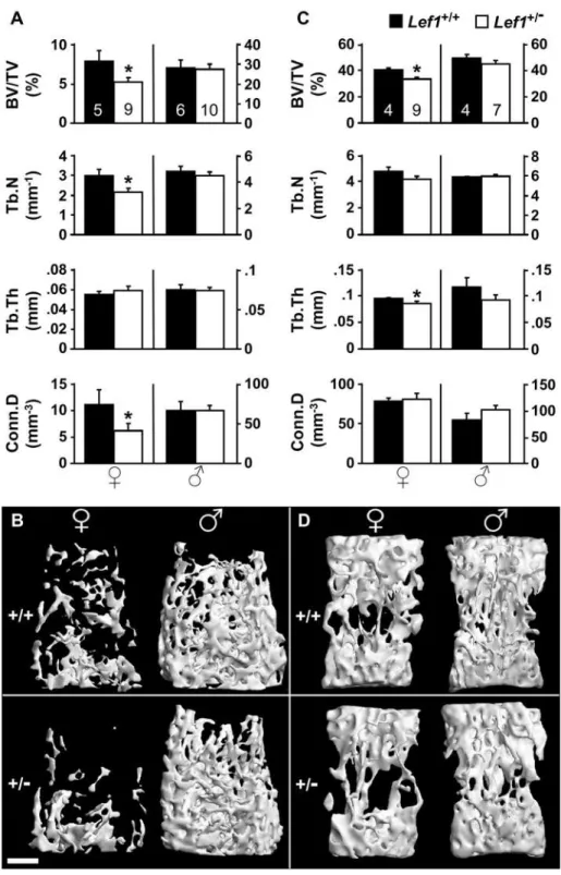
Micro-computed tomographic (mCT) analysis of 13-week old
Lef1 haploinsufficient female mice revealed a LBM phenotype compared to littermate controls (Figure 1). The trabecular bone volume density (BV/TV) measured in the distal femur (Figure 1A) and the vertebral body (Figure 1C) ofLef1haploinsufficient mice was 34% and 17% lower than the wild-type (WT) controls, respectively. In contrast to females, male mice showed no difference between Lef1+/2 and WT animals (Figure 1). The decrease in BV/TV due toLef1haploinsufficiency resembles the magnitude of trabecular bone loss due toLrp6haploinsufficiency [24].
Detailed analysis of the trabecular bone parameters in the 13-week old female mice revealed interesting site-specific responses to
Lef1 haploinsufficiency. While the decreased BV/TV at the vertebral bodies was attributable to thinning of trabeculae, the LBM at the distal femoral metaphysis was due to decreased trabecular number (Tb.N) and was also associated with decreased
connectivity density (Conn.D, Figure 1). Skeletal site-specific control of bone mass, both related and unrelated to the Wnt signaling pathway, has been previously observed [25,41–44], although the underlying mechanisms remain to be elucidated. A similar trabecular bone phenotype was observed in 17-week old
Lef1+/2females, and again males were unaffected. At 13 weeks of age, theLef1+/2females, but not males, also exhibited reduced cortical bone thickness (0.162 mm vs. 0.188 mm in wild-type females, p = 0.021), but the femoral length and mid-diaphyseal diameter were unaffected by the Lef1 gene dosage in either gender (data not shown). Additionally, we did not detect any skeletal abnormalities inLef1knockout newborns, as indicated by whole mount staining and histological analysis (Figure S1). The total body weight was similar inLef1heterozygous and gender/ age-matched WT controls (data not shown). Thus, Lef1
haploinsufficiency leads to a LBM phenotype specifically in females, demonstrating for the first time a role forLef1in bone metabolism.
Decreased bone formation inLef1+/2female mice Wnt signaling has been implicated in both promoting osteoblast [45] and attenuating osteoclast function [23]. Accordingly, Lef1
haploinsufficiency could lead to a LBM phenotype by either inhibiting bone formation or stimulating bone resorption. We therefore assessed trabecular bone formation and resorption in distal femoral metaphyses of 17-week old Lef1+/2 mice and littermate controls using vital calcein labeling and TRAP staining, respectively. As shown in Figure 2C, Lef1+/2 female mice exhibited a 25% lower bone formation rate (BFR) as compared to WT controls (Figure 2C), attributable mainly to decreased mineral apposition rate (MAR; Figure 2A), which represents the activity of the average osteoblast. Thus, the femaleLef1+/2LBM phenotype is attributable to reduced osteoblast function. In contrast, there was no indication for increased bone resorption inLef1+/2females because they had less, not more TRAP-positive cells compared to controls (Figure 2D). Importantly, the skeletal remodeling analysis in male mice revealed no difference between
Lef1+/2 and Lef1+/+
animals (Figure 2). On a side note, our findings demonstrate lower bone turnover in WT male compared to WT female mice (242.1% MAR;251.4% BFR and245.6% osteoclast number, p,0.05 for each parameter), which is consistent with previous reports [46,47]. The higher bone turnover in females compared to males may predispose the formers toLef1
haploinsufficiency-induced LBM (see below).
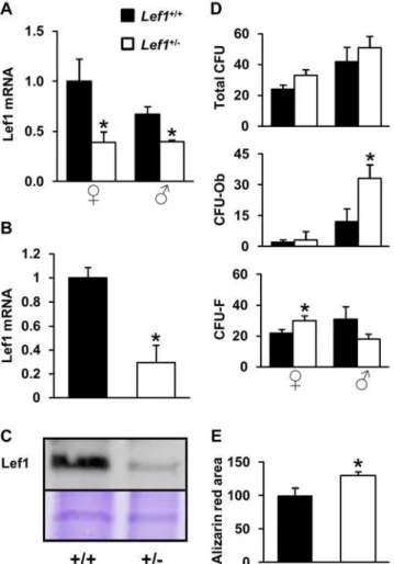
We next assessed the effects of Lef1 heterozygocity on Lef1 expression and on osteoblast differentiation in vitro. First, we confirmed that Lef1 expression was significantly reduced in bones and in newborn mouse calvarial osteoblast (NeMCO) cultures derived from Lef1 heterozygous compared to control mice (Figure 3A–3C). In mesenchymal stem cell (MSC) cultures derived from bone marrow of female mice, Lef1 haploinsufficiency increased the number of F, whereas the number of CFU-Ob was unchanged (Figure 3D). Interestingly, Lef1 haploinsuffi-ciency in male mice, which did not reduce bone mass in vivo
(Figure 1), was associated with an increase in bone marrow-derived CFU-Ob (Figure 3D).In vitroosteoblast differentiation as defined by mineralization in NeMCO cultures was accelerated by
Lef1haploinsufficiency (Figure 3E).
High bone mass (HBM) inGsk3bhaploinsufficient female mice
Alterations in components of the Wnt pathway other than Lef1 may also have stronger skeletal effects in females as compared to males. To address this notion, we compared the role of Gsk3b, a
negative regulator of the Wnt pathway, in femaleversusmale bone mass. Because theGsk3ß-null mice diein utero[48], we analyzed the trabecular bone in the distal femoral metaphysis of mice haploinsufficient for Gsk3ß. Indeed, female, but not male
Gsk3b+/2 mice exhibited a high bone mass (HBM) phenotype compared to WT littermates (Figure 4A). The elevated BV/TV
was attributable to increased trabecular number, and was associated with increased connectivity density (Figure 4), a mirror image of the respective Lef1+/2 LBM phenotype. Jointly, the gender-preferential effects of both Lef1 and Gsk3b haploinsuffi-ciency suggest that the skeleton is more sensitive to variations in Wnt signaling in females compared to males.
Figure 1. Low trabecular bone mass inLef1+/2female mice.(A,C)mCT analysis of the distal femoral (A) and the vertebral (C) trabecular bone of
Lef1+/+(black)
andLef1+/2(white)female (left) and male (right) 13-week old mice. BV/TV – trabecular bone volume density, Tb.N – trabecular number,
Tb.Th – trabecular thickness, Conn.D – connectivity density (Mean6SEM of 4–10 specimens as indicated within the bars at the top, * =p,0.05). (B,D)
mCT images of distal femoral (B) and vertebral (D) trabecular bone of female (left) and male (right) 13-week old mice with median BV/TV. Scale bar = 0.5 mm.
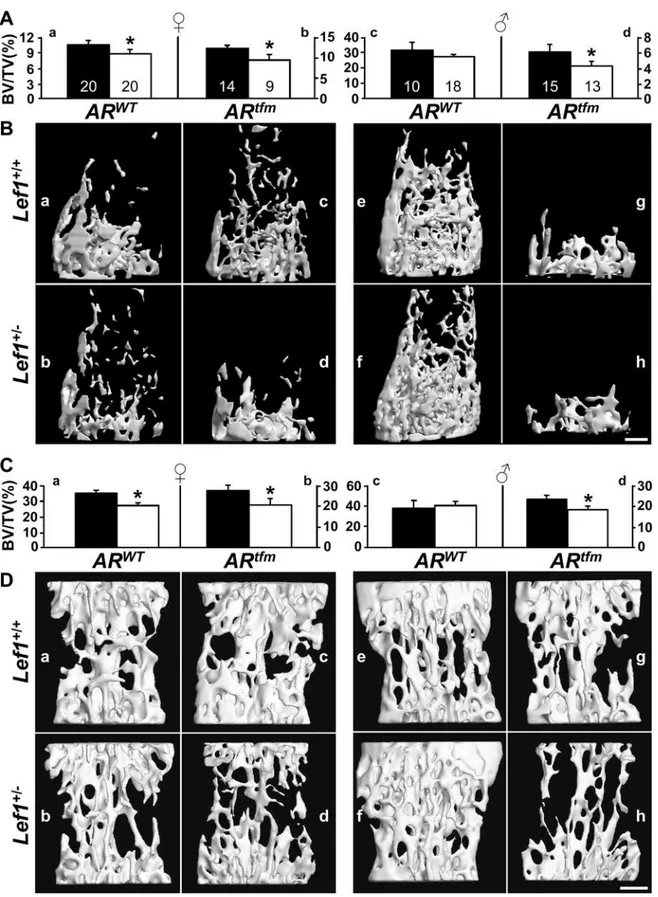
Androgen signaling protects againstLef1 haploinsufficiency
The female-preferential skeletal phenotype of theLef1+/2mice could be explained by a compensatory gene(s) on the Y chromosome, hypersensitization by estrogens, or protection by androgens. In support of the latter possibility, androgens can augment Wnt signaling [49,50], and even stimulate Lef1 expression in osteoblasts (Figure S2) similar to what has been observed in adipocytes [51]. To test the hypothesis that androgen signaling protects against Lef1 haploinsufficiency-induced LBM in vivo, we employed tfm male mice, in which androgen signaling is absent due to a naturally occurring mutation in the androgen receptor (AR) [52]. To generate
Lef1+/2;ARtfm
mice, Lef1+/2 males were bred with female tfm carriers. Because theARtfmallele is embedded in a tabby genomic sequence [53], we first analyzed the distal femora in offspring with wild type AR, but which are no longer on a pure C57BL/6 background. Similar to the original results (Figure 1), female
Lef1+/2 mice that partially carry the tabby genome exhibited a LBM phenotype (Figure 5Aa, and compare Figure 5Ba to 5Bb), while male mice on the same genetic background were protected (Figure 5Ac, and compare Figure 5Be to 5Bf). Remarkably, however, male tfm mice were vulnerable to Lef1 haploinsuffi-ciency, similar to females (Figure 5). Specifically, Lef1+/2;ARtfm mice had a 22% lower BV/TV as compared to their Lef1+/+
;
ARtfm counterparts (Figure 5Ad, and compare Figure 5Bg to 5Bh). Similar results were observed in the vertebral bodies (Figure 5C and 5D). Because estrogen levels intfmmales do not approach those of females [54], and because these mice still carry an intact Y chromosome, these results demonstrate that androgen signaling protects against Lef1 haploinsufficiency-induced LBM.
Aged females are resistant toLef1 haploinsufficiency-induced LBM
It has been previously suggested that androgen signaling can augment Wnt signaling in bone cells [49,50]. This could explain
how males are protected againstLef1 haploinsufficiency-induced LBM. Alternatively, androgens could also indirectly protect males by restraining bone turnover [54,55]. If low bone turnover protects male mice from Lef1 haploinsufficiency, then Lef1+/2 females may no longer display a low bone mass phenotype compared to WT females at ages older than 17 weeks, when bone turnover decreases [20,21]. Indeed,mCT analysis of 34-week old female mice revealed no difference between Lef1+/2 and WT females (Figure 6). At this age, the male skeleton was again unaffected byLef1haploinsufficiency (Figure 6). In summary,Lef1
haploinsufficiency induces LBM in a gender- and age-specific manner.
Figure 2. Low bone formation inLef1+/2female mice. Histomor-phometric analysis of the distal femoral metaphysis fromLef1+/+
(black) andLef1+/2(white) female (left) and male (right) 17-week old mice. (A)
mineral apposition rate (MAR), a surrogate for osteoblast activity; images show representative histological sections fromLef1+/+(left) and Lef1+/2(right) females; (B) mineralizing perimeter (Min.Peri), a surrogate
for osteoblast number; (C) bone formation rate (BFR); (D) trabecular bone osteoclast number (N.Oc/BS). Data represent mean6SEM of 4–9 specimens as indicated within the bars in A; * =p,0.05.
doi:10.1371/journal.pone.0005438.g002
Figure 3. Reduced Lef1 expression is not associated with impaired osteoblast differentiationin vitro. (A,B) Lef1 mRNA in tibia ofLef1+/2and control male and female mice (A) and in NeMCO
cultures (B) was assessed by RT-qPCR and corrected for the expression of GAPDH and rpL10A, respectively. Bars represent relative expression levels (Mean6SEM, n = 3). (C) Western blot analysis of Lef1 expression in NeMCO cultures. Equal loading was demonstrated by Coomassie blue staining (bottom panel shows a ,60 KDa Coomassie blue-stained
band). (D) Bone marrow mesenchymal cells from WT andLef1+/2mice
of each gender were cultured for 28 days, fixed and stained with Alizarin red. Colonies were manually counted in each of 6–11 independent cultures for each condition. Alizarin red-positive colonies were counted as osteoblastic colony-forming units (CFU-Ob), and the rest were considered fibroblastic CFU (CFU-F). (E) Day-20 NeMCO cultures from WT andLef1+/2mice were fixed and stained with Alizarin
red. Data represent mean mineralized area relative to WT6SEM in at least 3 cultures per condition. * =p,0.05vs.WT.
doi:10.1371/journal.pone.0005438.g003
Discussion
The present work demonstrates low bone mass in mice, in which oneLef1 gene copy is disrupted. Interestingly, bone mass was reduced in 13- and 17-week old females, but not in males of any age or in 34-week old females. This gender-specificity reflected neither protection by a Y chromosome-associated gene nor sensitization by estrogens, since Lef1+/2 male mice harboring a testicular feminization mutation (tfm) also displayed a LBM phenotype despite their having an intact Y chromosome and not having feminine estrogen levels [54]. Gsk3b+/2 mice displayed a mirror image of theLef1+/2 phenotype, namely increased bone mass in female mice only. In line with our findings, disruption of
Sfrp1, a Wnt antagonist, resulted in a female-preferential bone phenotype [29]. Thus, genetic alterations in components of the Wnt pathway appear to affect female more than male mice. That
Lrp5deficiency reduces bone mass equally in males and females [56] is not inconsistent with our conclusion in light of the recent work of Yadav etal.[57], who disputed the paradigm that places Lrp5 upstream of Gsk3b and Lef1 in osteoblasts. Plausibly, a genetic lesion at the level ofLef1itself compromises canonical Wnt signaling and is therefore more consequential in females than in males.
All mice that were sensitive toLef1 haploinsufficiency in our study, namely young females andtfmmales, are also characterized by a relatively high rate of bone turnover (Figure 2 and ref. [54]). In contrast, mice resistant toLef1haploinsufficiency—ARWTmales and aged females (Figure 1, Figure 5, Figure 6)—have a lower rate of bone turnover (Figure 2 and [20,21]). Thus, a unifying explanation for our observations is that bone turnover rate determines the skeletal response to genetic alterations in canonical Wnt signaling. Our favored interpretation of the gender-specific sensitivity toLef1haploinsufficiency is that androgens protect the skeleton from the potential deleterious effect of reduced Lef1 by restraining bone turnover [54,55].
We cannot rule out an alternative explanation whereby AR activity compensates for Lef1 haploinsufficiency via molecular interaction with the canonical Wnt pathway. In fact, it has been
shown that DHT stimulates Lef/Tcf-mediated transcription in osteoblasts [49,50]. This could occur via physical interaction of liganded AR withb-catenin [51] or with membrane residents such as Src [58], which could then impinge on the Wnt pathway through activation of the PI3 kinase/Akt/Gsk3baxis [50,59,60]. Alternatively, androgens could regulate the expression of either Lef1 itself (Figure S2 and [51]) or Wnt agonists and/or antagonists [61]. However, the normal bone phenotype observed in 34-week old Lef1+/2 female mice favors the hypothesis that androgen signaling, much like aging in females, overrides the skeletal sensitivity toLef1haploinsufficiency by restraining bone turnover. Obviously, the two explanations for AR-mediated protection against Lef1 haploinsufficiency – molecular interaction with the Wnt pathway and attenuation of bone turnover – are not mutually exclusive.
It remains to be examined to what extent the effect of Lef1 on bone formation is cell autonomous. In favor of cell autonomy is the reduced Lef1 expression inLef1+/2osteoblasts and the observed gender-dependent changes in the numbers of F and CFU-Ob in bone marrow-derived MSCs cultures (Figure 3D). Possibly,
Lef1 haploinsufficiency promotes premature osteoblast differenti-ation ([62,63] and Figure 3E). However, much like the effect of duodenal Lrp5 [57], the role of Lef1 in regulating bone formation may reside in cells other than osteoblasts. Cell type-specific knockout studies will be necessary to clarify this issue.
An intriguing, albeit speculative, extrapolation from our findings is that females reach lower peak bone mass than males because, in the absence of androgens, higher rate of bone turnover renders the young female skeleton more vulnerable to sub-optimal activity of canonical Wnt signaling and possibly other pathways. Other investigators reported on age- and gender-dependent bone phenotypes in mice with genetic alterations in different pathways. For example, osteoblast-specific disruption of BMP type-IA receptor leads to LBM in young mice but HBM in old mice [64]. Very similar age-dependent effects were reported forRunx2
haploinsufficiency in mice [20]. In addition, the strong anabolic effect of estrogen in youngRunx2+/2mice was almost completely abolished in aged mice [20]. With regard to gender specificity, and Figure 4. High trabecular bone mass inGsk3b+/2female mice.(A)mCT analysis of the distal femoral trabecular bone of 16 week oldGsk3b+/+
(black) andGsk3b+/2(white) female (left) and male (right) mice. Data represent mean
6SEM of 6–14 specimens as indicated within the bars at the top, * =p,0.05. (B)mCT images from female (left) and male (right) mice with median BV/TV. Scale bar = 0.5 mm.
Figure 5. Androgen signaling protects againstLef1haploinsufficiency-induced low bone mass.mCT analysis of the trabecular bone compartment in distal femora (A,B) and vertebral bodies (C,D) of 13-week old female (left) and male (right)Lef1+/+(black)
andLef1+/2(white)mice. ARtfmmales have no functional AR, whileARtfmfemales are carriers for the defectiveARallele. Data represent mean6SEM of 9–20 specimens as indicated within the bars at the top, * =p,0.05. (B) and (D) show respectivemCT images of female (left) and male (right) mice with median BV/TV. Mice in a, b, e and f carry the wild-type AR (ARWT); mice in c, d, g and h carry theARtfmallele; mice in a, c, e and g areLef1+/+; mice in b, d, f and h are Lef1+/2. Scale bar = 0.5 mm.
doi:10.1371/journal.pone.0005438.g005
in addition to the female-preferential response to genetic manipulation of Lef1, GSK3ß and Sfrp1, ablation of Cathepsin K
results in a 3-fold stronger effect in female compared to male mice [47]. Age- and gender-related variations in bone turnover may explain the differential skeletal responses to some of these and other genetic aberrations. Furthermore, hormonal and age-related variation in bone turnover may contribute to gender- and age-related susceptibility to osteoporosis and response to therapies.
Materials and Methods
Animals
Lef1+/2andGsk3b+/2mice and their controls, all on a C57BL/ 6 background, were generated by breedingLef1+/2[36] orGsk3b+/2 [26] mice with C57BL/6 mice from either Harlan Laboratories (Indianapolis, Indiana, USA) or the Ontario Cancer Institute (Toronto, Canada), respectively. Mice carrying the testicular feminization mutation (Tfm) (Jackson Laboratories, Bar Harbor, Maine, USA) on a C57BL/6J-A-Ta,6J. background were bred with theLef1+/2mice and F1 litters were examined. To measure the percentage of bone surface undergoing mineralization and the mineralization rate, mice were injected intraperitoneally with 15 mg/ kg of the fluorochrome calcein (Sigma-Aldrich, St. Louis, MO, USA) four days and again one day prior to sacrifice. One femur and the fifth lumbar vertebra (L5) from each mouse were dissected and fixed in 10% phosphate-buffered formalin (pH = 7.2) for 24 hours, and then stored in 70% ethanol. All experiments were approved by the Institutional Animal Care and Use Committee (IACUC) of the University of Southern California and of the University of Toronto.
Micro-computed tomography
Femora (one per mouse) and fifth lumbar vertebrae (L5) were examined as reported previously [65,66] using either ScancomCT 40 (Scanco Medical AG, Bru¨ttisellen, Switzerland), or Siemens MicroCAT II (Siemens Medical Solutions, Knoxville, TN, USA). Briefly, scans were performed at a 20-mm resolution in all three spatial dimensions. The mineralized tissues were differentially segmented by a global thresholding procedure [67]. Trabecular parameters in the secondary spongiosa of the distal femoral metaphysis included trabecular bone volume density (BV/TV), trabecular thickness (Tb.Th), trabecular number (Tb.N) and connectivity density (Conn.D). Cortical thickness, diaphyseal diameter, and medullary cavity diameter were determined in the mid-diaphyseal region. In L5 bodies, the entire trabecular bone compartment was analyzed. All morphometric parameters were determined by using a direct 3D approach [68]. Differences between groups were analyzed by student’st-test (two-tailed) and were considered significant whenp,0.05.
Histomorphometry
After mCT image acquisition, femora were embedded unde-calcified in polymethylmethacrylate (Technovit 9100, Heraeus Kulzer, Germany). Undeplasticized longitudinal 5-mm sections from the center of each bone were left unstained for dynamic histomorphometric measurements. To identify osteoclasts, con-secutive sections were deplasticized and stained with tartrate-resistant acid phosphatase (TRAP; Sigma-Aldrich, St. Louis, MO, USA) and counterstained with Mayer’s hematoxylin [69]. The morphometric analysis was performed using the Image-Pro Figure 6. Thirty four-week old female mice are insensitive toLef1haploinsufficiency.(A,B)mCT analysis of the distal femoral (A) and the vertebral (B) trabecular bone ofLef1+/+(black)
andLef1+/2(white)female (left) and male (right) 34-week old mice (Data represent mean
6SEM of 7–11 specimens as indicated within the bars at the top).
Discovery software (Media Cybernetics, Silver Spring, MD, USA). The following parameters were determined: mineral apposition rate (MAR), mineralizing perimeter (Min.Peri), bone formation rate (BFR) and osteoclast number (N.Oc/BS). The terminology and units used for these measurements were according to the convention of standardized nomenclature [70]. Statistical analysis was performed as above.
Tissue culture
NeMCO cultures were prepared from one day-old pups as described previously [71]. Cells were cultured in 6-well plates for Western blot analysis and in 12-well plates for RT-qPCR and mineralization assays. For the latter, osteogenic medium contain-ing ascorbic acid (50mg/mL) and b-glycerophosphate (10 mM) was initiated at confluence and alizarin red staining was performed at day 20. For MSC cultures, the cellular content of the bone marrow cavity from two femurs and two tibiae from each mouse was flushed using aMEM and passed through needles with decreasing diameters (down to 25G) to obtain a single cell suspension. Cells were then plated at 36106 per well in 6-well
plates and incubated for 3 days in aMEM (Invitrogen) supplemented with 15% FBS (Gemini Bio-Products, West Sacramento, CA). Starting at day 3, the MSC were cultured in osteogenic medium and stained with Alizarin red after 28 days.
Lef1 expression
Total RNA was extracted from freshly isolated tibiae of 10 week-old mice. Upon harvesting, one tibia per animal was stabilized in RNALater (Ambion, Austin, TX), homogenized in Trizol (Invitrogen), purified using 1-Bromo-3-Chloropropane and isopropanol, then rinsed in 70% ethanol. RNA from cells was extracted using Aurum Total RNA Mini Kit (Biorad, Hercules, CA). cDNA was produced using Superscript III First Strand cDNA synthesis kit (Invitrogen) and Real-Time PCR was performed using iQ SYBR green supermix (Biorad) and an Opticon 2 real time PCR machine (Biorad). Lef1 mRNA levels in tibiae and NeMCO cultures were corrected for GAPDH and ribosomal protein L10A (rpL10A) mRNA, respectively. Primers used for PCR are listed in Table 1. Western blot analysis of Lef1 in NeMCO cultures was performed essentially as previously described [71] using anti-Lef1 antibody from Cell Signaling (Danvers, MA) and secondary antibodies from Santa Cruz Biotechnology (Santa Cruz, CA).
Supporting Information
Figure S1 Lef12/2 mice have normal bone development. Histological evaluation of wild type (a–e) versus Lef12/2 (f–j) newborn mice. (a,b,f,g) Alizarin red/Alcian blue staining of craniofacial bones. (c,h) Alizarin red/Alcian blue staining of hind limb and vertebrae. (d,i) H-E staining of longitudinal femoral sections. (e,j) Toluidine blue staining of distal femoral growth
plates. Representative images are shown. No abnormality was detected in the Lef12/2 skeletons, except for the previously reported lack of teeth (f) (van Genderen et al. 1994, Genes Dev 8, 2691-703)
Found at: doi:10.1371/journal.pone.0005438.s001 (3.47 MB TIF)
Figure S2 DHT stimulates Lef1 expressionin vitro. MC3T3-E1 osteoblast cultures maintained in phenol-red freeaMEM supple-mented with 10% charcoal-stripped serum were treated with 30 nM DHT or 100 nM estradiol for 48 hours. Expression of the four members of the Lef/Tcf gene family was assessed by RT-qPCR and corrected for the expression of rpL10A. Bars represent expression levels in the presence of hormone relative to the ethanol vehicle, defined for each gene as 1.
Found at: doi:10.1371/journal.pone.0005438.s002 (0.88 MB TIF)
Acknowledgments
We thank Rudolf Grosschedl (University of Munich and Max-Planck-Institute, Germany) for theLef1-targeted mice.
Author Contributions
Conceived and designed the experiments: TN YG ES IB BF. Performed the experiments: TN YG JPC YS AT BC AD CL LK. Analyzed the data: TN YG JPC JT TK ES RM ES IB BF. Contributed reagents/materials/ analysis tools: TS LK JRW RM YC. Wrote the paper: TN YG ES IB BF. Co-directed the project: BF IB ES.
References
1. Miller JR, Moon RT (1996) Signal transduction through beta-catenin and specification of cell fate during embryogenesis. Genes Dev 10: 2527–2539. 2. Cadigan KM, Nusse R (1997) Wnt signaling: a common theme in animal
development. Genes Dev 11: 3286–3305.
3. Bhanot P, Brink M, Samos CH, Hsieh JC, Wang Y, et al. (1996) A new member of the frizzled family from Drosophila functions as a Wingless receptor. Nature 382: 225–230.
4. He X, Saint-Jeannet JP, Wang Y, Nathans J, Dawid I, et al. (1997) A member of the Frizzled protein family mediating axis induction by Wnt-5A. Science 275: 1652–1654. 5. Yang-Snyder J, Miller JR, Brown JD, Lai CJ, Moon RT (1996) A frizzled homolog functions in a vertebrate Wnt signaling pathway. Curr Biol 6: 1302–1306.
6. Pinson KI, Brennan J, Monkley S, Avery BJ, Skarnes WC (2000) An LDL-receptor-related protein mediates Wnt signalling in mice. Nature 407: 535–538.
7. Tamai K, Semenov M, Kato Y, Spokony R, Liu C, et al. (2000) LDL-receptor-related proteins in Wnt signal transduction. Nature 407: 530–535.
8. Wehrli M, Dougan ST, Caldwell K, O’Keefe L, Schwartz S, et al. (2000) arrow encodes an LDL-receptor-related protein essential for Wingless signalling. Nature 407: 527–530.
9. Hart MJ, de los Santos R, Albert IN, Rubinfeld B, Polakis P (1998) Downregulation of beta-catenin by human Axin and its association with the APC tumor suppressor, beta-catenin and GSK3 beta. Curr Biol 8: 573– 581.
Table 1.Primers for genotyping and RT–qPCR.
Genotyping
Lef1 LPP2.2 59TGTCTCTCTTTCCGTGCTAGTTC39
D8 59CCGTTTCAGTGGCACGCCCTCTCC39
Neo 59ATGGCGATGCCTGCTTGCCGAATA39
Sry Fwd 59TCATGAGACTGCCAACCACAG39
Rev 59CATGACCACCACCACCACCAA39
Tfm* Fwd 59GTGAAGCAGGTAGCTCTGGG39
Rev 59GTTCTCCAGCTTGATACGGG39
RT-qPCR
GAPDH Fwd 59CCAGAACATCATCCCTGCAT39
Rev 59CTTGCCCACAGCCTTGGCAGC39
rpL10A Fwd 59CGCCGCAAGTTTCTGGAGAC39
Rev 59CTTGCCAGCCTTGTTTAGGC39
Lef1 Fwd 59TGAGTGCACGCTAAAGGAGA39
Rev 59ATAATTGTCTCGCGCTGACC39
*PCR product was digested with
MwoI resulting in either a 137 bp (WT) or 182 bp (Tfm) band.
doi:10.1371/journal.pone.0005438.t001
10. Itoh K, Krupnik VE, Sokol SY (1998) Axis determination in Xenopus involves biochemical interactions of axin, glycogen synthase kinase 3 and beta-catenin. Curr Biol 8: 591–594.
11. Ikeda S, Kishida S, Yamamoto H, Murai H, Koyama S, et al. (1998) Axin, a negative regulator of the Wnt signaling pathway, forms a complex with GSK-3beta and beta-catenin and promotes GSK-GSK-3beta-dependent phosphorylation of beta-catenin. Embo J 17: 1371–1384.
12. Travis A, Amsterdam A, Belanger C, Grosschedl R (1991) LEF-1, a gene encoding a lymphoid-specific protein with an HMG domain, regulates T-cell receptor alpha enhancer function [corrected]. Genes Dev 5: 880–894. 13. Waterman ML, Fischer WH, Jones KA (1991) A thymus-specific member of the
HMG protein family regulates the human T cell receptor C alpha enhancer. Genes Dev 5: 656–669.
14. van de Wetering M, Oosterwegel M, Dooijes D, Clevers H (1991) Identification and cloning of TCF-1, a T lymphocyte-specific transcription factor containing a sequence-specific HMG box. Embo J 10: 123–132.
15. Korinek V, Barker N, Willert K, Molenaar M, Roose J, et al. (1998) Two members of the Tcf family implicated in Wnt/beta-catenin signaling during embryogenesis in the mouse. Mol Cell Biol 18: 1248–1256.
16. Bab I, Mu¨ller R, Hajbi-Yonissi C, Gabet Y (2007) Micro-Tomographic Atlas of the Mouse Skeleton. New York: Springer.
17. Neu CM, Manz F, Rauch F, Merkel A, Schoenau E (2001) Bone densities and bone size at the distal radius in healthy children and adolescents: a study using peripheral quantitative computed tomography. Bone 28: 227–232.
18. Macdonald H, Kontulainen S, Petit M, Janssen P, McKay H (2006) Bone strength and its determinants in pre- and early pubertal boys and girls. Bone 39: 598–608.
19. Glatt V, Canalis E, Stadmeyer L, Bouxsein ML (2007) Age-related changes in trabecular architecture differ in female and male C57BL/6J mice. J Bone Miner Res 22: 1197–1207.
20. Juttner KV, Perry MJ (2007) High-dose estrogen-induced osteogenesis is decreased in aged RUNX2(+/2) mice. Bone 41: 25–32.
21. Pantschenko AG, Zhang W, Nahounou M, McCarthy MB, Stover ML, et al. (2005) Effect of osteoblast-targeted expression of bcl-2 in bone: differential response in male and female mice. J Bone Miner Res 20: 1414–1429. 22. Glass DA 2nd, Karsenty G (2006) Molecular bases of the regulation of bone
remodeling by the canonical Wnt signaling pathway. Curr Top Dev Biol 73: 43–84.
23. Glass DA 2nd, Bialek P, Ahn JD, Starbuck M, Patel MS, et al. (2005) Canonical Wnt signaling in differentiated osteoblasts controls osteoclast differentiation. Dev Cell 8: 751–764.
24. Holmen SL, Giambernardi TA, Zylstra CR, Buckner-Berghuis BD, Resau JH, et al. (2004) Decreased BMD and limb deformities in mice carrying mutations in both Lrp5 and Lrp6. J Bone Miner Res 19: 2033–2040.
25. Bennett CN, Longo KA, Wright WS, Suva LJ, Lane TF, et al. (2005) Regulation of osteoblastogenesis and bone mass by Wnt10b. Proc Natl Acad Sci U S A 102: 3324–3329.
26. Kugimiya F, Kawaguchi H, Ohba S, Kawamura N, Hirata M, et al. (2007) GSK-3beta controls osteogenesis through regulating Runx2 activity. PLoS ONE 2: e837. doi:10.1371/journal.pone.0000837.
27. Morvan F, Boulukos K, Clement-Lacroix P, Roman Roman S, Suc-Royer I, et al. (2006) Deletion of a single allele of the Dkk1 gene leads to an increase in bone formation and bone mass. J Bone Miner Res 21: 934–945.
28. Li X, Liu P, Liu W, Maye P, Zhang J, et al. (2005) Dkk2 has a role in terminal osteoblast differentiation and mineralized matrix formation. Nat Genet 37: 945–952.
29. Bodine PV, Zhao W, Kharode YP, Bex FJ, Lambert AJ, et al. (2004) The Wnt antagonist secreted frizzled-related protein-1 is a negative regulator of trabecular bone formation in adult mice. Mol Endocrinol 18: 1222–1237.
30. Kamiya N, Ye L, Kobayashi T, Mochida Y, Yamauchi M, et al. (2008) BMP signaling negatively regulates bone mass through sclerostin by inhibiting the canonical Wnt pathway. Development 135: 3801–3811.
31. Smith E, Coetzee GA, Frenkel B (2002) Glucocorticoids inhibit cell cycle progression in differentiating osteoblasts via glycogen synthase kinase-3beta. J Biol Chem 277: 18191–18197.
32. Ohnaka K, Taniguchi H, Kawate H, Nawata H, Takayanagi R (2004) Glucocorticoid enhances the expression of dickkopf-1 in human osteoblasts: novel mechanism of glucocorticoid-induced osteoporosis. Biochem Biophys Res Commun 318: 259–264.
33. Leclerc N, Noh T, Cogan J, Samarawickrama DB, Smith E, et al. (2007) Opposing effects of glucocorticoids and Wnt signaling on Krox20 and mineral deposition in osteoblast cultures. J Cell Biochem.
34. Wang FS, Lin CL, Chen YJ, Wang CJ, Yang KD, et al. (2005) Secreted frizzled-related protein 1 modulates glucocorticoid attenuation of osteogenic activities and bone mass. Endocrinology 146: 2415–2423.
35. Armstrong VJ, Muzylak M, Sunters A, Zaman G, Saxon LK, et al. (2007) Wnt/ beta-catenin signaling is a component of osteoblastic bone cell early responses to load-bearing and requires estrogen receptor alpha. J Biol Chem 282: 20715–20727.
36. van Genderen C, Okamura RM, Farinas I, Quo RG, Parslow TG, et al. (1994) Development of several organs that require inductive epithelial-mesenchymal interactions is impaired in LEF-1-deficient mice. Genes Dev 8: 2691–2703.
37. Galceran J, Farinas I, Depew MJ, Clevers H, Grosschedl R (1999) Wnt3a2/2
like phenotype and limb deficiency in Lef1(2/2)Tcf1(2/2) mice. Genes Dev 13: 709–717.
38. Okamura RM, Sigvardsson M, Galceran J, Verbeek S, Clevers H, et al. (1998) Redundant regulation of T cell differentiation and TCRalpha gene expression by the transcription factors LEF-1 and TCF-1. Immunity 8: 11–20. 39. Staal FJ, Meeldijk J, Moerer P, Jay P, van de Weerdt BC, et al. (2001) Wnt
signaling is required for thymocyte development and activates Tcf-1 mediated transcription. Eur J Immunol 31: 285–293.
40. Galceran J, Miyashita-Lin EM, Devaney E, Rubenstein JL, Grosschedl R (2000) Hippocampus development and generation of dentate gyrus granule cells is regulated by LEF1. Development 127: 469–482.
41. Babij P, Zhao W, Small C, Kharode Y, Yaworsky PJ, et al. (2003) High bone mass in mice expressing a mutant LRP5 gene. J Bone Miner Res 18: 960–974. 42. Akhter MP, Wells DJ, Short SJ, Cullen DM, Johnson ML, et al. (2004) Bone
biomechanical properties in LRP5 mutant mice. Bone 35: 162–169. 43. Sawakami K, Robling AG, Ai M, Pitner ND, Liu D, et al. (2006) The Wnt
co-receptor LRP5 is essential for skeletal mechanotransduction but not for the anabolic bone response to parathyroid hormone treatment. J Biol Chem 281: 23698–23711.
44. Wu Y, Torchia J, Yao W, Lane NE, Lanier LL, et al. (2007) Bone microenvironment specific roles of ITAM adapter signaling during bone remodeling induced by acute estrogen-deficiency. PLoS ONE 2: e586. doi:10.1371/journal.pone.0000586.
45. Hill TP, Spater D, Taketo MM, Birchmeier W, Hartmann C (2005) Canonical Wnt/beta-catenin signaling prevents osteoblasts from differentiating into chondrocytes. Dev Cell 8: 727–738.
46. DeMambro VE, Clemmons DR, Horton LG, Bouxsein ML, Wood TL, et al. (2008) Gender-specific changes in bone turnover and skeletal architecture in igfbp-2-null mice. Endocrinology 149: 2051–2061.
47. Pennypacker B, Shea M, Liu Q, Masarachia P, Saftig P, et al. (2008) Bone density, strength, and formation in adult cathepsin K (2/2) mice. Bone. 48. Hoeflich KP, Luo J, Rubie EA, Tsao MS, Jin O, et al. (2000) Requirement for
glycogen synthase kinase-3beta in cell survival and NF-kappaB activation. Nature 406: 86–90.
49. Liu XH, Kirschenbaum A, Yao S, Liu G, Aaronson SA, et al. (2007) Androgen-induced Wnt signaling in preosteoblasts promotes the growth of MDA-PCa-2b human prostate cancer cells. Cancer Res 67: 5747–5753.
50. Liu XH, Kirschenbaum A, Yao S, Levine AC (2007) Androgens Promote Preosteoblast Differentiation via Activation of the Canonical Wnt Signaling Pathway. Ann N Y Acad Sci 1116: 423–431.
51. Singh R, Artaza JN, Taylor WE, Braga M, Yuan X, et al. (2006) Testosterone inhibits adipogenic differentiation in 3T3-L1 cells: nuclear translocation of androgen receptor complex with beta-catenin and T-cell factor 4 may bypass canonical Wnt signaling to down-regulate adipogenic transcription factors. Endocrinology 147: 141–154.
52. Lyon MF, Hawkes SG (1970) X-linked gene for testicular feminization in the mouse. Nature 227: 1217–1219.
53. Lyon MF (1961) Gene action in the X-chromosome of the mouse (Mus musculus L.). Nature 190: 372–373.
54. Vandenput L, Swinnen JV, Boonen S, Van Herck E, Erben RG, et al. (2004) Role of the androgen receptor in skeletal homeostasis: the androgen-resistant testicular feminized male mouse model. J Bone Miner Res 19: 1462–1470. 55. Matsumoto C, Inada M, Toda K, Miyaura C (2006) Estrogen and androgen
play distinct roles in bone turnover in male mice before and after reaching sexual maturity. Bone 38: 220–226.
56. Dubrow SA, Hruby PM, Akhter MP (2007) Gender specific LRP5 influences on trabecular bone structure and strength. J Musculoskelet Neuronal Interact 7: 166–173.
57. Yadav VK, Ryu JH, Suda N, Tanaka KF, Gingrich JA, et al. (2008) Lrp5 controls bone formation by inhibiting serotonin synthesis in the duodenum. Cell 135: 825–837.
58. Migliaccio A, Varricchio L, De Falco A, Castoria G, Arra C, et al. (2007) Inhibition of the SH3 domain-mediated binding of Src to the androgen receptor and its effect on tumor growth. Oncogene 26: 6619–6629.
59. Kang HY, Cho CL, Huang KL, Wang JC, Hu YC, et al. (2004) Nongenomic androgen activation of phosphatidylinositol 3-kinase/Akt signaling pathway in MC3T3-E1 osteoblasts. J Bone Miner Res 19: 1181–1190.
60. Smith E, Frenkel B (2005) Glucocorticoids inhibit the transcriptional activity of LEF/TCF in differentiating osteoblasts in a glycogen synthase kinase-3beta-dependent and -inkinase-3beta-dependent manner. J Biol Chem 280: 2388–2394. 61. Nantermet PV, Xu J, Yu Y, Hodor P, Holder D, et al. (2004) Identification of
genetic pathways activated by the androgen receptor during the induction of proliferation in the ventral prostate gland. J Biol Chem 279: 1310–1322. 62. Kahler RA, Galindo M, Lian J, Stein GS, van Wijnen AJ, et al. (2006)
Lymphocyte enhancer-binding factor 1 (Lef1) inhibits terminal differentiation of osteoblasts. J Cell Biochem 97: 969–983.
63. Kahler RA, Westendorf JJ (2003) Lymphoid enhancer factor-1 and beta-catenin inhibit Runx2-dependent transcriptional activation of the osteocalcin promoter. J Biol Chem 278: 11937–11944.
65. Bajayo A, Goshen I, Feldman S, Csernus V, Iverfeldt K, et al. (2005) Central IL-1 receptor signaling regulates bone growth and mass. Proc Natl Acad Sci U S A 102: 12956–12961.
66. Yirmiya R, Goshen I, Bajayo A, Kreisel T, Feldman S, et al. (2006) Depression induces bone loss through stimulation of the sympathetic nervous system. Proc Natl Acad Sci U S A 103: 16876–16881.
67. Ruegsegger P, Koller B, Muller R (1996) A microtomographic system for the nondestructive evaluation of bone architecture. Calcif Tissue Int 58: 24–29. 68. Hildebrand T, Laib A, Muller R, Dequeker J, Ruegsegger P (1999) Direct
three-dimensional morphometric analysis of human cancellous bone: microstructural
data from spine, femur, iliac crest, and calcaneus. J Bone Miner Res 14: 1167–1174.
69. Erlebacher A, Derynck R (1996) Increased expression of TGF-beta 2 in osteoblasts results in an osteoporosis-like phenotype. J Cell Biol 132: 195–210. 70. Parfitt AM, Drezner MK, Glorieux FH, Kanis JA, Malluche H, et al. (1987)
Bone histomorphometry: standardization of nomenclature, symbols, and units. Report of the ASBMR Histomorphometry Nomenclature Committee. J Bone Miner Res 2: 595–610.
71. Leclerc N, Noh T, Cogan J, Samarawickrama DB, Smith E, et al. (2008) Opposing effects of glucocorticoids and Wnt signaling on Krox20 and mineral deposition in osteoblast cultures. J Cell Biochem 103: 1938–1951.